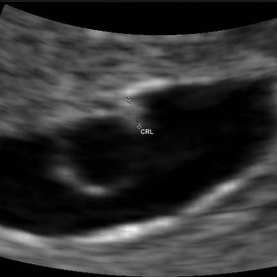
Baby ultrasound 18 weeks pregnant
Images and what to expect
Sometimes called an anatomy scan, an 18-week ultrasound can help with evaluating fetal development and detecting complications.
The 18th week of pregnancy is usually the earliest that a healthcare provider can do the anatomy scan.
During the 18-week ultrasound, a doctor or ultrasound technician will use an ultrasound machine to look at many different parts of the developing fetus, including the brain, heart, stomach, kidneys, skull, and genitals. It is usually possible to determine the sex of the fetus at this scan.
Ultrasounds work by sending sound waves into the body. Parts of the body, including the developing fetus, send these sound waves back. Based on how long it takes the waves to return to the ultrasound machine, the machine can determine how far away various body parts are and render a picture displaying this information.
In most cases, people do not need to do anything special to prepare for an anatomy scan.
During an 18-week ultrasound, an ultrasound technician or a doctor uses a transducer, which looks like a remote control. They apply a gel to the lower portion of the stomach and rub the transducer over the area to create clear images.
Sometimes, the provider also inserts the transducer into the vagina to get a clearer picture. Learn more about transvaginal ultrasounds here.
An ultrasound at 18 weeks usually takes longer than earlier ultrasounds, which doctors use to date the pregnancy. The provider may ask the woman to move, drink water, or use the bathroom to encourage the fetus to change positions.
Purpose
The doctor or technician will check a range of factors during this ultrasound, including:
- genital development and the sex of the fetus
- development of the skull and brain
- development of the heart, including whether the heart has four chambers
- development of organs, such as the kidneys, lungs, and intestines
- signs of cleft palate and other genetic anomalies
- development of the placenta, including whether it is in the right place
- amniotic fluid levels
The 18-week ultrasound provides many important pieces of information, including:
- Whether the fetus is developing normally: The healthcare provider can identify whether the baby is likely to need medical care right after birth.

- The overall health of the pregnancy: Problems with the placenta or amniotic fluid may mean that the pregnant woman needs to switch to a provider specializing in high risk pregnancies. In some cases — if the pregnant woman has placenta accreta, for example — the delivery may need to take place in an operating room or a specialized hospital.
- Delivery decisions: The anatomy scan may guide delivery decisions. For example, if a woman is considering a home birth, the anatomy scan may reveal whether this presents any particular risks.
Limitations
The 18-week ultrasound is not a perfect diagnostic tool. It cannot detect all congenital disabilities. Moreover, the images that it produces are not photos and may not provide fully accurate information. Sometimes, it is not possible to see all parts of the fetus. Small shadows and positioning issues can make diagnosing the health of the pregnancy challenging.
As the fetus is still relatively small at 18 weeks, people who choose to have an anatomy scan at this early point may not be able to get a clear image.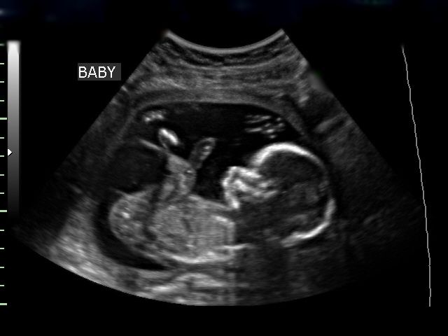 Their provider may recommend that they have a vaginal ultrasound or return later in the pregnancy if any images are unclear, or there are signs that there might be a problem with the fetus.
Traditional ultrasounds, sometimes called 2D ultrasounds, produce blurry and grainy images that may be hard for an untrained person to understand. While most people can make out larger shapes, such as the skull and torso, it is more difficult to see fine details on these ultrasounds.
People may see the fetus moving or kicking in the ultrasound. The sucking reflex develops around this age, so they may see the fetus sucking their thumb.
Many providers now routinely offer 3D ultrasounds. These scans use the same basic process as 2D ultrasounds, but they render a photo based on sounds from many different angles to create a composite image of the fetus. This technique makes it easier to see the fetus’s features, including smaller parts of the body, such as fingers, toes, and even genitals.
A 4D ultrasound offers even more precision. These scans take many images per second to create a very detailed depiction of the fetus. People can see the fetus move and sometimes even smile or suck their thumb.
As 4D ultrasounds offer so much more detail, doctors sometimes use them to assess whether a fetus’s behavior is normal. This assessment can provide useful information about development, especially when a doctor cannot get a clear image, or there are signs of an abnormality on a 2D or 3D ultrasound.
For many people, the 18-week ultrasound is the first chance to see the fetus up close and to learn the sex. For others, it can be a scary procedure, especially if they have worries about complications.
In most cases, the ultrasound offers reassurance that the fetus and pregnancy are healthy. Even when there is a problem with the ultrasound, subsequent ultrasounds may offer better images that ease any concerns. People should discuss the benefits, risks, and limitations of ultrasounds with a provider they trust.
20 Week Ultrasound (Anatomy Scan): What to Expect
Overview
What is the 20-week ultrasound?
A 20-week ultrasound, sometimes called an anatomy scan or anomaly scan, is a prenatal ultrasound performed between 18 and 22 weeks of pregnancy. It checks on the physical development of the fetus and can detect certain congenital disorders as well as major anatomical abnormalities. Your healthcare provider will use a 2D, 3D or even 4D ultrasound to take images of the fetus inside your uterus. The ultrasound technician, or sonographer, will take measurements and make sure the fetus is growing appropriately for its age. You may also learn the sex of the fetus at this appointment.
What is the 20-week anatomy scan looking for?
A 20-week ultrasound takes measurements of your fetal organs and body parts to make sure the fetus is growing appropriately. The scan also looks for signs of specific congenital disabilities or structural issues with certain organs.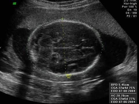
Some specific parts your provider will examine are the fetal:
- Heart.
- Brain, neck and spine.
- Kidneys and bladder.
- Arms and legs.
- Hands, fingers, feet and toes.
- Lips, chin, nose, eyes and face.
- Chest and lungs.
- Stomach and intestines.
The ultrasound technician will also:
- Listen to the fetal heart rate for abnormal rhythms.
- Check the umbilical cord for blood flow and where it attaches to the placenta.
- Look at the placenta to make sure it’s not covering your cervix (placenta previa).
- Check your uterus, ovaries and cervix.
- Measure the amount of amniotic fluid.
Several images are taken during this ultrasound. You will see the sonographer draw lines on the screen. This line acts as a ruler, documenting the sizes of organs and limbs. They compare these measurements against your due date. In some cases, you might hear you are measuring ahead, on track or behind your due date. If fetal measurements are within 10 to 14 days of the predicted due date, then the fetus is considered to be developing adequately. Your due date will not change unless the fetus measures outside of that time frame.
What does an ultrasound look like at 20 weeks?
It may be hard for you to identify what you’re looking at on an ultrasound. You will most likely spot the fetus's head, nose, arms and legs. Unlike your first trimester ultrasound when the fetus looked like a tiny cluster of cells, the fetus looks more like a real baby at 20 weeks. Ask your ultrasound technician if you are unsure what you are looking at. Keep in mind, your technician can’t interpret the results of the scan for you. However, if you ask what body part you’re looking at, they can usually answer.
Test Details
How do I prepare for my 20-week ultrasound?
There isn’t much to do to prepare for your appointment. This will be one of your longer appointments — around 45 minutes for just the ultrasound. If you are seeing your healthcare provider afterward, your appointment could last up to 75 minutes. Plan ahead by making any necessary arrangements for work or childcare. Some healthcare providers recommend eating or having a full bladder to make it easier to see the images and make the fetus more likely to move.
What can I expect at my 20-week ultrasound?
First, you will lie down on the exam table. Next, an ultrasonic gel is placed on your belly. Then, an ultrasound technician will move an ultrasound wand over different spots in your abdomen. They will take images and measurements of specific organs and parts by freezing the screen. You may also see them draw lines to measure the length of the fetus's limbs or the circumference of its head. This helps them evaluate the fetus's size for its gestational age. You will get to watch the fetus on a screen, one of the most exciting parts of pregnancy.
If the fetus is positioned in a way that makes it hard to take measurements, the technician might ask you to move around a little or take a drink of something sweet to make the fetus move.
Generally, your ultrasound technician will be quiet as they work. Don’t be alarmed if the conversation is minimal or if they aren't sharing results as they go. Your obstetrician will go over the results with you either immediately after the ultrasound or at a follow-up appointment in the next few days.
After all the images are taken and your ultrasound technician is happy with all the angles and measurements, you will be able to wipe the gel off your belly.
Make sure there is room on your refrigerator: Most ultrasound technicians will send you home with a few pictures as a keepsake.
Will a 20-week ultrasound tell me the sex of the fetus?
Yes, your fetus's external genitalia has developed enough to identify its sex. Depending on the position the fetus is in, your ultrasound technician may be able to see a penis or a labia. There is a slight chance that the fetus won't cooperate, and your ultrasound technician can’t get a clear picture of its genitals. If you don’t want to know the sex, speak up ahead of time so they don’t accidentally spoil the surprise.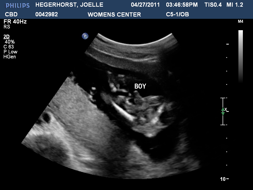 Most of the time, a prediction is made only when the technician is certain of sex.
Is a 20-week anatomy scan harmful?
No, having a 20-week ultrasound is safe. Studies have shown that ultrasound is not dangerous to you or the fetus. A 20-week prenatal ultrasound is considered medically necessary to detect potentially life-altering anomalies.
Results and Follow-Up
When will I know the results of my 20-week ultrasound?
Anatomy scans are usually a positive experience. In most cases, your healthcare provider will not find any major anomalies. Every healthcare facility has different processes, but you will likely meet with your obstetrician right after your 20-week scan. The ultrasound technician shares the images and any findings with them. Then, your obstetrician will do their own analysis before meeting with you and sharing results.
If they find something that doesn’t look right, they may recommend additional prenatal testing or treatment for a condition found during the scan.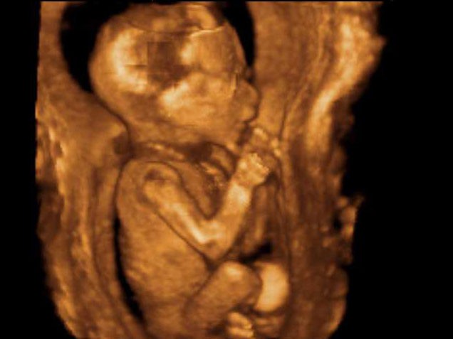 In some cases, they recommend a wait-and-see approach. This means during delivery, they will watch for signs of the suspected condition and determine treatment at that time.
What conditions can a 20-week ultrasound detect?
A 20-week ultrasound doesn’t find all congenital conditions. However, the scan can help detect several serious conditions:
- Anencephaly.
- Indicators for Down syndrome or trisomy 18 and trisomy 13.
- Cleft lip.
- Spina bifida.
- Congenital heart abnormalities.
- Renal agenesis (missing one or both kidneys).
- Gastroschisis (issue with the intestines).
- Omphalocele (type of abdominal wall issue).
- Skeletal dysplasia (problems with the bones).
- Diaphragmatic hernia (a hole in the diaphragm).
It’s important to note that the scan results are not a formal diagnosis of any condition. Instead, it’s usually an indicator that further tests are needed. If your healthcare provider is concerned with any scans, they will speak to you about your next steps.
Are there any conditions a 20-week ultrasound can’t identify?
While the 20-week scan can detect certain conditions, it can’t identify everything. The scan is meant to provide indicators, or markers, of serious health problems. There are some conditions you will not know about until your baby is born. For example, many heart abnormalities are not found until birth and you might not know if your baby has scoliosis.
Will I need further tests or ultrasounds?
If your healthcare provider is concerned about anything in your 20-week scan, they may recommend further tests. Some of these tests could include amniocentesis, chorionic villus sampling or additional ultrasounds with a perinatologist (physicians who specialize in high-risk pregnancies).
Speak with your healthcare provider early in your pregnancy to make sure you understand the types of noninvasive prenatal testing available to you. Several tests that indicate an increased risk for congenital anomalies can be done before 20 weeks of pregnancy.
Additional Details
What does a girl look like at a 20-week ultrasound?
One of the best ways to identify a girl on ultrasound is to look for three lines. However, it’s always best to leave the identification of sex up to experienced professionals.
What does a boy look like at a 20-week ultrasound?
Sometimes it’s the absence of the three lines that tell the ultrasound technician you have a boy in your belly. Other times you can see a protrusion coming from a round sack. This is the fetus's penis and testicles. It’s always best to let your healthcare providers determine fetal sex.
A note from Cleveland Clinic
It’s normal to be both excited and nervous about your 20-week ultrasound appointment. You will get to see the fetus and find out how it's developing. Talk to your healthcare provider about any concerns you have so they can offer reassurance and ease your worries. In most cases, the 20-week anatomy scan is a positive experience for soon-to-be parents.
fetal ultrasound at 18 weeks
- Fetal development by week
- Our apparatus
- other types...
- Pregnancy management
- Fetometry data at various times
The cost of ultrasound in the second trimester in the period from 14 to 26 weeks is 550 hryvnia. The price includes prenatal screening, biometric protocols, 3D/4D visualization. The cost of comprehensive prenatal screening according to PRISCA (ultrasound + free estriol + alpha-fetoprotein + beta-hCG with the calculation of the individual risk of chromosomal pathologies (for example, Down syndrome or Edwards syndrome) and developmental defects (for example, neural tube defect) - 1060 hryvnia.
At fetal ultrasound at 18 weeks of gestation the time for prenatal screening of the second trimester continues, more about which in fetal ultrasound at 17 weeks of gestation.
Changes occurring in the child
The bones of the fetus continue to grow rapidly and become stronger. With ultrasound of the fetus at 18 weeks of gestation, the weight of the fetus is determined, approximately 230 g. The calculation of the weight of the fetus is carried out by size with fetometry.
- BDP (biparietal size). With ultrasound of the fetus at 18 weeks of gestation, the biparietal size is 37-47 mm.
- LZ (frontal-occipital size). At 18 weeks of pregnancy 49-59 mm.
- OG (fetal head circumference). At 18 weeks of pregnancy, the head circumference corresponds to 131-161 mm.
- OB (abdominal circumference of the fetus) - at 18 weeks of pregnancy is 102 mm 104 -144 mm.
Normal measurements of long bones on fetal ultrasound at 18 weeks gestation:
- Femur 23-31 mm,
- Humerus 15-21mm,
- Forearm bones 17-23 mm,
- Lower leg bones 23-31 mm.
Meconium continues to form - the original feces in the intestines of the fetus. It consists of undigested residues of amniotic fluid, which the fetus actively swallows, and secretion products of the digestive tract.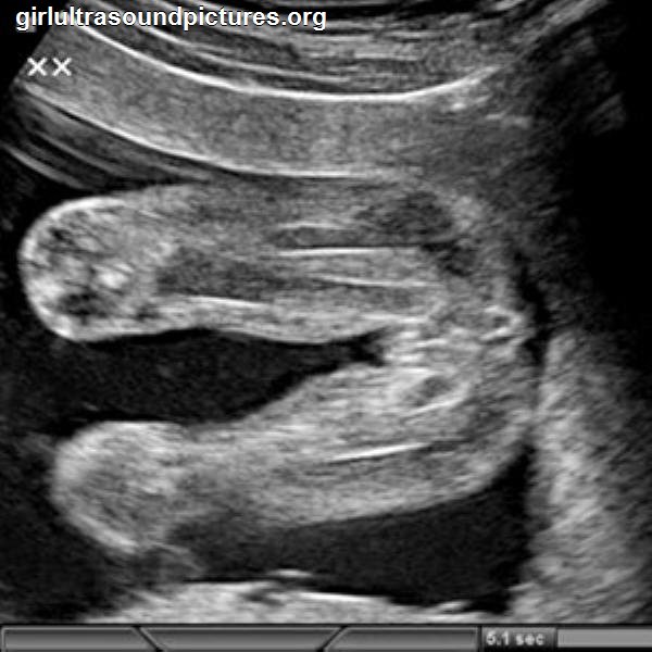 The child normally carries out his first act of defecation only after childbirth, usually on the next day. The discharge of meconium before childbirth (with the so-called meconium amniotic fluid) indicates an episode of intrauterine suffering of the child (asphyxia). With meconium waters, constant monitoring of the state of the child in utero is carried out to determine the adequate tactics of delivery. With ultrasound of the fetus, it is not possible to judge the color and quality of amniotic fluid. The color and nature of amniotic fluid can be assessed when they are discharged or during a special procedure - amnioscopy. With amnioscopy on a gynecological chair through the cervix, it is possible to conduct a visual assessment of the water through the fetal bladder without violating its integrity. If you are carrying a boy, the prostate gland (prostate) begins to form in his small pelvis. A fetal ultrasound at 18 weeks gestation does not show a normal fetal prostate.
Maternal and fetal blood do not mix, which provides powerful protection of the fetus in utero from many negative environmental factors and the internal maternal environment. But still, some substances pass through this barrier - antibodies, low molecular weight drugs, etc. The main food of the child is carried out through the umbilical cord. The baby also receives oxygen through the umbilical cord. A white substance - a cheese-like lubricant - covers the skin of the fetus and protects it in the aquatic environment. During childbirth, this lubricant will contribute to the normal progress of the fetus through the birth canal.
The fetus actively moves, pushes with legs and arms, sucks fingers, rubs eyes with fists. All this can be seen during fetal ultrasound at 18 weeks of pregnancy , especially when used, especially when using 3D, 4D ultrasound during pregnancy. Half of pregnant women already feel the movements of their baby.
On ultrasound of the fetus at 18 weeks of pregnancy you can see a delightfully yawning mouth. The facial muscles of the fetus are actively working, the child trains all innate reflexes in utero in order to successfully undergo physiological adaptation after childbirth.
Some important physiological processes cannot be monitored by fetal ultrasound at 18 weeks of gestation. These include the development of the fetal nervous system. The nerves in the fetus are now covered with myelin, a special substance that ensures the rapid transmission of nerve impulses from nerve to nerve. Neural connections are becoming more ordered, multifaceted and more complex. Many sensitive centers are formed in the brain: sight, touch, hearing, taste, smell. The hearing of the fetus at 18 weeks becomes more acute, the fetus is able to hear and respond with jerks to many sound signals from the mother's body, for example, to hear a rapid heartbeat when the mother is anxious, hiccups.
Changes in the mother's body
Have you noticed "pregnancy marks" (dark spots on your face)? Try to avoid direct sunlight, which will make the pigmentation even darker and more pronounced. Suffering from back pain? The uterus has now grown to the size of a small melon. The fundus of the uterus is now under your navel. This circumstance changes the center of gravity, so the ligaments and nerves of the spine undergo loads of an unusual nature, which provokes pain. Another important circumstance: everyone can already notice that you are pregnant, unless, of course, you are overweight. The cardiovascular system now has a lower reactivity. You need to avoid sudden changes in body position and sudden movements so that fainting does not develop. Fresh air is very important. Ventilate the room regularly, try to stay outside more away from cars and industrial exhaust gases, because you breathe for two!
But eating for two is not necessary and even dangerous! Overeating will be reflected in excessive weight gain, which is fraught with complications of pregnancy: gestational pyelonephritis, gestational diabetes mellitus, preeclampsia. In addition, it will be much more difficult to return to shape, the skin will stretch, and the former forms will be lost forever. When recruiting within acceptable limits, you can quickly restore your previous figure after childbirth, the chest will remain attractive.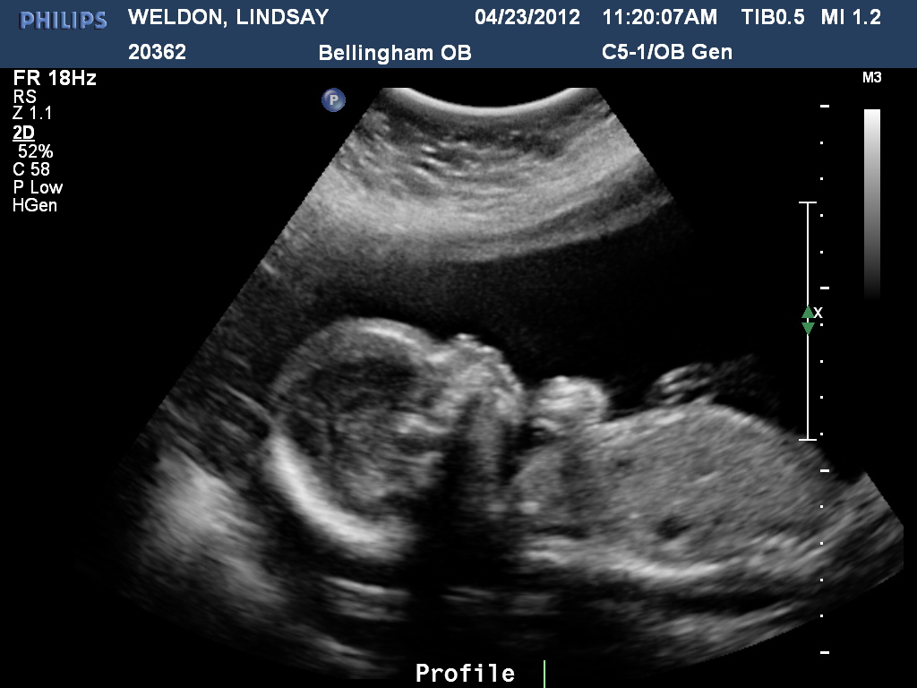
Watch for urinary tract infections. It is necessary to observe hygiene, regularly do urine tests, drink enough fluids. Urinary tract infection is dangerous because it can lead to a serious complication - intrauterine infection of the fetus. The proximity of the reproductive and urinary systems causes their close relationship. With changes in the general analysis of urine, in the presence of elevated body temperature, clarifying analyzes are made: urinalysis according to Nechiporenko, bacterial culture of urine to determine the causative agent of infection. Genitourinary infections during exacerbation during pregnancy are subject to mandatory antibacterial treatment!
read more: 19th week of pregnancy
- Hydrotubation (echohydrotubation): study of the patency of the fallopian tubes (ultrasonic hysterosalpingoscopy)
- Transvaginal
- Ovaries
- Uterus
- Mammary glands
Female ultrasound
- Duplex Scan
- Cerebral vessels
- Neck vessels (duplex angioscanning of the main arteries of the head)
- Veins of the lower extremities
Vascular ultrasound
- Transrectal (trusion): prostate
- Scrotum (testicles)
- Vessels of the penis
Male ultrasound
- Appendicitis
- Abdomen
- Gallbladder
- Stomach
- Intestines
- Bladder
- Soft tissue
- Pancreas
- Liver
- Kidney
- Joints
- Thyroid
- Echocardiography (ultrasound of the heart)
Organ ultrasound
- Varicose veins: ultrasound diagnosis of varicose veins
- Hypertension: Ultrasound diagnosis of hypertension
- Thrombosis: ultrasound diagnosis of vein thrombosis
- Ultrasound diagnosis of chronic pancreatitis
- for kidney stones
- for cholecystitis
Ultrasound diagnostics of diseases
- Hip joints in newborns (if hip dysplasia is suspected)
Pediatric ultrasound
what is happening, the development of pregnancy and fetus
Week by week
17 - 20 weeks of pregnancy
Elena Gevorkova
Obstetrician-gynecologist, Moscow
17th week
BABY
The body length of the fetus at this period is 12-13 cm, and its weight reaches 150 g by the end of the 17th week.
the process of this week is to build the muscle mass of the baby. Various muscle groups are strengthened, which is manifested by increased motor activity of the fetus. Movements become more varied and complex. Frequent movements of the arms and legs not only train the muscles, but also contribute to the development of joints - knee and elbow. The handles are clenched into fists and are unclenched infrequently - to capture the umbilical cord or when sucking a finger. Until this time, the fetal head practically did not move, it was lowered low, the chin was tightly pressed to the chest. By the 17th week, the muscles of the back and neck are strengthened so much that the fetal head can rise to an almost vertical position.
All internal organs continue to improve, their structure becomes more complex, and their functional abilities expand. In other words, intensive preparation continues, allowing the internal organs to begin full-fledged work immediately after the birth of the child.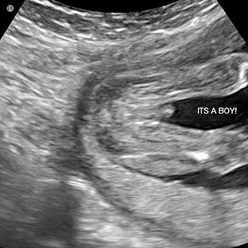
Important changes are taking place in the baby's cardiovascular system. Around the heart there are many nerve plexuses that form the conduction system of the heart. Its uniqueness lies in the fact that the heart does not require external signals to work. Each internal human organ obeys the central nervous system - the brain, which sends a command, and the organ works according to the signal "from above". The peculiarity of the functioning of the heart lies in the fact that the command signals that cause it to contract arise in itself. This autonomous mode allows the heart to work throughout life, day and night, regardless of the activity of the central nervous system.
The formation of the fetal respiratory system continues. The respiratory muscles of the lungs are strengthened. Breathing movements become more and more intense - contractions that simulate inhalation and exhalation, which allows you to strengthen the muscles of the chest. At a period of 17 weeks, the alveolar apparatus of the lungs is completely formed.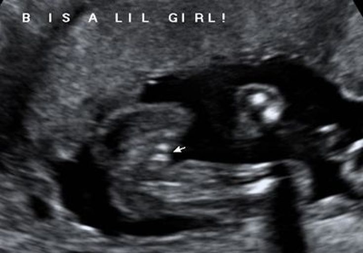 Alveoli are the end parts of the respiratory apparatus in the lung, having the form of tiny bubbles, in which, after the birth of the baby, gas exchange will occur: oxygen from the air will enter the blood vessels of the lungs, and carbon dioxide will be released into the lumen of the alveoli. Each breath will saturate the baby's blood with oxygen. From 17 weeks, the fetal alveoli are in a collapsed state. The first breath of the baby will fill the lungs with air, and the alveoli will expand widely.
MOM-to-be
The tummy continues to grow, and the bottom of the uterus is in the middle between the pubic symphysis (pubis) and the navel. Weight gain by this period is about 5 kg. An increase in the total volume of the mother's blood is natural, since the fetus needs intensive nutrition. The expectant mother feels this as a rapid heartbeat, as more blood passes through the heart of a pregnant woman than usual, and this requires additional heart contractions. An increased heartbeat - tachycardia - may not be felt at all, and may be a source of discomfort for a woman. If you experience tachycardia, you should report it to your gynecologist or cardiologist to determine if it is normal or a symptom of a heart condition such as an arrhythmia.
Another manifestation of increased blood volume (CBV) in pregnant women can be nosebleeds and increased bleeding of the gums. This is due to the increased load on thin-walled vessels - capillaries located in the mucous membranes of the nasal and oral cavities. Vitamins come to the rescue, the timely appointment of which will strengthen the walls of blood vessels. Hygiene procedures will also require caution: the use of toothbrushes with soft bristles is recommended. If minor nosebleeds appear, you can rinse the nasal passages with a salt solution using AQUAMARIS, PHYSIOMER, SALIN preparations or prepare the solution yourself by adding 1 teaspoon of salt to a glass of water. The salt solution will compress the walls of the capillaries of the mucosa, thereby helping to stop the bleeding.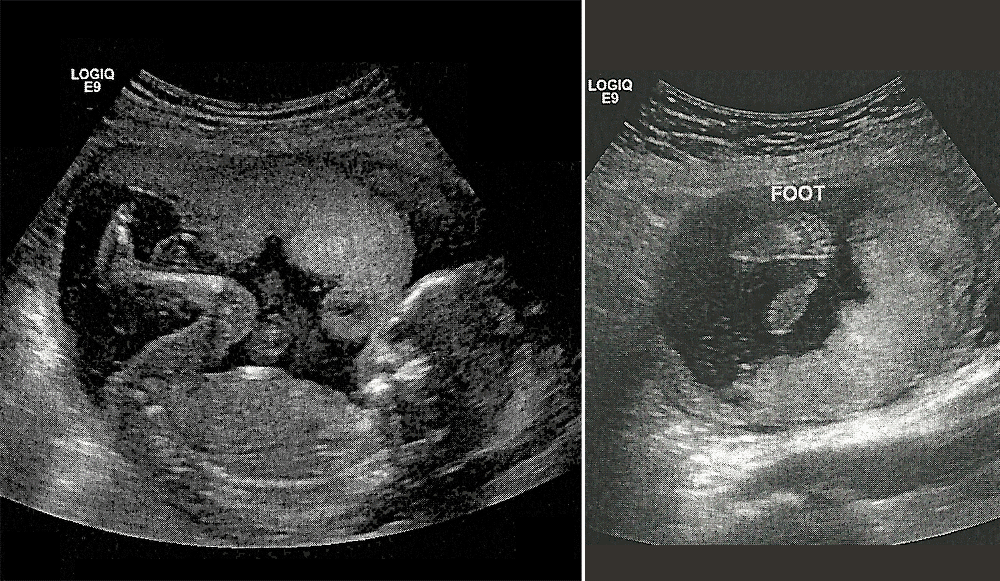 For more severe bleeding, see a doctor.
18th week
BABY
At the 18th week the length of the fetus is 20 cm, and the weight reaches 200-250 g.
During this period, the structure of the brain becomes more complicated, its total mass increases. Nerve fibers that braid organs and tissues are covered with a special fatty sheath - myelin. The myelin sheath provides a high speed of nerve impulses. In the future, this will allow a person to instantly respond to external stimuli, for example, pull his hand away from a hot or sharp object at the slightest contact with it.
The appearance of the fetus approaches the appearance of a newborn child, facial features become more and more distinct, ears straighten out, which until this time were tightly pressed to the head.
The middle ear is being formed and the neural transmission pathway responsible for auditory perception is being improved. In other words, from this period the baby is able to perceive external stimuli and respond to excessively loud sounds - knocking doors, screams, car signals, etc. The noises of the mother's body (intestinal peristalsis, heartbeat, blood flow through large vessels, etc.) are a completely normal environment for the fetus, moreover, they train his auditory perception.
The fetal endocrine system functions very intensively. Starting from the period of 18-19 weeks, the release of "children's" hormones is so great that even the mother's body is able to provide. For example, if the mother had an insufficiency of adrenal or thyroid hormones, then at this time it is quite possible to compensate for this condition.
Expectant MOM
18 weeks of pregnancy is the period when you can feel the movements of the fetus. Usually, in primiparas, these sensations occur later - at 19— 20th week. As a rule, at earlier terms (at 16-18 weeks), movements are felt by multiparous or thin women who have an insignificant layer of subcutaneous fat.
At first these will be episodic, short-term, weak sensations. After some time, the movements are felt more and more clearly. Their nature can be different: light pushes, touches, rolling or twitching. The first movements of the baby are a very anticipated and exciting moment in the life of pregnant women. For the first time, the expectant mother receives a response to her condition, “proof” of the connection between her and the baby.
Fetal movements are very chaotic, varied. The motor activity of the fetus increases during its wakefulness and is practically absent during sleep. Most of the time the fetus sleeps. Movement can become more active if the expectant mother is experiencing emotional stress. In this case, the level of stress hormones in the pregnant woman's body rises and the fetus, receiving them with the mother's blood, experiences similar sensations, which is manifested by an increase in its activity. Also, the baby may respond to an insufficient supply of oxygen to the mother's body if the pregnant woman does not move much, spends insufficient time in the fresh air, or is in a forced position for a long time (for example, driving a car).
19th week
BABY
At the 19th week the fetus is 25 cm tall and weighs 250-300 g. , and the fetal head significantly slows down its growth. Due to this, the baby changes outwardly and no longer looks disproportionate.
The relief of the fetal body changes somewhat and acquires roundness due to the accumulation of subcutaneous fat. This is the so-called brown fat of newborns, which is laid down for a period of 19-20 weeks and after the birth of a child, it performs the function of thermoregulation - it protects against overheating and hypothermia. Gradually, this fat disappears, and in adults it remains in the form of lumps in the thickness of the cheeks, in the region of the kidneys, shoulder blades, and armpits.
At this time, an interesting event occurs - the laying of molars (permanent) teeth. They are located under the rudiments of milk teeth in the thickness of the dental plate - this is a special formation from which the maxillo-dental complex is formed and which disappears by the full term of pregnancy.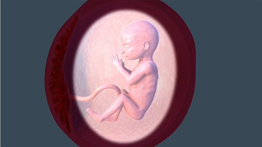
The most important process of the 19th week of fetal development is rightly considered the maturation of the brain. Separate units of the brain (neurons) form connections with each other with the help of special processes. Cells seem to cling to each other, thereby providing the possibility of a variety of pathways for the passage of impulses and the synchronism of the brain. There is a formation of brain structures responsible for perception - taste, sight, smell, hearing, touch.
Interesting changes occur in the hematopoietic system of the fetus. The spleen is involved in the formation of blood cells. It produces mainly "white" blood cells - leukocytes. Their main task is to protect the body from external and internal factors - toxins, microbes, etc. Thus, the composition of the blood of the fetus changes. Until deadline 19-20 weeks, the blood of the fetus consisted exclusively of erythrocytes - cells of the "red" blood, but now it is close in composition to the blood of a newborn.
Future MOM
The tummy continues to grow rapidly, and by this time the bottom of the uterus is 1.5-2 cm below the navel.
By the 19th week, the weight of the pregnant woman increases by 3-6 kg. This increase is distributed between the placenta, amniotic fluid, uterus and fetus, and it accounts for only a tenth of the total weight.
Rapid growth of the uterus may cause discomfort. Most often this is due to the fact that when you change the position of the body, when walking or bending, pain is felt in the lateral sections. Its nature is different: the pain can be pulling or sudden, acute, occur only on one side or on both. This is how the ligaments are stretched. These are special strands that are attached to the side walls of the uterus and pelvic bones and fix the uterus. The uterus is "suspended" with the help of this ligamentous apparatus. When there is a change in its position, the ligaments are stretched, and this can cause pain.
Such "ligamentous" pains are normal and do not require treatment - sometimes it is enough to take antispasmodics that relax the uterus and eliminate pain. But you need to inform your gynecologist about this condition and undergo an examination to exclude pregnancy complications - uterine hypertonicity, the threat of interruption. In this case, treatment should be prescribed - possibly in a hospital setting.
20th week
BABY
Fetal length reaches 28 cm, weight increases to 340 g.
Until the 20th week, the length of the baby was measured from the crown to the coccyx, after this period, the concept of growth will also include the length of the body and legs.
The fetus is active, makes many different movements: pushes off the walls of the uterus, somersaults, grabs the umbilical cord with its handles, feels itself. The facial expressions of the baby become more pronounced: he frowns, smiles, closes his eyes.
The skin of the fetus thickens, completely covered with downy hairs and cheese-like grease. The largest amount of lubricant accumulates in the folds, which prevents skin irritation during mechanical friction. Also, the lubricant has bactericidal properties, and during childbirth, it provides better gliding of the fetus along the mother's birth canal.
The gastrointestinal tract is developing intensively. The fetus constantly swallows amniotic fluid in small portions, which washes the stomach and intestines and trains peristalsis - reducing bowel movements.
From the 20th week, the process of amniotic fluid processing begins, a prototype of future digestion. The fetus produces the original feces - meconium, a sticky substance of a dark green color. Meconium consists of epithelial cells, mucus, bile and water. It accumulates in the intestinal lumen of the fetus and normally comes out on the first day after birth.
Future MOM
20 weeks complete the first half of pregnancy. Starting from this period, the load on the mother's body increases due to the intensive growth of the fetus.
The tummy increases in size, the height of the fundus of the uterus reaches the level of the navel. The growing uterus begins to put pressure on the lungs, pregnant women experience the first symptoms of shortness of breath.
Some increase in urination may occur. The reason for this is mechanical irritation due to compression of the bladder by the growing uterus. In many cases, there is an increase in the volume of vaginal discharge. The extra blood flow to the genitals causes an increase in vaginal secretion, which appears as a mucus or white discharge. Normally, they should not have a smell and be accompanied by discomfort in the vagina - itching, burning, etc. It is necessary to monitor the nature of the discharge and consult a doctor for any changes - with the appearance of yellow, thick, curdled discharge, an unpleasant odor, etc. Such whites can be a sign of trouble - thrush, microflora disorders, exacerbation of genital infections. During pregnancy, the tendency to inflammation of the vagina - colpitis - increases, as the mucosa changes its composition and becomes more "attractive" to microbes.