When do birthmarks show up
Birthmarks - Better Health Channel
Actions for this page
Summary
Read the full fact sheet- Any mark that is present on the skin at birth, or that develops soon afterwards, is called a birthmark.
- Birthmarks are common – many children have a mark of some sort.
- Occasionally, a birthmark may be a sign of other problems or diseases.
Any mark that is present on the skin at birth, or that develops soon afterwards, is called a birthmark. They are common and many children have a mark of some sort. Most are harmless and some go away as the child grows. Occasionally, a birthmark may be a sign of other problems or diseases. Check with your doctor if you are not sure, especially if the mark changes unexpectedly.
Causes of birthmarks
In most cases, the cause of a birthmark is unknown. They are not caused by mothers doing something wrong during pregnancy. They happen by chance.
The occurrence of birthmarks may be inherited. Some marks may be similar to marks on other family members, but most are not. Red birthmarks are caused by an overgrowth of blood vessels. Blue or brown birthmarks are caused by pigment cells (melanocytes).
Types of birthmarks
There are various different types of birthmarks including:
- naevus flammeus, also known as a stork bite or stork mark
- Mongolian spots
- haemangioma of infancy, also known as a strawberry mark or strawberry naevus
- café au lait spots
- congenital melanocytic naevus.
Naevus flammeus or ‘stork bite’ mark
Other names for ‘stork bite’ mark include salmon patch or macular stain. The typical characteristics include:
- They are pink, flat and irregular-shaped marks.
- The skin is not thickened and you cannot feel any difference when you touch the mark.

- They are usually on the nape of the neck, eyelids, forehead and sometimes the sides of the nose and on the top lip.
- Nearly half of all babies have a ‘stork bite’ mark.
- The marks usually disappear by 12 months of age, if not earlier.
- The mark at the back of the neck may stay for longer, but it is usually covered by hair and out of sight.
- Occasionally, marks on the forehead, side of the nose and upper lip may persist longer.
Mongolian spots
The typical characteristics of Mongolian spots include:
- They are bluish, irregular flat patches.
- They are mainly found on the back and bottom, although any area can be affected.
- There is no thickening or change to the feel of the skin.
- They are more common in babies from Asian and African ethnic groups who have darker skin colouring.
- They are harmless and become less obvious as the child grows.
- They can be confused with bruises.

Haemangioma of infancy or ’strawberry mark’
Another name for a strawberry mark is haemangioma of infancy. The typical characteristics include:
- They are red, raised and lumpy areas.
- They usually appear at around one to four weeks of age, then get bigger – sometimes quite quickly – for a few months.
- They stop growing between six and 12 months of age, then gradually disappear over the next few years.
- The skin of the birthmark is as strong as any other skin. It might rarely bleed if knocked hard or scratched, or develop an ulcer on the surface and need to be treated.
- Sometimes, the strawberry mark may grow on the face, near the eye. If it pushes on the eye, it needs urgent treatment or the child may not develop normal vision.
It is not possible to predict exactly how big a strawberry mark will grow before it stabilises and eventually starts to disappear. If diagnosed within the first weeks of life, laser therapy will most likely make the marks disappear. While expanding, a number of medications can be used to stop its growth.
If the haemangioma has already stopped growing, then no treatment may be necessary. Some will regress and disappear spontaneously by the age of two years, about 60 per cent by five years, and 90 to 95 per cent by nine years. I If they are large, disfiguring, block vision or start to ulcerate, they should be referred to a dermatologist for immediate treatment.
Café au lait spots
- Café au lait macules are flat, roughly oval-shaped light brown (milk-coffee coloured) spots.
- They may be present at birth or appear in early childhood.
- Many children have one or two of these – they are not signs of a health problem.
If your child has more than three or four spots, check with your doctor, as they can sometimes indicate a rare disease, such as neurofibromatosis.
Melanocytic naevus (moles)
The typical characteristics of melanocytic naevus (congenital and acquired), also known as moles, include:
- A congenital melanocytic naevus is a brown spot that is present at birth or in the first year of life.
- Acquired melanocytic naevi are much more common and develop in childhood from around the age of two.
- Some are large dark brown, blue or black birthmarks that sometimes grow dark hairs.
- Some are raised and lumpy, while others are flat and irregular in shape.
It is very rare for melanoma cancer to develop in such lesions later in life and even rarer in childhood. A sign that cancer may be developing includes changes of the skin on or around the mark.
Capillary malformation (port wine stain)
The typical characteristics include:
- They are flat, large or small areas of skin that are pink or purple in colour
- They are usually present from birth and the colour gets darker (from pale pink to deep red-purple) as the child grows.

- They may thicken and become lumpier around and after puberty.
- The marks are often on the face, sometimes just on one side. They may have a clear edge along the middle of the face, but they can appear anywhere on the body.
- Some of these marks, particularly on the face and leg, can be associated with other problems.
Treatment for birthmarks
Most birthmarks are harmless but permanent. The only ones that fade with time are Mongolian spots and haemangiomas of infancy.
Treatment may include:
- Haemangioma of infancy – sometimes, a strawberry mark will grow over an eye or may block one side of the nose, ulcerate or cause other problems. In these cases, they can be treated with a medication called propranolol.
- Melanocytic naevus – sometimes, these will need to be removed surgically, because the child finds the mark distressing. Others that start to grow, often during the teenage years, need to be carefully watched.
 If the mark changes, get it checked by your doctor.
 - Capillary malformations – these don’t go away by themselves. It is best to have expert advice about laser treatment early, because their appearance can affect a child’s feelings about themselves. Laser therapy may give good results.
Birthmarks may be a symptom
Very occasionally, port-wine-stain birthmarks may indicate the presence of a rare underlying disorder, including:
- Sturge-Weber syndrome – symptoms include a port-wine-stain birthmark on the upper eyelid and forehead, and abnormalities of the brain (and sometimes of an eye).
- Klippel-Trenaunay-Weber syndrome – symptoms include a port-wine-stain birthmark on the leg (usually). The bones, muscles and other tissue near the birthmark grow larger than the other normal limb.
- Your GP (doctor)
- Dermatologist
- Paediatrician
- The Australasian College of Dermatologists Tel.
 (02) 8765 0242
Where to get help
- Storch, C. and Hoeger, P. 2010, 'Propranolol for infantile haemangiomas: insights into the molecular mechanisms of action'. British Journal of Dermatology, 163: 269–274. More information here.
This page has been produced in consultation with and approved by:
This page has been produced in consultation with and approved by:
Give feedback about this page
Was this page helpful?
More information
Content disclaimer
Content on this website is provided for information purposes only. Information about a therapy, service, product or treatment does not in any way endorse or support such therapy, service, product or treatment and is not intended to replace advice from your doctor or other registered health professional. The information and materials contained on this website are not intended to constitute a comprehensive guide concerning all aspects of the therapy, product or treatment described on the website. All users are urged to always seek advice from a registered health care professional for diagnosis and answers to their medical questions and to ascertain whether the particular therapy, service, product or treatment described on the website is suitable in their circumstances. The State of Victoria and the Department of Health shall not bear any liability for reliance by any user on the materials contained on this website.
Reviewed on: 01-06-2020
Birthmarks - Better Health Channel
Actions for this page
Summary
Read the full fact sheet- Any mark that is present on the skin at birth, or that develops soon afterwards, is called a birthmark.
- Birthmarks are common – many children have a mark of some sort.
- Occasionally, a birthmark may be a sign of other problems or diseases.
Any mark that is present on the skin at birth, or that develops soon afterwards, is called a birthmark. They are common and many children have a mark of some sort. Most are harmless and some go away as the child grows. Occasionally, a birthmark may be a sign of other problems or diseases. Check with your doctor if you are not sure, especially if the mark changes unexpectedly.
Causes of birthmarks
In most cases, the cause of a birthmark is unknown. They are not caused by mothers doing something wrong during pregnancy. They happen by chance.
The occurrence of birthmarks may be inherited. Some marks may be similar to marks on other family members, but most are not. Red birthmarks are caused by an overgrowth of blood vessels. Blue or brown birthmarks are caused by pigment cells (melanocytes).
Types of birthmarks
There are various different types of birthmarks including:
- naevus flammeus, also known as a stork bite or stork mark
- Mongolian spots
- haemangioma of infancy, also known as a strawberry mark or strawberry naevus
- café au lait spots
- congenital melanocytic naevus.

Naevus flammeus or ‘stork bite’ mark
Other names for ‘stork bite’ mark include salmon patch or macular stain. The typical characteristics include:
- They are pink, flat and irregular-shaped marks.
- The skin is not thickened and you cannot feel any difference when you touch the mark.
- They are usually on the nape of the neck, eyelids, forehead and sometimes the sides of the nose and on the top lip.
- Nearly half of all babies have a ‘stork bite’ mark.
- The marks usually disappear by 12 months of age, if not earlier.
- The mark at the back of the neck may stay for longer, but it is usually covered by hair and out of sight.
- Occasionally, marks on the forehead, side of the nose and upper lip may persist longer.
Mongolian spots
The typical characteristics of Mongolian spots include:
- They are bluish, irregular flat patches.
- They are mainly found on the back and bottom, although any area can be affected.

- There is no thickening or change to the feel of the skin.
- They are more common in babies from Asian and African ethnic groups who have darker skin colouring.
- They are harmless and become less obvious as the child grows.
- They can be confused with bruises.
Haemangioma of infancy or ’strawberry mark’
Another name for a strawberry mark is haemangioma of infancy. The typical characteristics include:
- They are red, raised and lumpy areas.
- They usually appear at around one to four weeks of age, then get bigger – sometimes quite quickly – for a few months.
- They stop growing between six and 12 months of age, then gradually disappear over the next few years.
- The skin of the birthmark is as strong as any other skin. It might rarely bleed if knocked hard or scratched, or develop an ulcer on the surface and need to be treated.
- Sometimes, the strawberry mark may grow on the face, near the eye.
 If it pushes on the eye, it needs urgent treatment or the child may not develop normal vision.
It is not possible to predict exactly how big a strawberry mark will grow before it stabilises and eventually starts to disappear. If diagnosed within the first weeks of life, laser therapy will most likely make the marks disappear. While expanding, a number of medications can be used to stop its growth.
If the haemangioma has already stopped growing, then no treatment may be necessary. Some will regress and disappear spontaneously by the age of two years, about 60 per cent by five years, and 90 to 95 per cent by nine years. I If they are large, disfiguring, block vision or start to ulcerate, they should be referred to a dermatologist for immediate treatment.
Café au lait spots
- Café au lait macules are flat, roughly oval-shaped light brown (milk-coffee coloured) spots.
- They may be present at birth or appear in early childhood.

- Many children have one or two of these – they are not signs of a health problem.
If your child has more than three or four spots, check with your doctor, as they can sometimes indicate a rare disease, such as neurofibromatosis.
Melanocytic naevus (moles)
The typical characteristics of melanocytic naevus (congenital and acquired), also known as moles, include:
- A congenital melanocytic naevus is a brown spot that is present at birth or in the first year of life.
- Acquired melanocytic naevi are much more common and develop in childhood from around the age of two.
- Some are large dark brown, blue or black birthmarks that sometimes grow dark hairs.
- Some are raised and lumpy, while others are flat and irregular in shape.
It is very rare for melanoma cancer to develop in such lesions later in life and even rarer in childhood. A sign that cancer may be developing includes changes of the skin on or around the mark.
Capillary malformation (port wine stain)
The typical characteristics include:
- They are flat, large or small areas of skin that are pink or purple in colour
- They are usually present from birth and the colour gets darker (from pale pink to deep red-purple) as the child grows.
- They may thicken and become lumpier around and after puberty.
- The marks are often on the face, sometimes just on one side. They may have a clear edge along the middle of the face, but they can appear anywhere on the body.
- Some of these marks, particularly on the face and leg, can be associated with other problems.
Treatment for birthmarks
Most birthmarks are harmless but permanent. The only ones that fade with time are Mongolian spots and haemangiomas of infancy.
Treatment may include:
- Haemangioma of infancy – sometimes, a strawberry mark will grow over an eye or may block one side of the nose, ulcerate or cause other problems.
 In these cases, they can be treated with a medication called propranolol.
 - Melanocytic naevus – sometimes, these will need to be removed surgically, because the child finds the mark distressing. Others that start to grow, often during the teenage years, need to be carefully watched. If the mark changes, get it checked by your doctor.
- Capillary malformations – these don’t go away by themselves. It is best to have expert advice about laser treatment early, because their appearance can affect a child’s feelings about themselves. Laser therapy may give good results.
Birthmarks may be a symptom
Very occasionally, port-wine-stain birthmarks may indicate the presence of a rare underlying disorder, including:
- Sturge-Weber syndrome – symptoms include a port-wine-stain birthmark on the upper eyelid and forehead, and abnormalities of the brain (and sometimes of an eye).
- Klippel-Trenaunay-Weber syndrome – symptoms include a port-wine-stain birthmark on the leg (usually).
 The bones, muscles and other tissue near the birthmark grow larger than the other normal limb.
 - Your GP (doctor)
- Dermatologist
- Paediatrician
- The Australasian College of Dermatologists Tel. (02) 8765 0242
Where to get help
- Storch, C. and Hoeger, P. 2010, 'Propranolol for infantile haemangiomas: insights into the molecular mechanisms of action'. British Journal of Dermatology, 163: 269–274. More information here.
This page has been produced in consultation with and approved by:
This page has been produced in consultation with and approved by:
Give feedback about this page
Was this page helpful?
More information
Content disclaimer
Content on this website is provided for information purposes only. Information about a therapy, service, product or treatment does not in any way endorse or support such therapy, service, product or treatment and is not intended to replace advice from your doctor or other registered health professional. The information and materials contained on this website are not intended to constitute a comprehensive guide concerning all aspects of the therapy, product or treatment described on the website. All users are urged to always seek advice from a registered health care professional for diagnosis and answers to their medical questions and to ascertain whether the particular therapy, service, product or treatment described on the website is suitable in their circumstances. The State of Victoria and the Department of Health shall not bear any liability for reliance by any user on the materials contained on this website.
Reviewed on: 01-06-2020
Baby birthmarks, recommended medical supervision. Lakhta Junior in St. Petersburg
Let's talk about children's birthmarks, their possible and most common options.
“Like a baby,” we envy about the most healthy, satiny, velvety, uniform skin. But the idea that any spot on the skin of a baby is obviously a pathology is nothing more than a myth.
Birthmarks are called so because they are often visible already in the newborn, although sometimes they appear at later stages of development. The color, size, shape of a birthmark can be very different. Some of these marks disappear over time, others become brighter and/or remain permanently.
Much and completely justified attention is paid today to the dangers that moles carry in themselves in adulthood and old age. On the contrary, most children's moles do not pose any danger.
Vascular ("stork's mark", "angel's kiss", hemangiomas, venous malformations, etc.). The cause of such spots are problems with small blood vessels, usually transient. Vascular spots are distinguished by a red-violet color in one shade or another; may rise slightly above the surface of the skin or be flat.
Age spots (“café au lait”, melanocystosis, Mongolian spots, etc.). Formed by various skin cells; look, as a rule, brown-gray, usually do not rise above the skin.
Again, in most cases, both main varieties of birthmarks are quite safe and harmless. And yet, only a doctor can judge whether it is possible to confine oneself to observation or whether the stain is to be removed for medical reasons. This type of consultation is strictly required.
Let's take a closer look at common types of birthmarks.
Stork Mark (stork peck, stork mark, etc.).
This is the name in many countries for reddish-pink spots that a newborn may have on the back of the head or back of the neck. As a rule, such spots do not rise above the surface and have an uneven shape. Most often, the stork mark occurs in children with fair skin.
The explanation that this is a trace of the "transportation" of the baby by the stork, older brothers and sisters, as a rule, is accepted unconditionally, and even with delight. "Stork peck" usually disappears without a trace by 18 months of age. Otherwise, especially if the stain causes noticeable discomfort, it is removed (for example, using minimally invasive laser technology).
Port-wine stain
Port-wine stain or, in medical terminology, flaming nevus - flat capillary moles, outwardly and really resembling a puddle of spilled purple-red wine. They occur with a frequency of 3-5 cases per 1000 newborns. With age, they can increase and acquire a darker shade.
Treatment is not usually required, but the skin in this area tends to become dry and irritated. Sometimes they resort to the method of laser removal, which, however, does not always completely eliminate the cosmetic defect.
"Strawberry hemangioma"
Obsolete name for infantile hemangioma, a bright red congenital benign vascular tumor that rises above the surface of the skin and has a characteristic surface texture. It occurs in about 5% of newborns, more often in girls, twins and premature babies.
Usually localized on the face, head or chest; as a rule, decreases with age and finally disappears in the preschool period. It is possible to remove a hemangioma, especially if it is large and / or located near the eyes, nose, mouth. Both minimally invasive surgical and conservative methods of removal are practiced.
Venous malformations
These are blue-violet spots that can sometimes appear on the skin, especially during moments of intense crying. Venous malformations (lit. "malformations") are always congenital in nature, but sometimes remain almost invisible until adolescence. They can be located on any part of the body. Compared to other types of birthmarks, they are rare - in one case in about five thousand.
Coffee with milk
Localized abnormal pigmentation that occurs in 30% of people. The color of such a spot is usually light brown or beige, which is reflected in the name. The edges are usually even, the perimeter is sometimes darker than the inner area. In most cases, a café au lait pigment spot is quite harmless, but it can enlarge and/or darken with age. Such spots are subject to observation in dynamics, in some cases they can be removed.
Moles proper
Well-known round or oval freckle-like formations. The color varies from light brown or pink-red to almost black. Sizes in individual cases also vary widely. They can be both flat and protruding above the surface.
Moles should be inspected regularly to prevent any damage (mechanical, chemical, etc.). If a mole begins to bleed, change color, shape, appearance and any other characteristics, you should see a specialist as soon as possible - a dermatologist or oncologist at the Lakhta Junior Children's Clinic in St. Petersburg.
Birthmarks are beautiful and dangerous
12/29/2017
200457
Since ancient times, moles and birthmarks have received special attention. The most famous folk sign on this subject says that if a person without a mirror can see a lot of moles on himself, he is considered lucky.
Surely even today there will be those who believe in the karmic influence of birthmarks on a person's destiny.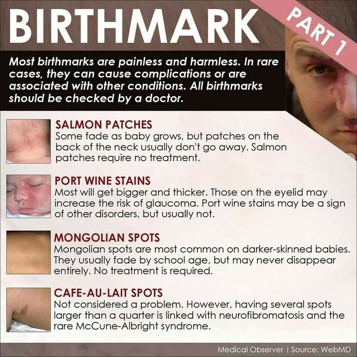 Skeptics will decide that it is not worth paying attention to. Deputy chief physician of the GBU RO "OKKVD" for organizational and methodological work, dermatovenereologist Mikhail Zhuchkov is in a hurry to convince: it's worth it, and how! “Each pigmented neoplasm on the surface of the skin,” he says, “is not just an ornament, but a full-fledged tumor.”
Take a picture of your mole!
We must immediately make a reservation: any congenital mole during its life is doomed to turn into the most terrible malignant tumor of the skin - melanoma, which grows from cells that produce pigment (melanocytes). This transformation occurs during the life of the pigment formation, which can reach 110-150 years, or maybe more. A person, due to objective reasons, often does not live up to this transformation. But it may also happen that some age spots are in a hurry to "live" and acquire "malignancy" much earlier, that is, during a person's life.
Moles are divided into two main types: congenital and acquired. From the names it follows that congenital birthmarks are those with which a person is born, acquired ones occur during life. But their character is different. To begin with, it is worth learning to distinguish the first from the second. This is as easy as shelling pears: if the mole is less than a centimeter, then it is acquired. Exceptions certainly exist, but they are quite rare.
Acquired moles first appear as a brown spot that does not rise above the surface of the skin. Then the pigment cells go deep into the skin. There, in the process of their destruction, the so-called "epidermal growth factors" are released - substances that make the mole grow. Over time, an intangible speck of dark brown color begins to rise, without changing in total size, to lose shades of brown. Over the course of life, this mole can become benign if every one of the pigment cells leaves it. Then it can be removed in any cosmetic way. But to determine the presence of pigment in a mole is possible only with the help of a special procedure - confocal epiluminescent dermatoscopy. The combination of words for the layman is frightening. In fact, it is a simple procedure.
Look inside the mole
Using a special microscope that is placed directly on the mole, the dermatoscope, you can look inside the formation. A research method that allows you to look deep into the skin without making incisions with a scalpel and without using any "irradiation" is called dermatoscopy. This technique is already about a century old, but it has become relatively recent to be used to determine the malignancy of birthmarks. It was first used in clinical practice for the diagnosis of one of the most severe diseases of the connective tissue - systemic scleroderma. And only a few years later it became obvious that it was in this way that many questions regarding moles could be answered: is there a threat in this or that pigmented formation?
Before the widespread introduction of dermatoscopy into practice, the only way to know the truth was to remove the birthmark with a scalpel and conduct a subsequent histological examination. Moreover, today it has become possible with a certain degree of probability to predict the risk of developing malignant neoplasms that develop against the background of birthmarks.
Tricolor and ugly ducklings
Any changes in congenital moles: the appearance of shades of black, peripheral growth, discoloration, is a reason for an urgent appeal for dermato-oncological care. Abroad, in almost all EU countries, congenital moles are removed in a planned manner before the child reaches the age of 14. The procedure is performed by dermato-oncologists. Strange as it may seem, there is no such specialty in Russia yet.
But foreign experience in self-control of moles could well be useful. Abroad, people come to an appointment with a dermato-oncologist with photographs of their pigmentation. The benefits of this are enormous. Studies show that self-monitoring can detect melanoma 2-2.5 years earlier. Therefore, you should remember rule number one: to independently compare changes on your own skin is a simple matter, but effective.
Here is a short set of rules for observing birthmarks from Mikhail Zhuchkov :
- Each mole should rise and fade, but not grow in width.
- The appearance of shades of black is a cause for concern. This indicates that the pigment cells do not go deep, but approach the surface.
- The "colors of the Russian flag" should not appear: shades of white, blue, red. It doesn't have to be a tricolor. But, white-pink, blue-violet and so on are the characteristic colors of melanoma. As well as two or more shades of brown.
- Spontaneous bleeding is also a reason for an extraordinary examination.
There is also a generally accepted rule that is available to an ordinary person who does not have special knowledge in dermatology and dermato-oncology. It tells you which acquired mole should be observed first. It turns out that all moles satisfy the ABCD rule, which includes the assessment of a pigmented skin neoplasm according to 4 parameters:
A - pigment spot asymmetry;
B - border roughness;
C - color unevenness;
D - diameter more than 6 mm.
Mikhail Valeryevich recommends paying attention to one more nuance. A mandatory examination of moles that demonstrate the "ugly duckling syndrome", that is, they differ in their development dynamics from their "brothers", is necessary. For example, if the birthmark did not begin to rise above the surface of the skin at a time when the rest rushed up.
Attention, deletion!
How to get rid of moles? It is necessary to contact the oncology clinic, where the objectionable mole will be removed with a scalpel within healthy tissue and a few barely noticeable stitches will be applied. Dermatologists remove only those moles from which the pigment is completely removed, that is, those that no longer pose any oncological danger. Other birthmarks are the prerogative of oncologists. In no case should you do this in beauty salons and other non-specialized places! Finding out everything about your mole without an incision is possible only with the help of a dermatoscopy procedure.


