Whats a transvaginal ultrasound
Transvaginal ultrasound: MedlinePlus Medical Encyclopedia
URL of this page: //medlineplus.gov/ency/article/003779.htm
To use the sharing features on this page, please enable JavaScript.
Transvaginal ultrasound is a test used to look at a woman's uterus, ovaries, tubes, cervix and pelvic area.
Transvaginal means across or through the vagina. The ultrasound probe will be placed inside the vagina during the test.
You will lie down on your back on a table with your knees bent. Your feet may be held in stirrups.
The ultrasound technician or doctor will introduce a probe into the vagina. It may be mildly uncomfortable, but will not hurt. The probe is covered with a condom and a gel.
- The probe transmits sound waves and records the reflections of those waves off body structures. The ultrasound machine creates an image of the body part.
- The image is displayed on the ultrasound machine. In many offices, the patient can see the image also.
- The provider will gently move the probe around the area to see the pelvic organs.
In some cases, a special transvaginal ultrasound method called saline infusion sonography (SIS) may be needed to more clearly view the uterus.
You will be asked to undress, usually from the waist down. A transvaginal ultrasound is done with your bladder empty or partly filled.
In most cases, there is no pain. Some women may have mild discomfort from the pressure of the probe. Only a small part of the probe is placed into the vagina.
Transvaginal ultrasound may be done for the following problems:
- Abnormal findings on a physical exam, such as cysts, fibroid tumors, or other growths
- Abnormal vaginal bleeding and menstrual problems
- Certain types of infertility
- Ectopic pregnancy
- Pelvic pain
This ultrasound is also used during pregnancy.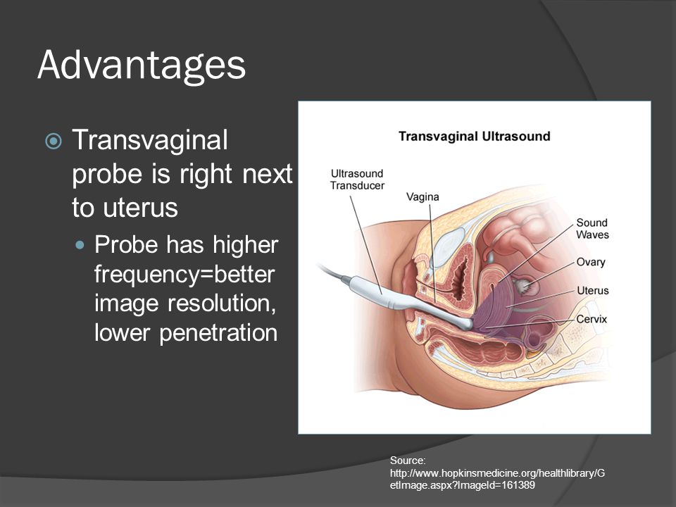
The pelvic structures or fetus is normal.
An abnormal result may be due to many conditions. Some problems that may be seen include:
- Birth defects
- Cancers of the uterus, ovaries, vagina, and other pelvic structures
- Infection, including pelvic inflammatory disease
- Benign growths in or around the uterus and ovaries (such as cysts or fibroids)
- Endometriosis
- Pregnancy outside of the uterus (ectopic pregnancy)
- Twisting of the ovaries
There are no known harmful effects of transvaginal ultrasound on humans.
Unlike traditional x-rays, there is no radiation exposure with this test.
Endovaginal ultrasound; Ultrasound - transvaginal; Fibroids - transvaginal ultrasound; Vaginal bleeding - transvaginal ultrasound; Uterine bleeding - transvaginal ultrasound; Menstrual bleeding - transvaginal ultrasound; Infertility - transvaginal ultrasound; Ovarian - transvaginal ultrasound; Abscess - transvaginal ultrasound
- Ultrasound in pregnancy
- Female reproductive anatomy
- Uterus
- Transvaginal ultrasound
Brown D, Levine D.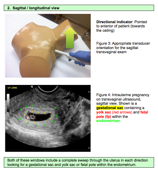 The uterus. In: Rumack CM, Levine D, eds. Diagnostic Ultrasound. 5th ed. Philadelphia, PA: Elsevier; 2018:chap 15.
Coleman RL, Ramirez PT, Gershenson DM. Neoplastic diseases of the ovary: screening, benign and malignant epithelial and germ cell neoplasms, sex-cord stromal tumors. In: Lobo RA, Gershenson DM, Lentz GM, Valea FA, eds. Comprehensive Gynecology. 7th ed. Philadelphia, PA: Elsevier; 2017:chap 33.
Dolan MS, Hill C, Valea FA. Benign gynecologic lesions: vulva, vagina, cervix, uterus, oviduct, ovary, ultrasound imaging of pelvic structures. In: Lobo RA, Gershenson DM, Lentz GM, Valea FA, eds. Comprehensive Gynecology. 7th ed. Philadelphia, PA: Elsevier; 2017:chap 18.
Updated by: John D. Jacobson, MD, Professor of Obstetrics and Gynecology, Loma Linda University School of Medicine, Loma Linda Center for Fertility, Loma Linda, CA. Also reviewed by David Zieve, MD, MHA, Medical Director, Brenda Conaway, Editorial Director, and the A.D.A. M. Editorial team.
Transvaginal Ultrasound: Purpose, Procedure, and Results
What is a transvaginal ultrasound?
An ultrasound test uses high-frequency sound waves to create images of your internal organs. Imaging tests can identify abnormalities and help doctors diagnose conditions.
A transvaginal ultrasound, also called an endovaginal ultrasound, is a type of pelvic ultrasound used by doctors to examine female reproductive organs. This includes the uterus, fallopian tubes, ovaries, cervix, and vagina.
“Transvaginal” means “through the vagina.” This is an internal examination.
Unlike a regular abdominal or pelvic ultrasound, where the ultrasound wand (transducer) rests on the outside of the pelvis, this procedure involves your doctor or a technician inserting an ultrasound probe about 2 or 3 inches into your vaginal canal.
There are many reasons why a transvaginal ultrasound might be necessary, including:
- an abnormal pelvic or abdominal exam
- unexplained vaginal bleeding
- pelvic pain
- an ectopic pregnancy (which occurs when the fetus implants outside of the uterus, usually in the fallopian tubes)
- infertility
- a check for cysts or uterine fibroids
- verification that an IUD is placed properly
Your doctor might also recommend a transvaginal ultrasound during pregnancy to:
- monitor the heartbeat of the fetus
- look at the cervix for any changes that could lead to complications such as miscarriage or premature delivery
- examine the placenta for abnormalities
- identify the source of any abnormal bleeding
- diagnose a possible miscarriage
- confirm an early pregnancy
In most cases, a transvaginal ultrasound requires little preparation on your part.
Once you’ve arrived at your doctor’s office or the hospital and you’re in the examination room, you have to remove your clothes from the waist down and put on a gown.
Depending on your doctor’s instructions and the reasons for the ultrasound, your bladder might need to be empty or partially full. A full bladder helps lift the intestines and allows for a clearer picture of your pelvic organs.
If your bladder needs to be full, you have to drink about 32 ounces of water or any other liquid about one hour before the procedure begins.
If you’re on your menstrual cycle or if you’re spotting, you have to remove any tampon you’re using before the ultrasound.
When it’s time to begin the procedure, you lie down on your back on the examination table and bend your knees. There may or may not be stirrups.
Your doctor covers the ultrasound wand with a condom and lubricating gel, and then inserts it into your vagina. Make sure your provider is aware of any latex allergies you have so that a latex-free probe cover is used if necessary.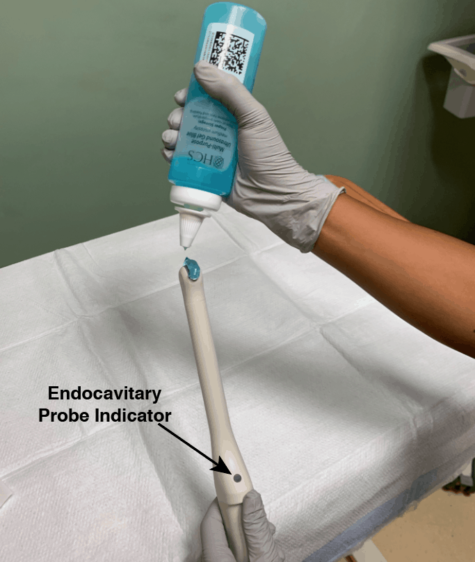
You might feel some pressure as your doctor inserts the transducer. This feeling is similar to the pressure felt during a Pap smear when your doctor inserts a speculum into your vagina.
Once the transducer is inside of you, sound waves bounce off your internal organs and transmit pictures of the inside of your pelvis onto a monitor.
The technician or doctor then slowly turns the transducer while it’s still inside of your body. This provides a comprehensive picture of your organs.
Your doctor may order a saline infusion sonography (SIS). This is a special kind of transvaginal ultrasound that involves inserting sterile salt water into the uterus before the ultrasound to help identify any possible abnormalities inside the uterus.
The saline solution stretches the uterus slightly, providing a more detailed picture of the inside of the uterus than a conventional ultrasound.
Although a transvaginal ultrasound can be done on a pregnant woman or a woman with an infection, SIS cannot.
There are no known risk factors associated with transvaginal ultrasound.
Performing transvaginal ultrasounds on pregnant women is also safe, for both mother and fetus. This is because no radiation is used in this imaging technique.
When the transducer is inserted into your vagina, you’ll feel pressure and in some cases discomfort. The discomfort should be minimal and should go away once the procedure is complete.
If something is extremely uncomfortable during the exam, be sure to let the doctor or technician know.
You might get your results immediately if your doctor performs the ultrasound. If a technician performs the procedure, the images are saved and then analyzed by a radiologist. The radiologist will send the results to your doctor.
A transvaginal ultrasound helps diagnose multiple conditions, including:
- cancer of the reproductive organs
- routine pregnancy
- cysts
- fibroids
- pelvic infection
- ectopic pregnancy
- miscarriage
- placenta previa (a low-lying placenta during pregnancy that may warrant medical intervention)
Talk with your doctor about your results and what type of treatment, if any, is necessary.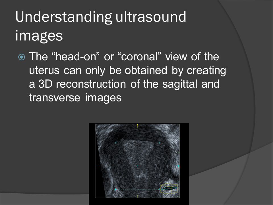
There are virtually no risks associated with a transvaginal ultrasound, although you might experience some discomfort. The entire test takes about 30 to 60 minutes, and the results are typically ready in about 24 hours.
If your doctor is unable to get a clear picture, you might be called back to repeat the test. A pelvic or abdominal ultrasound is sometimes done before a transvaginal ultrasound depending on your symptoms.
If you experience too much discomfort from a transvaginal ultrasound and can’t tolerate the procedure, your doctor may perform a transabdominal ultrasound. This involves your doctor applying gel to your stomach and then using a handheld device to view your pelvic organs.
This approach is also an option for children when pelvic images are needed.
vaginal and transvaginal - MEDSI
What is vaginal ultrasound?
Vaginal (transvaginal) pelvic ultrasound is performed by inserting a special device equipped with a transducer into the vagina.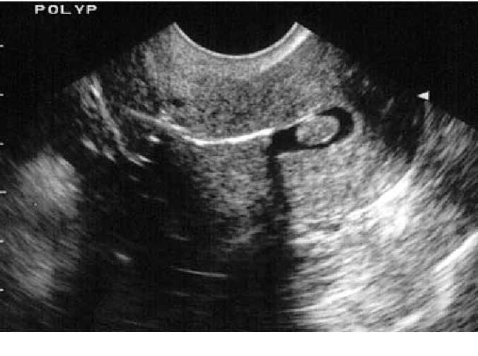
The device is a rod with a handle, which is made of plastic, about 10-12 centimeters long and up to three centimeters in diameter. A special groove can be built into it to insert a needle for taking biopsy material.
Examination allows to determine the presence of pathologies, neoplasms or diseases in the following female genital organs:
- Uterus
- Fallopian tubes
- Ovaries
- Cervix
It is considered the most effective for the study of these parts of the reproductive system, as it allows you to identify various health problems in the patient at an early stage. Ultrasound of the small pelvis with a sensor is able to show the presence of deviations already at a time when other studies do not show any problem areas.
How is the procedure?
The examination is organized as follows:
- The patient must remove clothing from the lower part of the body (from the waist down)
- She settles down on a special couch in the same way as during a regular gynecological examination
- The doctor prepares the transducer: puts an individual condom on it, lubricates it with a special gel for the procedure
- The physician then inserts the device shallowly into the patient's vagina
- To get a complete picture of the state of the organs, he can move the sensor from side to side
- All data is recorded and processed by the doctor
The gel is needed to facilitate the penetration of the transducer (and thereby reduce the likelihood of negative sensations) and enhance the ultrasonic effect by increasing the conductivity.
This type of examination lasts no more than 10 minutes. It is painless and gives the most complete picture even when an abdominal ultrasound shows nothing or cannot be done.
When is a pelvic ultrasound probe needed?
There are symptoms in which the doctor must refer the patient to a transvaginal examination:
- Pain in the lower abdomen (not related to the menstrual cycle)
- Neoplasm suspected
- Too short or too long period of menstrual bleeding or lack thereof
- Impossibility of pregnancy
- Bleeding other than menstruation
- The presence of violations of the patency of the fallopian tubes
- Nausea, vomiting and weakness with bleeding from the vagina
Doctors recommend using this type of examination for preventive purposes, since not every ailment can have symptoms at an early stage, just as pregnancy in the first trimester may not manifest itself with classic symptoms (nausea, etc.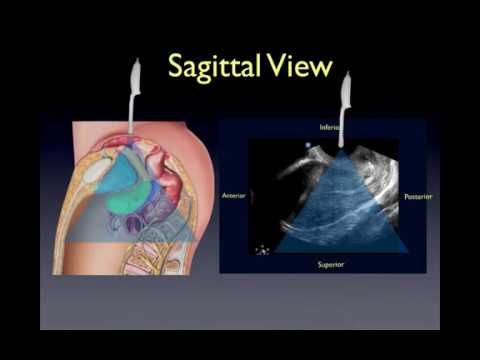 ).
).
In this case, vaginal ultrasound is used for:
- Infertility diagnostics
- The need to determine the presence of changes in the size of the ovaries and uterus
- Pregnancy diagnostics
- Pregnancy control (first trimester only)
- General supervision of the uterus, fallopian tubes and ovaries
A pelvic ultrasound with two probes can be performed at the same time. In this case, an abdominal ultrasound examination is performed first, and then a transvaginal one. The use of two types of analysis at once is necessary to detect violations in the highly located organs of the small pelvis.
What does a vaginal ultrasound show?
This examination allows to evaluate the following parameters of the organs of the reproductive system:
- The size of the uterus. In normal condition, it should be about seven centimeters long, six wide and 4.2 in diameter. If it is significantly less or more, then this indicates the presence of pathology
- Echogenicity.
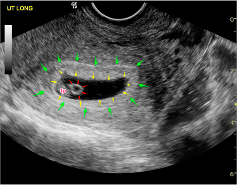 The structure of the organs should be homogeneous, uniform, have clearly defined, well-visible edges
 - General picture of the internal organs. The uterus should be slightly tilted forward. And the fallopian tubes may be slightly visible, but should not be clearly visible without the use of contrast agent
Diagnosable diseases
Transvaginal ultrasound can detect a number of diseases and problems in the reproductive system at an early stage. It allows you to detect:
- Fluid and pus in the uterus and fallopian tubes. The cause of their appearance may be infections, viruses, mechanical damage
- Endomentriosis - excessive growth of cells of the inner layer of uterine tissues into other layers and organs. It can occur due to inflammatory processes, damage (surgery, abortion), the appearance of neoplasms, disruption of the endocrine system, too frequent use of certain drugs and substances
- Myoma is a benign neoplasm in the tissues of the uterus or its cervix.
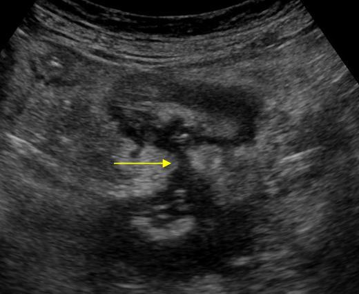 May occur due to chronic diseases, frequent abortions, hormonal disorders, constant stress, pathologies, overweight, with hereditary predisposition
 - Cysts and polycystic ovaries are tumors filled with fluid. Occur in endocrine disorders, chronic diseases of the genitourinary system
- A variety of polyps on the walls of the uterus - benign formations in the endometrium of the organ. They can reach several centimeters in diameter. Their appearance may be associated with polycystic disease, chronic diseases, mastopathy, fibroma
- Inflammation and enlargement of organs can occur due to both infection and trauma
- Cystic skid - appears instead of a full-fledged embryo in the process of conception, filled with fluid. It occurs due to duplication of male chromosomes with the loss of female chromosomes, sometimes due to the fertilization of an egg that does not contain a nucleus. This disease is rare
- Fetal development disorders during pregnancy
- Malformations and pathologies in the development of the fallopian tubes: obstruction, spiral or too long tubes, blind passages, duplication of organs
- An ectopic pregnancy occurs when an egg, after fertilization, attaches itself outside the tissues of the uterus.
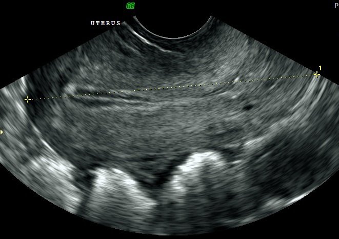 Occurs due to blockage of the fallopian tubes, congenital anomalies in them, as well as after the inflammatory process, abortion
 - Cancer - a malignant tumor in various organs:
- Uterus
- Ovaries
- Cervix
- Chorionepithelioma is a malignant neoplasm that occurs during or after pregnancy from chorion cells (the membrane of the embryo attached to the wall of the uterus)
Steps to prepare for the exam
No special preparation is required for a pelvic ultrasound with a transducer, but there are several mandatory requirements:
Doctors also recommend using such a study on certain days of the cycle, depending on which organ and for what purpose it is necessary to diagnose:0012
It is important to remember about personal hygiene before the examination, use wet and other wipes.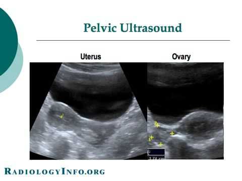
If you plan to perform a pelvic ultrasound with two sensors, then you should pay attention to the preparation for the abdominal examination.
This includes:
- Diet at least three days prior to the examination to reduce the likelihood of symptoms of flatulence and bloating
- The last meal must be finished by six o'clock in the evening on the eve of the analysis
- It is recommended to take an enema after a meal
- If there is still a risk of flatulence, special drugs should be used to reduce gas formation
- Drink at least 400 ml of water one hour before the examination
The diet involves the exclusion of a number of products from the diet:
- Sweets
- Flour (bread, biscuits and other)
- Legumes
- Cabbage
- Milk and dairy products
- Uncooked vegetables and fruits
- Coffee and strong tea
- Carbonated drinks
- Fast food
- Fatty foods (meat, fish, oils)
You can eat cereals cooked with water, low-fat boiled beef, poultry and fish, hard cheeses. It is recommended to drink loosely brewed lightly sweetened tea.
It must be remembered that since fluids are required before the abdominal examination, the bladder must be emptied before the transvaginal examination.
Contraindications
Vaginal ultrasound has a small number of contraindications:
- It is never performed if the patient is a virgin, so as not to violate the integrity of the hymen. In this case, it is possible to perform a transrectal examination for such a patient, in which the probe is inserted into the rectum
- Examination should not be performed during the second or third trimester of pregnancy because it may cause preterm labor or uterine contractions before the expected date of delivery
- This test is not used if the patient is allergic to latex
- If the patient has epilepsy because the examination requires her to lie still
Do not delay treatment, see a doctor right now:
- Ultrasound
- Ultrasound of the pelvic organs for a child
- Hospitalization and transportation
Gynecological ultrasound of the pelvic organs
Ultrasound of the pelvis still remains one of the most popular procedures among the population, as this study is carried out quickly, painlessly, and at the same time provides maximum information about the state of the internal reproductive organs of a woman.
Our hospital uses the following methods:
• Transabdominal ultrasound of the pelvic organs - examination of the internal organs through the anterior abdominal wall. The procedure is performed with a full bladder and allows you to determine the size of the genital organs, their structure and the presence of large pathological formations (tumors, cysts).
• Transvaginal ultrasound - examination using a special probe inserted directly into the vagina. The method allows you to examine the structure of organs in more detail, determine the size, shape and structural features of pathological formations.
• Combined - transabdominal scanning with a full bladder and after emptying the bladder, the transition to transvaginal examination.
• Transrectal - a highly informative method of research, carried out using a sensor inserted into the rectum. This diagnostic method is indispensable when examining girls who do not live sexually.
Indications for ultrasound of the pelvic organs
- Diagnosis of pregnancy at an early stage.
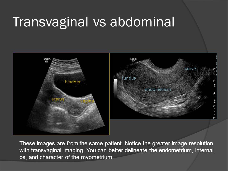
- Ultrasound of the small pelvis in women should be performed for menstrual irregularities (delayed menstruation, early onset of menstruation, bleeding in the middle of the cycle), with heavy or scanty menstruation, in the absence of menstruation, with various vaginal discharges, with pain in the lower abdomen, with the appearance of discharge during menopause.
- With the help of gynecological ultrasound, various diseases are detected: from inflammatory gynecological diseases to benign and malignant tumors of the uterus and ovaries (including endometriosis, salpingo-oophoritis, ovarian cysts, endometritis, etc.).
- Ultrasound of the uterus enables early diagnosis of uterine fibroids.
- Pelvic ultrasound is widely used to monitor the ovarian follicular apparatus in the treatment of infertility and pregnancy planning.
- Ultrasound examination of the pelvis is prescribed when taking contraceptive and hormonal drugs, in the presence of an intrauterine contraceptive (“spiral”) to monitor and detect complications.

- Ultrasound during pregnancy (obstetric ultrasound) allows you to monitor the normal development of the fetus and timely detect pathology.
- In urology, pelvic ultrasound is necessary to identify the causes of urinary disorders, urinary incontinence and pathology of the urethra (urethra).
Folliculometry is a diagnostic ultrasound method that allows you to determine the day of ovulation and the quality of the phases of the menstrual cycle. The method involves ultrasonic monitoring of the maturation of the follicle, the growth of the endometrium and the presence of signs of ovulation in the corresponding phases of the cycle.
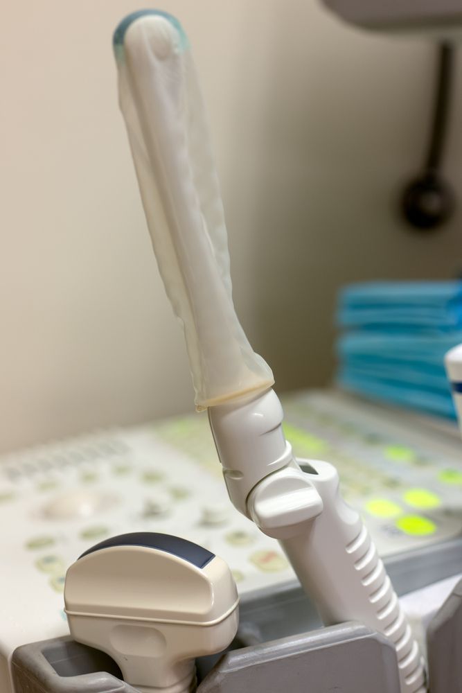
As a rule, the first time this examination is prescribed for 4-5-6 days from the beginning of the menstrual cycle or immediately after the end of menstruation. In this early phase, there is a simultaneous development of several follicles, one of which then begins to outstrip the growth of others. To see subsequent changes, ultrasound is repeated 1-2 days before the onset of signs of ovulation, or until the next menstruation if ovulation has not occurred. More often it happens like this: 1st study on the 4-6th day of MC, 2nd study on the 12-14th day of MC, 3rd study - before the expected menstruation 3-5 days before.
The fact of ovulation during dynamic ultrasound confirms:
• fixation of a mature follicle;
• his disappearance;
• the appearance in the space behind the uterus of free fluid;
• formation of a corpus luteum in place of a mature follicle;
As a result of the examination, the following results can be obtained:
• Normal ovulation - the processes of growth and development of the follicle proceed within the normal range.


