Umbilical cord cover
Umbilical cord care in newborns Information | Mount Sinai
Cord - umbilical; Neonatal care - umbilical cord
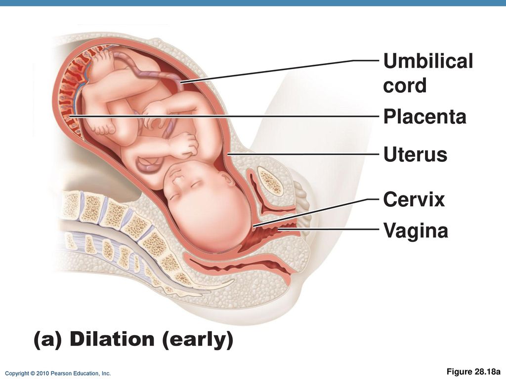
The umbilical cord connects the baby to the mother's placenta. During fetal development in the womb, the umbilical cord is the lifeline to the baby supplying nutrients. After birth, the cord is clamped and cut. Eventually between 1 to 3 weeks the cord will become dry and will naturally fall off. During the time the cord is healing it should be kept as clean and as dry as possible.
A sponge bath is the best way to clean your baby until the umbilical cord falls off. To give a sponge bath, dip a soft cloth in the warm water and wring out the excess. If needed, a mild soap can be used in the water. Wipe the baby's skin gently starting from the area of the baby's head and work your way down to the rest of the body. Pay special attention to the skin creases and diaper area. Rinse your baby with clean warm water and dry him or her completely.
Information
When your baby is born the umbilical cord is cut and there is a stump left. The stump should dry and fall off by the time your baby is 5 to 15 days old. Keep the stump clean with gauze and water only. Sponge bathe the rest of your baby, as well. Do not put your baby in a tub of water until the stump has fallen off.
Let the stump fall off naturally. Do not try to pull it off, even if it is only hanging on by a thread.
Watch the umbilical cord stump for infection. This does not occur often. But if it does, the infection can spread quickly.
Signs of a local infection at the stump include:
- Foul-smelling, yellow drainage from the stump
- Redness, swelling, or tenderness of the skin around the stump
Be aware of signs of a more serious infection. Contact your baby's health care provider immediately if your baby has:
- Poor feeding
- Fever of 100.
 4°F (38°C) or higher
 - Lethargy
- Floppy, poor muscle tone
If the cord stump is pulled off too soon, it could start actively bleeding, meaning every time you wipe away a drop of blood, another drop appears. If the cord stump continues to bleed, call your baby's provider immediately.
Sometimes, instead of completely drying, the cord will form pink scar tissue called a granuloma. The granuloma drains a light-yellowish fluid. This will most often go away in about a week. If it does not, call your baby's provider.
If your baby's stump has not fallen off in 4 weeks (and more likely much sooner), call you baby's provider. There may be a problem with the baby's anatomy or immune system.
Esper F. Postnatal bacterial infections. In: Martin RJ, Fanaroff AA, Walsh MC, eds. Fanaroff and Martin's Neonatal-Perinatal Medicine.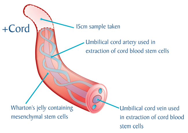 11th ed. Philadelphia, PA: Elsevier; 2020:chap 48.
Nathan AT. The umbilicus. In: Kliegman RM, St. Geme JW, Blum NJ, Shah SS, Tasker RC, Wilson KM, eds. Nelson Textbook of Pediatrics. 21st ed. Philadelphia, PA: Elsevier; 2020:chap 125.
Taylor JA, Wright JA, Woodrum D. Newborn nursery care. In: Gleason CA, Juul SE, eds. Avery's Diseases of the Newborn. 10th ed. Philadelphia, PA: Elsevier; 2018:chap 26.
Wesley SE, Allen E, Bartsch H. Care of the newborn. In: Rakel RE, Rakel DP, eds. Textbook of Family Medicine. 9th ed. Philadelphia, PA: Elsevier; 2016:chap 21.
Last reviewed on: 12/10/2021
Reviewed by: Neil K. Kaneshiro, MD, MHA, Clinical Professor of Pediatrics, University of Washington School of Medicine, Seattle, WA. Also reviewed by David Zieve, MD, MHA, Medical Director, Brenda Conaway, Editorial Director, and the A.D.A.M. Editorial team.
Umbilical Cord | Newborn Nursery
-
Normal Umbilical Cord
-
Meconium Stained Umbilical Cord
-
Umbilical Hernia
-
Skin Irritation from Dry Cord
-
Periumbilical Erythema
-
Omphalitis
-
Umbilical Cord Hematoma
-
Umbilical Cord Hemangioma
-
Wharton's Jelly Cyst
Normal Umbilical Cord
This infant is 7 hours old.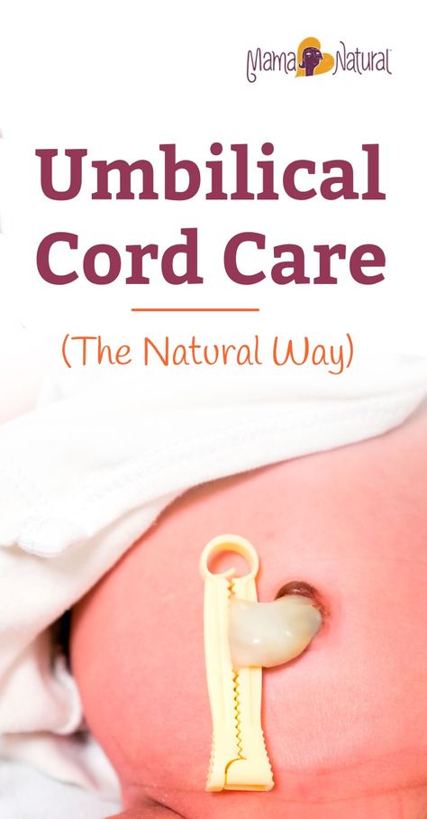 The cord is plump and pale yellow in appearance. One of the umbilical arteries is visible protruding from the cut edge. A normal cord has two arteries (small, round vessels with thick walls) and one vein (a wide, thin-walled vessel that usually looks flat after clamping).
photo by Janelle Aby, MD
Normal Umbilical Cord
Looking at the cut edge more clearly shows the normal vessels of the umbilical cord. The two arteries are to the left and the vein, with a spot of blood in its large lumen, is on the right.
photo by Janelle Aby, MD
Normal Umbilical Cord
The dark stripes within the cord in this picture are examples of intravascular clots -- a normal finding in newborns. In some cases, the vessels are so full of clotted blood that all three may be clearly identified as they wind around through the umbilical stump.
photo by Janelle Aby, MD
Normal Umbilical Cord
This infant is 19 hours old. The cord is beginning to dry and darken as it makes its transition into a non-functioning organ.
photo by Janelle Aby, MD
Normal Umbilical Cord
After a couple of days, the cord is a stiff, dry stump. The bulge of skin around the edge is a normal variant and does not represent an abnormality.
photo by Janelle Aby, MD
Normal Umbilical Cord
Just minutes after the cord falls off, some of the remaining moist debris is still visible on the skin. A spot of blood or a slight amount of moist, yellow material may be present on the diaper or clothing after cord separation. Any bleeding or discharge that persists should be evauated, as this is not a normal finding.
photo by Janelle Aby, MD
Meconium Stained Umbilical Cord
This cord is also about 7 hours old at the time of the photo, but the normal light yellow color is not visible, even though the cord is still plump. This cord was stained by the presence of meconium in utero, which gives it a dark green color. When an infant shows signs of meconium staining, it is evidence that meconium has been present in the amniotic fluid for some time.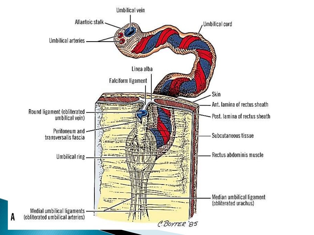 In addition to the umbilical cord, meconium staining is also frequently seen on the nails.
photo by Janelle Aby, MD
Umbilical Hernia
When the umbilical ring is weak or large, an umbilical hernia can result. With increased abdominal pressure (the infant was crying for this photo), a bulge of intra-abdominal contents through the ring can be seen. This does not require treatment, as most hernias of this type resolve spontaneously during the first year of life. Complications, such as strangulation of bowel, are extremely rare. Surgical correction is only considered for those who have large defects that are still open at several years of age.
photo by Janelle Aby, MD
Umbilical Hernia
In this view, the hernia has been reduced with slight digital pressure on the bulged area. The infant is now quiet, and intra-abdominal pressure low, so the hernia is no longer visible. Parents are often concerned about the degree of elevation of the bulge, but the severity of the hernia is determined solely by the size of the umbilical ring. The visibility of the bulge is related to intra-abdominal pressure and is therefore constantly changing.
photo by Janelle Aby, MD
Skin Irritation from Dry Cord
The redness superior to the umbilical cord in this photo is simply irritation from the hard, dry umbilical stump rubbing against the abdomen. Because omphalitis (an infection of the cord itself) is a dangerous condition, redness in this area should be carefully evaluated. However, the localized nature of the redness here, along with the fact that it is somehat removed from the actual insertion of the cord and appears very superficial is reassuring that infection is not the underlying etiology.
photo by Janelle Aby, MD
Periumbilical Erythema
When "dry cord care" is the standard practice (no application of alcohol or triple dye), there are a number of infants who have a small rim of erythema around the drying cord. Although this should be monitored to be sure progression does not occur to omphalitis, this minimal redness is thought to be related to the normal wbc infiltration that occurs naturally in the process of cord separation. These otherwise well infants do not need evaluation.
photo by Janelle Aby, MD
Omphalitis
For comparison, this is the appearance of a full-blown case of omphalitis. The progression of erythema over time can be clearly seen from the circles marked on the abdomen. The first circle was drawn less than 12 hours after redness was initially seen, and the second circle was drawn several hours after that. Omphalitis of this degree can be fatal, even with aggressive antibiotic therapy, so infection in this area should always be taken seriously.
photo by JoDee Anderson, MD
Umbilical Cord Hematoma
Umbilical hematomas are fortunately very rare (incidence approximately 1:5000). They may be spontaneous or related to trauma from prenatal procedures or birth, but they are caused by rupture of the umbilical vessels, usually the umbilical vein. When the rupture occurs in utero, fetal distress is common, and up to 50% of cases result in fetal death.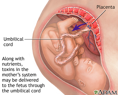 The etiology is thought to be either hypoxia secondary to the venous mass occluding the umbilical vessels or exanguination. When noted as a physical finding in an otherwise well newborn, the hematoma is expected to resolve spontaneously and requires no additional evaluation.
photo by David A Clark, MD
Umbilical Cord Hematoma
Here is another example of an umbilical hematoma. Risk factors for this condition include short cord, vilamentous insertion, cord prolapse or torsion, traction on the cord, chorio-amnionitis, or post-dates delivery leading to thinning of the cord components with secondary aneurysm formation. In this case, the infant had no distress either before or after birth, so no further evaluation was needed.
photo by Janelle Aby, MD
Umbilical Cord Hematoma
This is the underside view of the cord in the previous photo. From this angle, the bruised appearance of the internal structures can be more easily seen. Spontaneous resolution and normal cord separation is expected.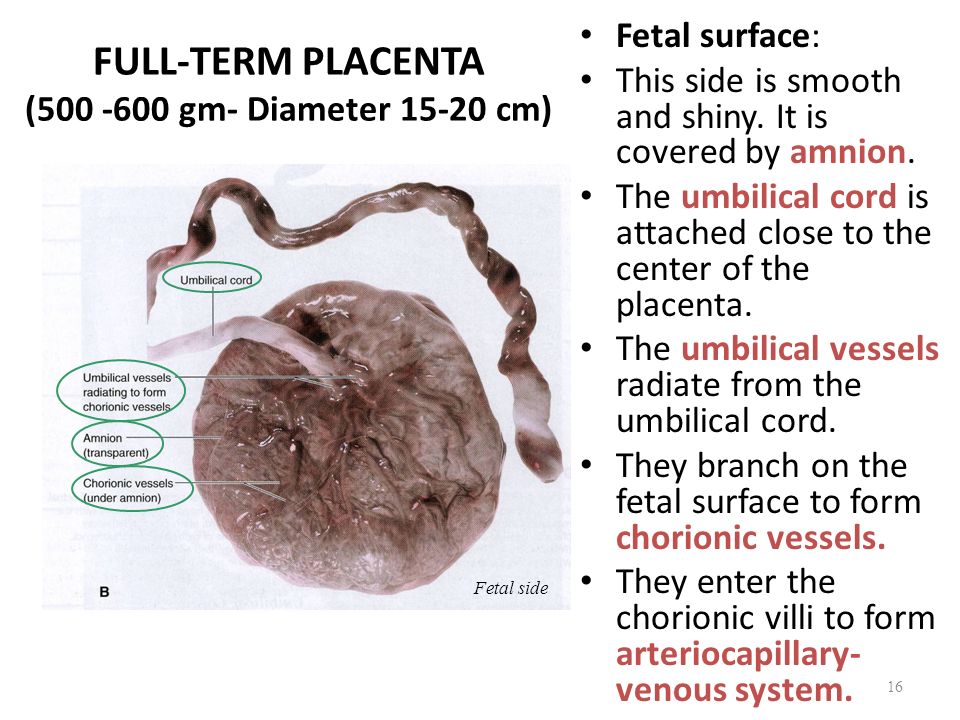
photo by Janelle Aby, MD
Umbilical Cord Hemangioma
A very rare anomaly of the cord, hemangiomas can be quite serious. Large hemangiomas can comprise the vasculature or completely obstruct flow in the cord in utero or lead to high output cardiac failure. Fetal deaths have been reported.
photo by Janelle Aby, MD
Wharton's Jelly Cyst
Also known as a "false cyst" of the cord, a Wharton's jelly cyst is an area where liquefaction of the jelly has occured. Up to 20% of infants with this condition have associated anomalies.
photo by David A Clark, MD
Wharton's Jelly Cyst
With backlighting, the homogeneous nature of the "cyst" can be seen.
photo by David A Clark, MD
Wharton's Jelly Cyst
Here is another example of a Wharton's jelly cyst. Again, the affected area is translucent and homogeneous in appearance. Although some infants with this finding have associated anomalies, this patient was otherwise well.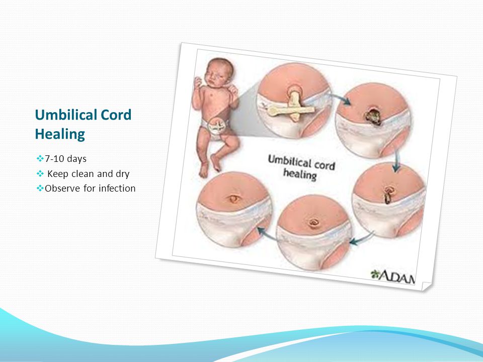
photo by Janelle Aby, MD
Wharton's Jelly Cyst
This is the same cord as in the previous photo. Viewed from the side, one can see the difference in appearance of the "cystic" area as compared with the normal cord. By the following morning, the distended area had completely deflated as the cord was starting to dry and could no longer be distinguished from the rest of the umbilical cord.
photo by Janelle Aby, MD
Neonatal Physical Findings Atlas
What are umbilical clamps for? — Articles
The umbilical clamp avoids the complications that can occur when cutting off the umbilical cord in newborns. The umbilical anchor for babies is easy to use, sterile and environmentally friendly.
After the birth of a child, the umbilical cord, through which the fetus feeds throughout the entire gestation period, becomes unnecessary for him. The umbilical cord is cut, and an umbilical clamp is applied to the place of the cut, called the "stump".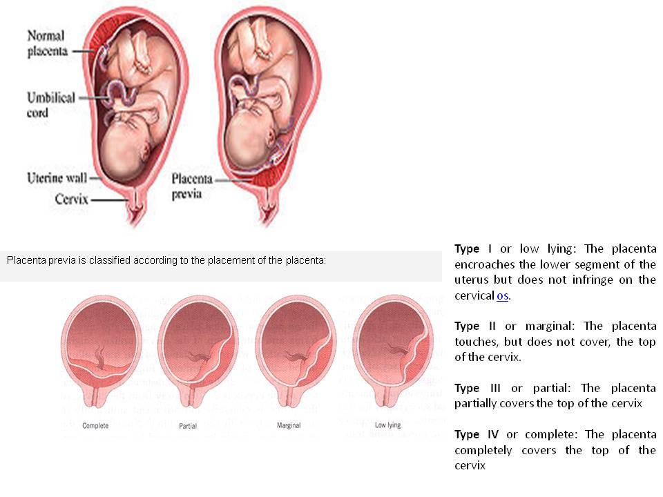 This manipulation is done right in the delivery room.
Design and purpose of the umbilical clamp
The umbilical clamp consists of two elements, the inner part of which has a slightly curved shape. Both parts are interconnected by a ring into a movable structure. The product is made of durable and resilient polypropylene, medically tested, non-toxic and safe in every respect.
The main purpose of the product is to clamp the umbilical cord (ligature fixation of vessels) of a child after his birth. Reliable and strong compression is due to the presence of atraumatic teeth on the inner surfaces of the jaws, which prevent sliding along the soft tissue of the umbilical cord. The branches have longitudinal grooves, the presence of which is necessary for the uniform distribution of the jelly-like mass present in the umbilical cord and surrounding the blood vessels.
After the doctor fixes the clamp, the device remains in one position until the dried navel falls off. To ensure that the doctor's fingers do not slip on the outer surfaces of the branches at the moment of fixation, the latter are provided with corrugation.
The clip has a special lock that prevents accidental opening. Each product undergoes an ethylene oxide sterilization process and is packaged in an individual bag. Umbilical clips are not intended for multiple use - they are disposable products that must be disposed of.
How to treat the navel with a clip
Umbilical clips are used in perinatal centers and all maternity hospitals. The purpose of using the product is to stop bleeding in the umbilical vessels and prevent umbilical infections. The umbilical clamp ensures proper healing of the navel and prevents the development of complications.
After cutting and processing the umbilical cord, it is subjected to medical treatment. As a result, a two-centimeter process remains from the long umbilical cord. Then they put a plastic clip on it. Previously, metal fixators were used for these purposes, but they quickly fell out of use because they were unsafe and could injure the child.
After the wound heals, a full-fledged navel will form in its place. However, in order for the healing process to proceed without complications, the stump must be properly processed. Daily competent treatment of the navel and hygiene ensures that the process dries quickly.
New mothers should not be afraid of the presence of clothespins on the child's body. Processing the navel with a clamp is easy.
How to do it right:
- wash hands thoroughly with soap; 90,031 babies are placed on their backs;
- the entire area around the navel and the umbilical cord itself is treated with hydrogen peroxide;
- clamp also needs to be disinfected with the same solution;
- further, the navel is treated with brilliant green.
This procedure must be done daily. After about 7-10 days, the shoot will become completely dry and fall off on its own. Along with it, the plastic clip will also disappear, which can be thrown away or kept as a keepsake, just like tags from pens and the first booties of a baby are kept.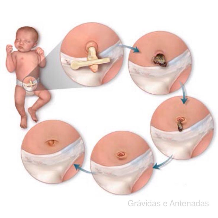
The umbilical wound is treated in the same way. Additionally, picking up a cotton swab, carefully collect the moisture left from the peroxide solution with it. The condition of the navel must be monitored until it is completely healed. After water procedures, it is necessary to check whether the navel is oozing, whether the skin on the umbilical ring has reddened. If it is found that the navel is inflamed, this should be reported to the doctor immediately.
Delayed Cord Clamping & Cord Blood Banking
As more and more research highlights the benefits of delayed cord cutting, it is understandable that parental concerns about cord blood collection are many. We propose to consider this issue in more detail and understand how the delay in cutting the umbilical cord is compatible with the collection of cord blood stem cells to protect the health of your baby in the future.
Delayed cord cutting is the process of clamping and/or cutting the umbilical cord after the blood has stopped pumping or after the placenta has been delivered.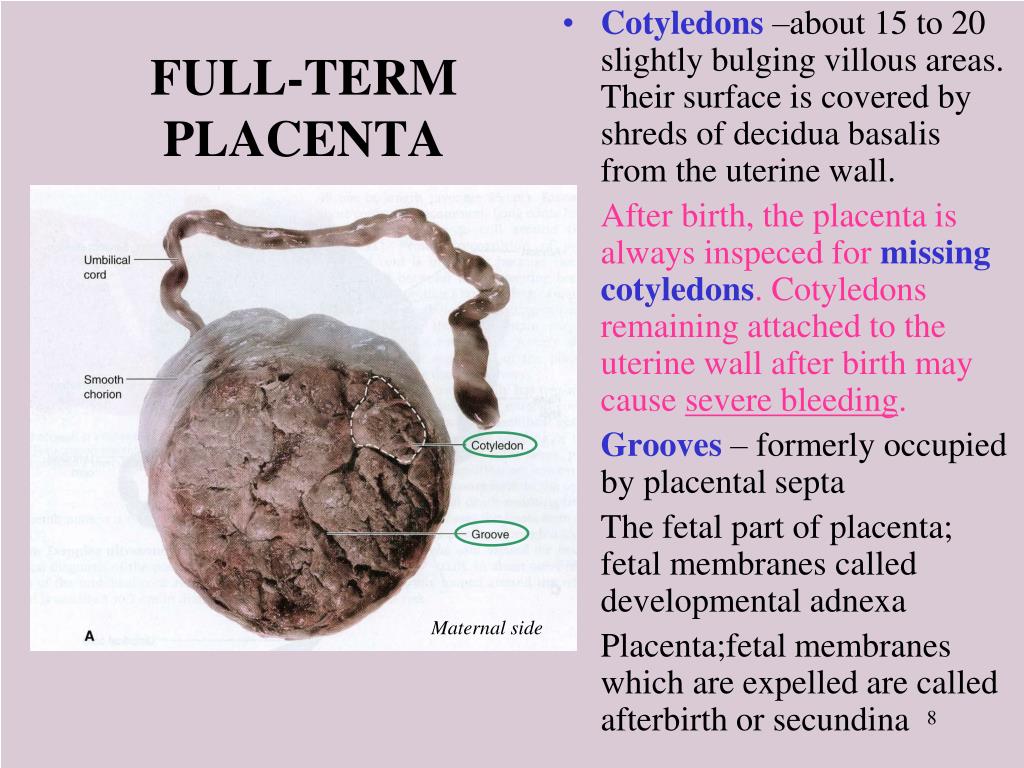 This ensures that your child receives an adequate volume of blood, including a full complement of red blood cells, stem cells and immune cells.
The latest recommendations from the World Health Organization (WHO) recommend delayed cord cutting (for most newborns) within the first 30-60 seconds after birth. During this time, approximately 80% of cord blood returns to the baby. This helps protect against disorders such as iron deficiency anemia and vascular instability. To learn more about cord blood storage, also visit the Parent's Guide to Cord Blood Foundation.
Working Together...
Delayed Cord Cutting and Cord Blood Stem Cell Collection
There is a common myth that cord blood collection prevents a newborn baby from receiving the benefits of delayed cord cutting. In fact, sometimes as little as 15 ml of blood is enough to obtain stem cells, which is all that is needed compared to the approximately 200 ml of blood left in the umbilical cord and placenta after the baby is born.