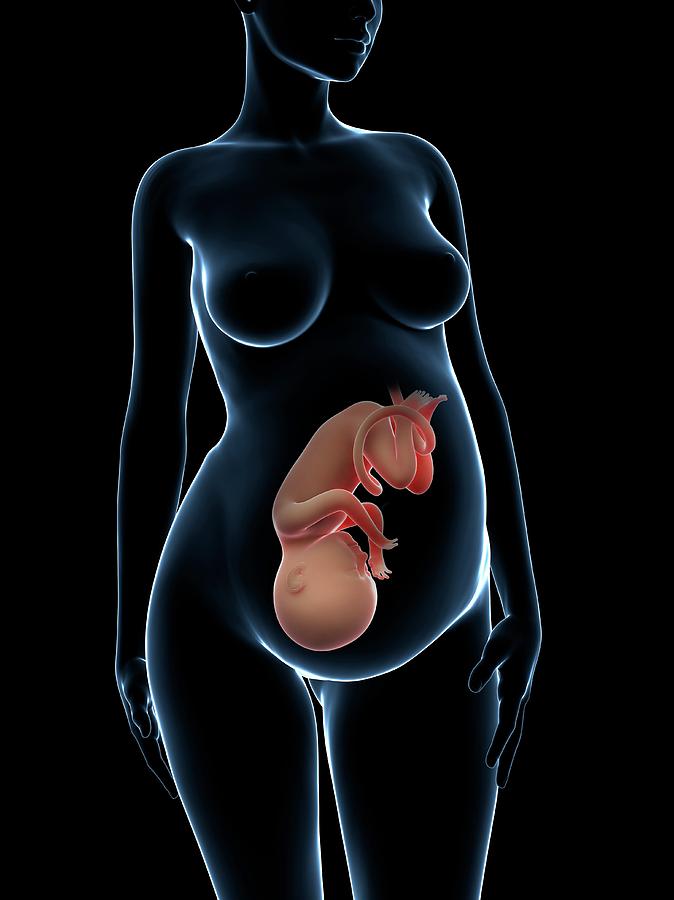
Skeleton of a pregnant woman
Ancient Skeletons of Woman and Fetus Hint at Childbirth Death 3,700 Years Ago
A jar and a reddish pot lay in the grave next to the woman's remains. (Image credit: Ministry of Antiquities)Archaeologists in Egypt recently unearthed a grim discovery: the skeleton of a young woman dating to about 3,700 years ago who was in the final weeks of her pregnancy when she died.
And she was buried with the unborn fetus still inside her body.
The tiny skeleton was head-down within the woman's pelvis — a position typically seen in the third trimester — suggesting that she may have died following the onset of labor, officials with Egypt's Ministry of Antiquities said in a statement on Nov 14. [The 8 Most Grisly Archaeological Studies]
An international team of experts from Yale University and the University of Bologna in Italy found the remains. They uncovered the skeletons in a cemetery at the dig site Kom Ombo in Aswan, a city in southern Egypt located about 530 miles (852 kilometers) from Cairo. The graveyard was used between 1750 B.C. and 1550 B.C. by nomadic people who traveled north into the region from Nubia, Secretary General of Egypt's Supreme Council of Antiquities Mostafa Waziri said in the statement.
Other recent discoveries at Kom Ombo include a statue of a cobra-crowned sphinx, engravings of a warrior pharaoh and a stone head representing the Roman emperor Marcus Aurelius.
In the recently excavated gravesite, the woman's body was curled inward and wrapped in a leather shroud. Scientists estimated that she was around 25 years old when she died. Archaeologists inspected her pelvic bones and discovered abnormalities that may have stemmed from an old fracture that was improperly set or that had healed incorrectly. This may have contributed to the woman's difficulties during childbirth, ultimately leading to her death and that of her fetus, Waziri said.
Beads made from ostrich eggshell hint at the buried woman's social status. (Image credit: Ministry of Antiquities)Objects found in the grave included a pottery jar, a container that was colored red on the outside and black on the inside in the style of pots made in ancient Nubia, and beads made from the shell of an ostrich egg. Some unworked shell material was included near the body, possibly indicating that the woman was a bead maker, according to the statement.
All of these funerary objects were likely included to honor the dead woman and to show the respect of her family and loved ones, ministry representatives said.
- The 25 Most Mysterious Archaeological Finds on Earth
- Move Over, 'Tomb Raider': Here Are 11 Pioneering Women Archaeologists
- 10 'Barbaric' Medical Treatments That Are Still Used Today
Originally publishedon Live Science.
Mindy Weisberger is a Live Science editor for the channels Animals and Planet Earth. She also reports on general science, covering climate change, paleontology, biology, and space. Mindy studied film at Columbia University; prior to Live Science she produced, wrote and directed media for the American Museum of Natural History in New York City. Her videos about dinosaurs, astrophysics, biodiversity and evolution appear in museums and science centers worldwide, earning awards such as the CINE Golden Eagle and the Communicator Award of Excellence.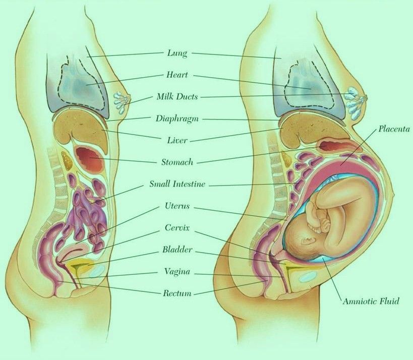 Her writing has also appeared in Scientific American, The Washington Post and How It Works Magazine.
Rare Find at King Solomon's Mines: Ancient Pregnant Woman's Remains
Remains of the pregnant woman's fetus bones in her pelvis can be seen in this photo. She was in her first trimester when she died. (Image credit: Photo courtesy Central Timna Valley Project )The skeleton of a pregnant woman, dating back around 3,200 years, has been found near a temple dedicated to the Egyptian goddess Hathor at a place that was once called King Solomon's Mines, archaeologists recently announced.
Located in the Timna Valley in Israel, ancient Egyptians and others in the region used the mines for copper mining. Early archaeologists and explorers believed that King Solomon, an ancient Israeli ruler, controlled the Timna mines. However, many scholars now think the claim is unlikely.
Archaeologists discovered the pregnant woman's skeleton buried in a tumulus (a tomb covered by rocks) near Hathor's temple. The people worshipped Hathor — the goddess of love, pleasure and maternity — at Timna, and considered her to be the protector of the miners. [See Photos of the Burial and Skeletal Remains in Timna Valley]
At the time the pregnant woman lived, Egypt controlled the mines at Timna, suggesting she was Egyptian. In addition, she may have been a singer at the Hathor temple, said Erez Ben-Yosef, the director of the Central Timna Valley Projectand a senior lecturer in archaeology at Tel Aviv University. She was buried with beads whose design is similar to those found at the Hathor temple, Ben-Yosef told Live Science.
An examination of her remains indicates she was in her early 20s and in the first trimester of her pregnancy when she died. The cause of her death is unknown.
The woman likely accompanied one of the mining expeditions sent to the Timna Valley to extract copper; she would have served in the Hathor temple while mining operations were underway. The rituals and ceremonies performed at the temple were important, since Hathor was thought to protect the miners.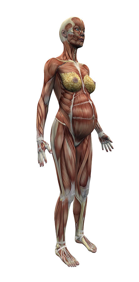
It's not known whether the woman traveled to Timna from Egypt while she was pregnant or whether she was impregnated while serving at the Hathor temple, Ben-Yosef said. "Probably she wouldn't have traveled if she knew she was pregnant, but this is only a guess.
Egypt's power in the Timna area weakened in the century after the woman died, and Egypt eventually lost control of the mines to other groups in the region.
Other findings in Timna Valley include preserved leftovers from a mining camp called Slaves' Hill. Those leftovers suggested the metalworkers ate meals of sheep and goat, as well as pistachios, grapes and fish during the 10th century B.C. More recently at Slaves' Hill, scientists revealed the remains of a sophisticated gatehouse and donkey stables that suggested the settlement had a highly organized defense system. In another discovery, archaeologists reported in 2013 that they had dug up artifacts in the valley that dated to the time of the biblical King Solomon.
Other research is underway to get a better understanding of the history of Timna.
Original article on Live Science.
Owen Jarus is a regular contributor to Live Science who writes about archaeology and humans' past. He has also written for The Independent (UK), The Canadian Press (CP) and The Associated Press (AP), among others. Owen has a bachelor of arts degree from the University of Toronto and a journalism degree from Ryerson University.
The body of a woman during pregnancy
Structural and functional changes in the body of a pregnant woman are aimed at achieving the following main goals:
- ensuring an adequate supply of the growing body of the fetus with oxygen, nutrients and the evacuation of the products of its vital activity from the body of the fetus;
- preparation of the mother's body for the process of childbirth and breastfeeding.
Since these goals are the normal physiological tasks of the human reproduction process, the changes in the woman's body during pregnancy should be considered as natural and physiological. On the other hand, since all the systems of a woman's body work during this period in a more intense mode, a point of view has recently appeared that considers pregnancy as a kind of "strength test" of the mother's body. According to this concept, pregnancy reveals "weak links" in a woman's body, which can lead to the development of pregnancy pathology. nine0003
Let's consider these changes according to the body systems. At the same time, this will make it possible to formulate some preventive measures that prevent the development of pathology in the event that such a system is a “weak link”.
Changes in body weight
Undoubtedly, one of the most noticeable changes that occur in a pregnant woman is a change in body weight. By the end of pregnancy, a woman's weight increases by about 10-12 kg. This value is distributed as follows: fetus, placenta, membranes and amniotic fluid - approximately 4.0 - 4.5 kg, uterus and mammary glands -1.0 kg, blood - 1.5 kg, intercellular (tissue) fluid - 1 kg , an increase in the mass of adipose tissue of the mother's body - 4 kg. nine0003
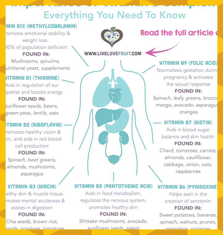
Obviously, such an increase in the weight of the woman herself, as well as the process of development and growth of the body of the fetus, place increased demands on the nutrition of the pregnant woman. Along with an adequate intake of proteins, fats and carbohydrates, it is usually recommended to supplement the woman's diet with iron preparations (necessary for the synthesis of maternal and fetal red blood cells), vitamins and calcium preparations (building the fetal bone skeleton).
The question often arises - what weight gain should be considered normal, and what is excessive? It all depends on the initial weight of the woman before pregnancy. And not so much on weight, but on the ratio of weight and height, expressed by the so-called body mass index (BMI). BMI is calculated using the formula:
BMI \u003d Weight (kg) / Height 2 (m 2 )
2 . Women with an index from 20.0 to 26.0 are considered proportionately built. If the index exceeds 26.0, then these are women with signs of obesity, and if the BMI is less than 20.0, then the woman has a nutritional deficiency.
The approach to weight gain in pregnant women according to BMI is as follows. Significantly greater weight gain is allowed in undernourished women than in normal-nourished women (eg, weight gain of 15-18 kg should not be considered pathological in them). The approximate estimated value of weight gain during pregnancy for women with a normal physique is 10-12 kg. And for women with signs of obesity, weight gain should be less than for women in the two previous groups and not exceed 10 kg. nine0003
Women who smoke during pregnancy have less overall weight gain than non-smokers. This weight deficit is mainly formed due to a smaller increase in the weight of the fetus, placenta and amniotic fluid. Children born to smoking mothers weigh 250 g less than non-smoking women, which directly indicates the obvious negative effect of smoking on intrauterine development of the fetus.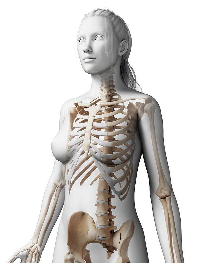
Let's consider the changes in the main vital systems of a woman's body during pregnancy.
Respiratory system
An increase in the blood concentration of the hormone of pregnancy - progesterone leads to additional relaxation of the smooth muscles of the bronchial wall and an increase in the lumen of the airways. Increasing requirements for the supply of oxygen to a growing fetus is expressed in an increase in tidal volume (the amount of air inhaled in one respiratory movement) and respiratory rate per minute. This leads to an increase in the so-called "minute ventilation" indicator by 30-40%, which significantly covers the oxygen demand of the pregnant woman's body increased to 15-20%. The fetal requirement is approximately 30% of the total increase in oxygen consumption of the body of a pregnant woman. An additional 10% falls on the needs of the placenta, and the rest goes to cover the increased work of the woman's body systems due to pregnancy. nine0003
Cardiovascular system
This system takes on the main burden of increasing the blood supply to the pregnant uterus and delivering oxygen and nutrients to it. Oxygen entering through the lungs must bind to its carrier - hemoglobin contained in red blood cells - erythrocytes. Therefore, an increase in oxygen transport to the uterus and maternal tissues is impossible without a corresponding increase in blood volume. The volume of circulating blood (blood contained in the vessels) during pregnancy increases by 40-55%, which in absolute terms is approximately 1.5 liters. This increase in the mass of pumped blood leads to a significant increase in the work of the heart. This is done both by increasing (by 30%) the stroke volume of the heart (the amount of blood ejected by the heart into the aorta in one contraction), and by increasing the heart rate by 15-20%. To ensure the circulation of such a significant volume of blood through the vascular system, the resistance of the latter is reduced by about 25%. Against the background of an increase in the stroke volume of the heart, heart rate and circulating blood volume, the diameters of the output tracts of the ventricles and large vessels no longer provide a relatively laminar (layered) blood flow and generate significant turbulence (the presence of eddies) of the blood flow.
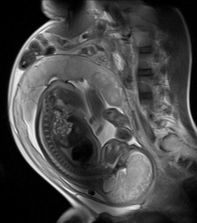 This is manifested in the appearance of the so-called physiological systolic noise - sound vibrations that occur when a blood stream passes through these holes. Thus, in the majority (80%) of healthy pregnant women, starting from the second trimester of pregnancy, a systolic heart murmur can be heard, which is completely normal. nine0003
An increase in the lumen of the spiral arteries of the uterus and an increase in blood flow through the uterus leads to a decrease in the total peripheral resistance of the vascular system. Therefore, many pregnant women experience a decrease in both systolic and diastolic blood pressure.
If the heart copes relatively well with an increase in the minute volume of blood circulation, then the vascular system finds itself in much more stressful conditions of functioning. Indeed, a 50% larger volume of blood must be accommodated in the existing volume of the vascular system. And the most vulnerable in this situation is the venous system. The arterial system, which delivers oxygenated and nutrient-rich blood, operates under conditions of relatively high pressure. We know its normal values, It is 120 mm Hg at the moment of ejection of blood into the aorta (systole) and 70 mm Hg in diastole (when the heart fills with blood for the next systole). The pressure in the venous system is much less - it is about 10 mm Hg. nine0003
To the right of the spine, everyone (both men and women) has a large venous vessel - the inferior vena cava, which collects blood from the lower extremities, uterus and internal organs of the pelvis. After the 20th week of pregnancy, the weight of the uterus, containing the growing fetus, placenta and amniotic fluid, reaches a significant value. Therefore, if the woman at this time is in a horizontal position lying on her back, the uterus can cause partial compression of the inferior vena cava. This, in turn, leads to an increase in blood pressure below the site of clamping, additional vasodilation and deterioration of blood outflow from the lower extremities, uterus and rectum. Obviously, it is these events that can contribute to or cause the development of a fairly common complication in pregnant women - varicose veins of the lower extremities and rectum (hemorrhoids).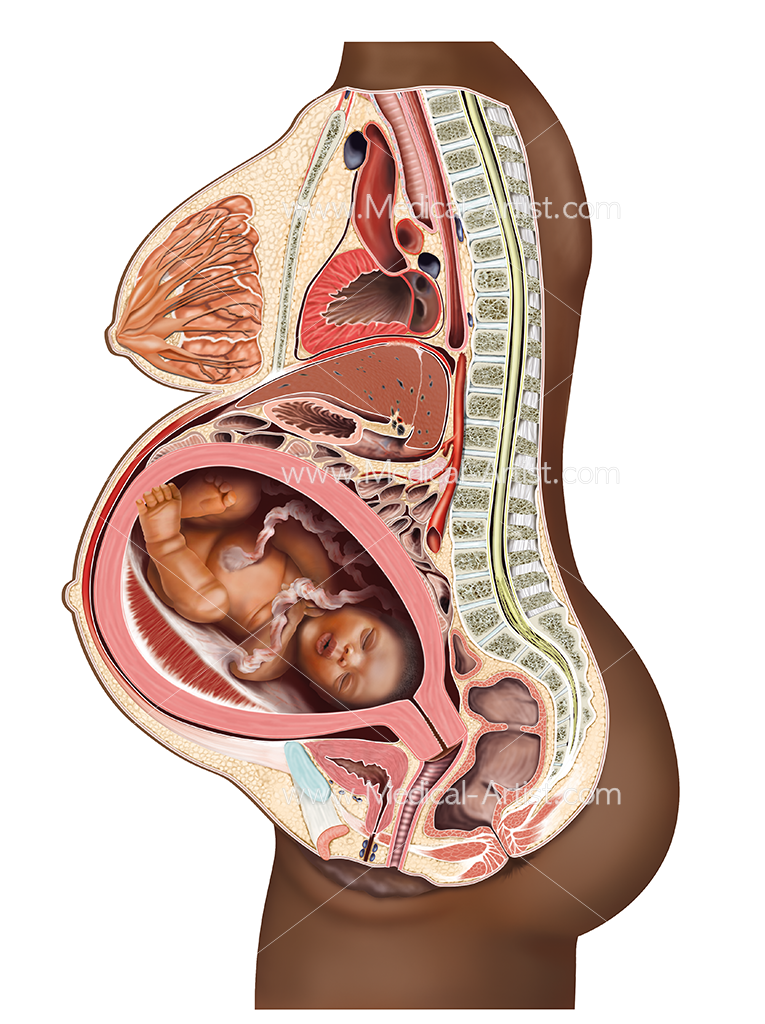 nine0003
In this regard - small practical recommendations:
1. Pregnant women (after 20 weeks) are categorically contraindicated for any physical exercises on the back (especially those accompanied by lifting the legs).
2. It is advisable to sleep in a lying position on your side - if possible on the left (because the inferior vena cava runs on the right) and put a pillow between your legs. The latter contributes to the optimal outflow of blood from the uterus and pelvic organs. Of course, many women find it hard to sleep on their side for the rest of their pregnancy. Therefore, a pregnant woman should have as many pillows in bed as she needs. You can put a couple of pillows under your back on one side so that the uterus deviates slightly to the side and does not press vertically on the vena cava. At the same time, it is useful to have a special pillow that a woman will put under her stomach, which will ensure a comfortable position for the uterus. nine0003
3. Daily exercise reduces the risk of these complications.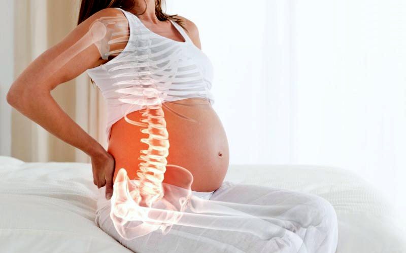 The veins of our lower extremities have valves that reduce the pressure of the blood column on the walls of the lower sections of the veins of the legs in an upright position. When a person walks, the contraction of the muscles surrounding the vessels promotes blood flow to the heart and unloads the venous system of the legs.
Changes in the blood system
As noted, one of the most significant adaptive changes in the body of a pregnant woman is an increase in the volume of circulating blood. The volume of the liquid component of the blood (plasma) of the blood begins to increase already in the first trimester (6-10 weeks) and reaches its peak by 30-34 weeks. A good correlation has been shown between the increase in maternal plasma volume and estimated fetal weight. The mass of erythrocytes also increases starting from the 10th week, but the rate of this increase is somewhat less and reaches only 18%-30% of the initial level. Thus, the liquid part of the blood increases in a greater proportion than the mass of red blood cells. This leads to a physiological decrease in blood viscosity and the content of formed elements per unit of its volume, which is expressed in a decrease in hematocrit - an indicator that reflects the percentage volume of cellular elements from the total blood volume, from about 42% to 37%. Therefore, during pregnancy, it is necessary to ensure sufficient intake of proteins and especially iron preparations for adequate synthesis of red blood cells. It was shown that in pregnant women who received iron supplements during pregnancy, an increase in the mass of erythrocytes reached 30%, and in those who did not receive only 18%. nine0003
During pregnancy, the number of white blood cells - leukocytes - may slightly increase. The number of platelets (platelets that provide blood clotting) usually does not change significantly. However, blood clotting increases, which provides a state of so-called hypercoagulability.
Changes in the excretory system
The work of the kidneys ensures not only the excretion of metabolic products (urea) from the body, but is also extremely important for maintaining a normal level of blood pressure, regulating the exchange of water and electrolytes (potassium, sodium, chlorine ions) between the vessels and tissues.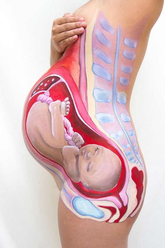 An increase in the volume of circulating blood and blood flow through the kidneys of a pregnant woman is also expressed in changes in their structure. Already starting from 10-12 weeks, there is some expansion of the pyelocaliceal complex (the system of cavities that collect urine in the kidney). It is believed that at this early stage of pregnancy, changes in the kidneys are associated with an increase in the synthesis of progesterone (pregnancy hormone) in the woman's body. Then, due to the increase in the size of the uterus and the possible compression of the ureters by it, further expansion of the pyelocaliceal complex of the kidneys can be observed. Progesterone also slightly reduces the tone of the smooth muscle cells of the bladder wall and increases its capacity. All of these things make a woman less resistant to a possible ascending urinary tract infection. If inflammatory changes in the kidneys occurred before pregnancy, then under these conditions, an exacerbation of inflammatory kidney diseases is more likely.
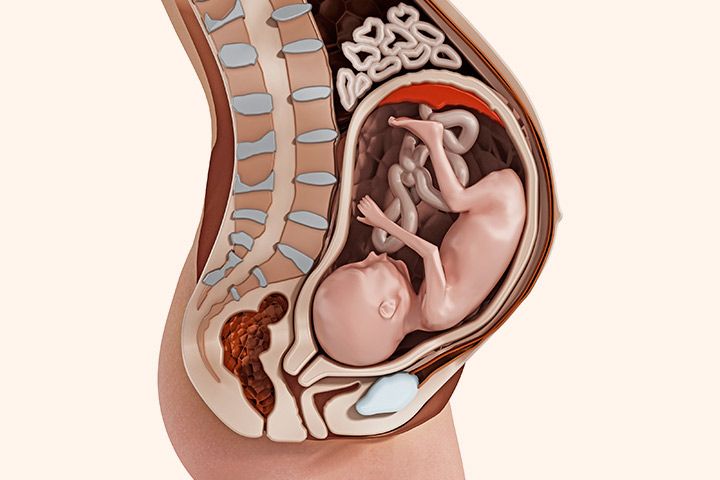 In late pregnancy, the uterus can put significant pressure on the bladder, causing frequent urination. In some women, against the background of such changes, symptoms of urinary incontinence may be observed. nine0003
Corresponding to changes in blood flow through the kidneys, renal filtration also increases (up to 50%). Due to increased filtration through the kidneys, some women with uncomplicated pregnancy may experience a small concentration of sugar in the urine. However, protein during a physiological pregnancy in the urine should not be detected. The appearance of protein in the urine can be the result of either inflammatory processes in the kidneys or preeclampsia.
As noted above, during pregnancy, the volume of blood circulating in the vascular system of a woman increases by 1.5 liters. It is quite natural that part of the fluid, under the influence of increased hydrostatic pressure, passes from the vascular system to the tissues. Therefore, at the end of a normal pregnancy, a woman may have slight swelling. nine0003
Pregnant women's water intake is an important issue. Consider the principle of the kidneys. Urine production consists of two stages: filtration and reabsorption. At the first stage, in the cells of the kidneys - nephrons, under the influence of a certain level of arterial pressure, the liquid part of the blood - plasma - is filtered through the thinnest membranes. Only blood cells and proteins do not pass through these membranes. All other substances (necessary and unnecessary) freely cross these membranes and form the so-called "primary urine". Such urine is produced in the body of an adult about 150 liters per day. Obviously, we cannot lose this amount of fluid. Therefore, the second stage of urine formation is reabsorption - reabsorption. At the same time, from the primary urine passing through the network of thin tubules, the absorption of useful substances into the blood begins: such as potassium ions, glucose, partially sodium ions and other substances. Together with these substances, the absorption of water occurs.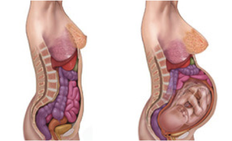 Substances that are unnecessary for the body - slags (urea) are not absorbed and remain in the urine. As a result of this process, the concentration of urine and a kind of compression of toxins in a much smaller volume (1.5 - 2.0 liters) occur. The work of concentrating urine is quite intense and requires a certain expenditure of energy. It is obvious that the restriction of fluid intake by a pregnant woman puts her kidneys in rather stressful conditions in terms of urine concentration. After all, the kidneys have to allocate not only the daily amount of toxins formed in the mother's body, but also the toxins that are filtered through the placenta from the body of the fetus. Therefore, current recommendations for fluid intake by a healthy pregnant woman suggest that a pregnant woman should consume at least 2 liters of fluid per day. Moreover, dehydration is much more dangerous for the mother and fetus than a hypothetical increase in water intake, which is well regulated by thirst. nine0003
Changes in the gastrointestinal tract
Changes in the digestive system are also largely the result of the influence of an increased concentration of progesterone in the blood. In pregnant women, saliva production is slightly increased. This reduces motility (peristaltic contractile activity) of the entire gastrointestinal tract. The time of evacuation of food from the stomach increases. In some women, there is a decrease in the tone of the sphincter (muscle sphincter) between the stomach and esophagus, which may be the cause of reflux (reflux of gastric contents into the esophagus), accompanied by heartburn. This negative phenomenon is partially offset by a decrease in the synthesis of hydrochloric acid and an increase in mucus production. However, if a woman has heartburn, then the right solution is to take any antacids - drugs that reduce acidity such as Rennie or baking soda. nine0003
Decreased intestinal motility may manifest as constipation. Therefore, during pregnancy, women need to pay special attention to the diet and the mandatory use of foods high in fiber that stimulates intestinal motility (black bread, fruits, vegetables).
Significant changes occur with the activity of the gallbladder.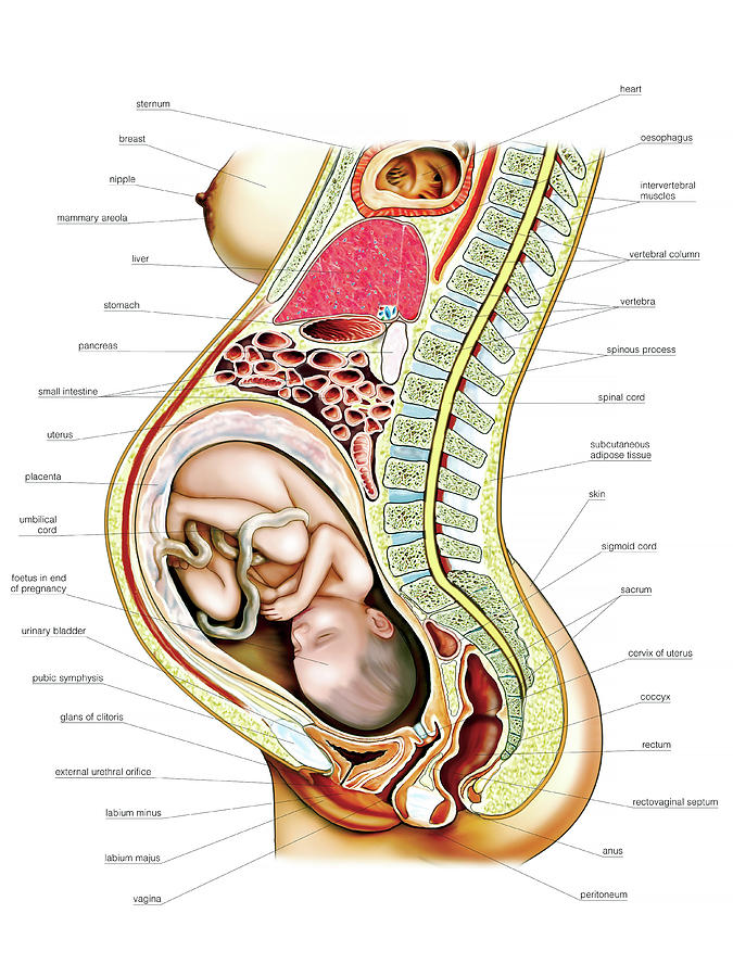 A decrease in the motor activity of the gallbladder, squeezing it with an enlarged uterus in the later stages and a change in the chemical composition of bile (an increase in cholesterol concentration) can contribute to the formation of gallstones. nine0003
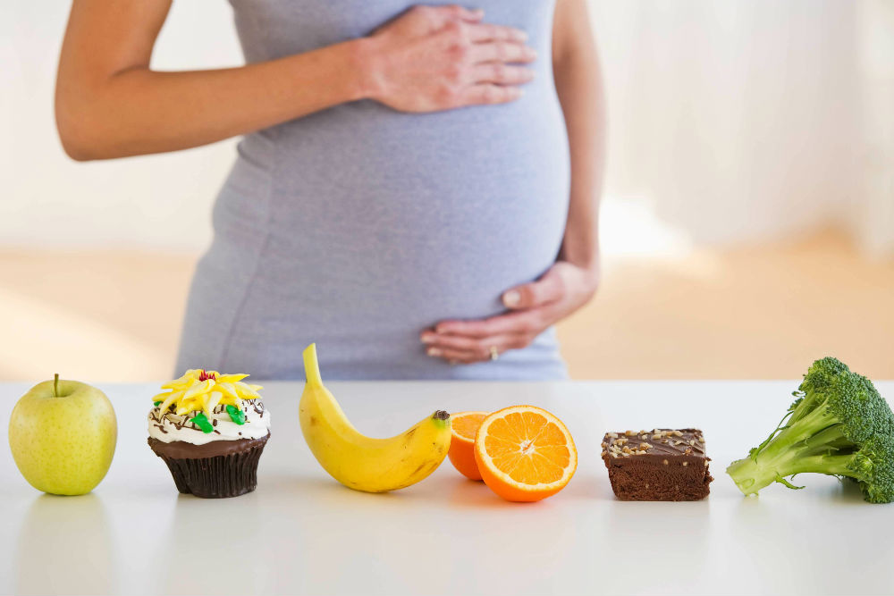
A few words about the diet
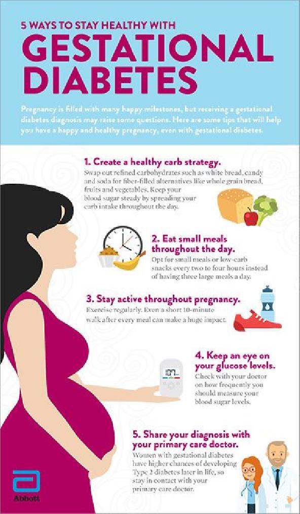
Taking into account the above-mentioned changes in motility and direct mechanical pressure on the digestive organs by the growing uterus, frequent fractional meals are considered the most rational for a pregnant woman. It is advisable to have approximately 6 meals per day. Given that the body of the fetus is formed mainly from proteins, these food components play a special role in the diet of a pregnant woman. In the gastrointestinal tract of a pregnant woman, proteins are broken down, digested and absorbed. It should be noted that amino acids are absorbed - the elementary components that make up proteins. The source of amino acids in the body of the mother and fetus can be proteins of a very different nature. These are primarily proteins of animal origin: lean meat (beef, pork), poultry meat. Fish is very useful, especially fatty varieties of sea fish containing omega-3 fatty acids, which play a positive role in the prevention of preeclampsia. An important source of protein is eggs, milk and dairy products. Proteins of plant origin (nuts, soy) are very useful for the mother and the unborn child. nine0003
In addition to their significant nutritional value, proteins have another important property. Proteins have been shown to somewhat slow down the absorption of carbohydrates from food, the main energy substrate for a growing organism. Thanks to this property, proteins smooth out the sharp peaks in the increase in the concentration of glucose in the mother's blood after a meal and make the concentration of carbohydrates in the blood of a pregnant woman more stable, which contributes to a more even flow of these substances to the fetus.
Skeletal and muscular system changes
An increase in the concentration of the hormones relaxin and progesterone in the blood contributes to the leaching of calcium from the skeletal system.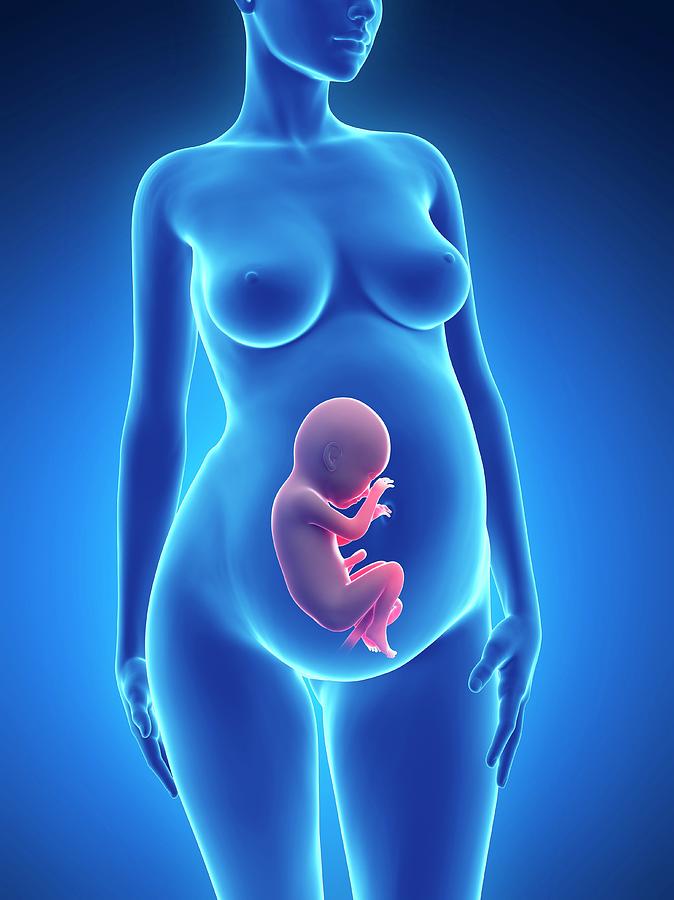 This accomplishes two goals. On the one hand, this helps to reduce the rigidity of the joints between the bones of the pelvis (especially the pubic joint) and increase the elasticity of the pelvic ring. Increasing the elasticity of the pelvis is of great importance in increasing the diameter of the internal bone ring in the first stage of labor and further reducing the resistance of the birth tract to fetal movement in the second stage of labor. Secondly, calcium, washed out of the mother's skeletal system, is used to build the skeleton of the fetus. nine0003
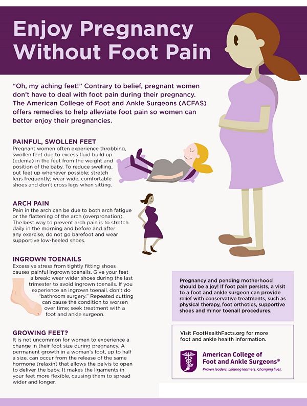
It should be noted that calcium compounds are washed out not only from the bones, but from all the bones of the maternal skeleton (including the bones of the foot and spine). As shown earlier, a woman's weight increases during pregnancy by 10 -12 kg. This additional load against the background of a decrease in bone stiffness can cause foot deformity and the development of flat feet. A shift in the center of gravity of the body of a pregnant woman due to an increase in the weight of the uterus can lead to a change in the curvature of the spine and the appearance of pain in the back and pelvic bones. Therefore, for the prevention of flat feet, pregnant women are advised to wear comfortable shoes with low heels. It is advisable to use insoles that support the arch of the foot. For the prevention of back pain, special physical exercises are recommended that can unload the spine and sacrum, as well as wearing a comfortable bandage. Despite an increase in calcium loss by the bones of the skeleton of a pregnant woman and an increase in their elasticity, the structure and density of bones (as is the case with osteoporosis in older women) does not change. nine0003
Skin changes
The skin of a pregnant woman is mainly influenced by three types of hormones. These are: female sex hormones (estrogens), pregnancy hormone (progesterone) and melanin-stimulating hormone. The latter hormone contributes to the accumulation of melanin in the skin. The increase in the content of this substance in the skin is responsible for its darkening during sunburn.
Due to the complex action of these hormones, many pregnant women notice an increase in the pigmentation of certain areas of the skin.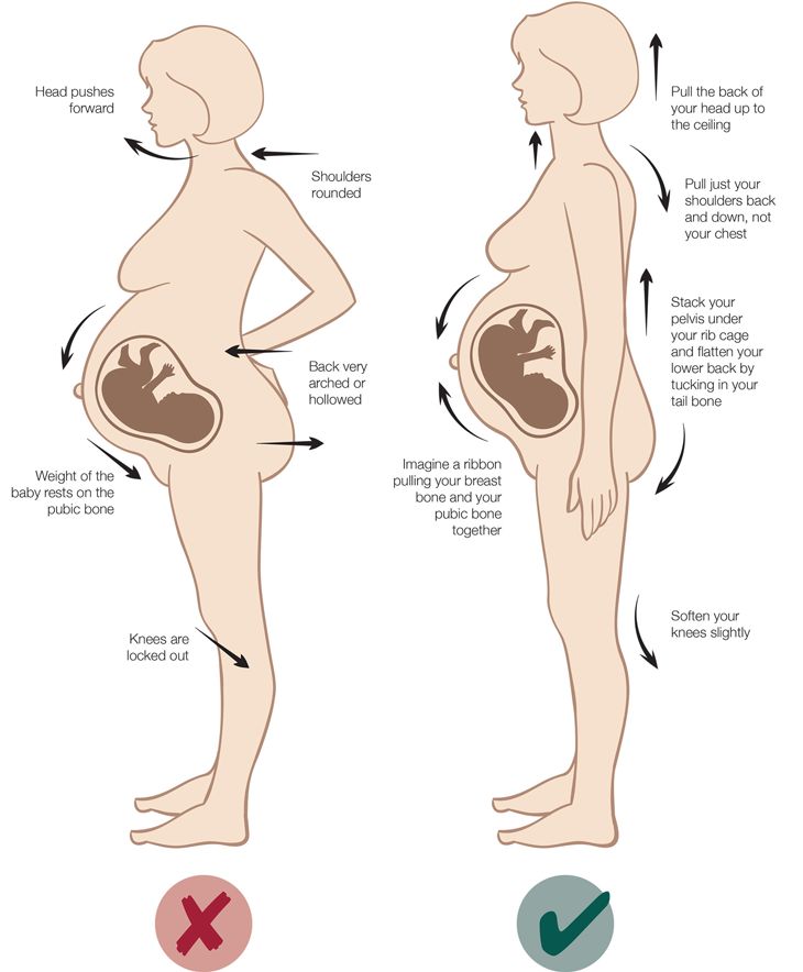 Among them: halos of the nipples of the mammary glands, skin areas around the navel, midline of the abdomen and perineum. In some women, changes in facial pigmentation have a rather characteristic appearance, referred to as the “pregnant mask”. Stimulates pigmentation of birthmarks. Therefore, pregnant women are not recommended to sunbathe at this time, and tanning in solariums using intense ultraviolet radiation is simply contraindicated. nine0003
Many women develop structural changes in areas of the skin that are most exposed to stretching during pregnancy. These include the lateral surfaces of the abdomen, mammary glands and thighs. These changes appear in the form of so-called "stretch marks" or, as they are called in Latin, stria gravidarum (pregnancy bands). Stretch marks on the skin are mostly mechanical in nature and currently cannot be prevented or eliminated by any type of medical or hormonal treatment. nine0003
Changes in the mammary glands
Changes in the mammary glands to prepare them for breastfeeding begin in the earliest stages of pregnancy.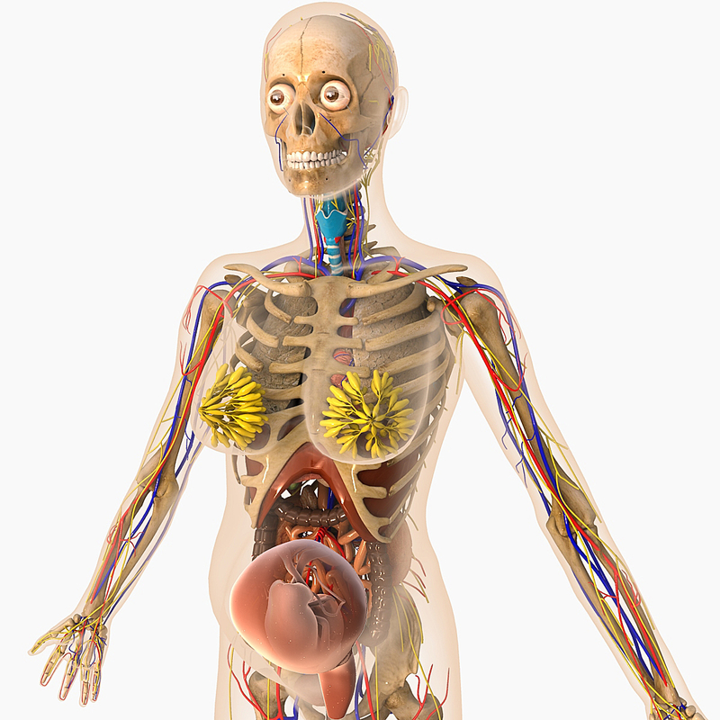 An increase in the production of progesterone by the placenta and an increase in the concentration of the second hormone that affects lactation, prolactin, leads to the growth of glandular cells that produce milk. At the same time, under the influence of estrogen, the growth of the milk ducts is activated, supplying milk from the glandular cells to the nipple. This increase in the cell mass of the mammary glands requires a greater blood supply. Therefore, blood flow to the mammary glands increases. Often, a pronounced vascular network is visible around the mammary glands. The areolas of the nipples increase in diameter and become darker. Gradually, the production of the precursor of milk - colostrum begins. This is a light, thick liquid that is released in the amount of a few drops when pressing on the nipple at the end of pregnancy. nine0003
Conclusion
Summarizing the facts concerning changes in a woman's body during pregnancy, it is worth emphasizing that these changes reflect the processes of physiological adaptation of the mother's body to the process of intrauterine development of the fetus.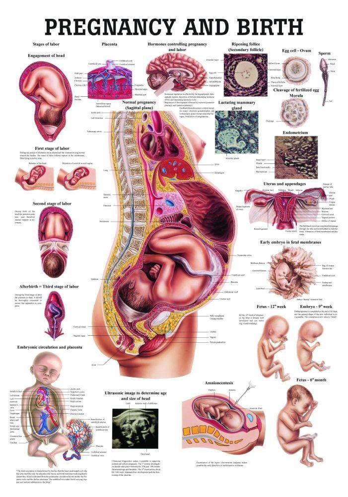 Therefore, measures aimed at preventing the pathology of pregnancy should be, first of all, natural, physiological. This is a proper and balanced diet, smoking cessation, a sufficient level of physical activity and fluid intake. In a healthy woman, such approaches ensure the normal course of pregnancy and adequate preparation of the mother's body for childbirth and breastfeeding. nine0003
Tsyvyan Pavel Borisovich, Professor,
head of the Partner childbirth center, (343) 374-31-20
Source: http://eka-mama.ru/matter/pregnancy/39712.html
Physiological changes in the body during pregnancy
From the very first days of pregnancy, a woman's body undergoes profound transformations. These transformations are the result of the coordinated work of almost all body systems, as well as the result of the interaction of the mother's body with the child's body. During pregnancy, many internal organs undergo significant restructuring. These changes are adaptive in nature, and, in most cases, are short-lived and completely disappear after childbirth.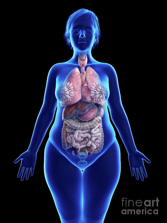 Consider the changes in the basic systems of the vital activity of a woman's body during pregnancy. nine0003
The respiratory system during pregnancy works hard. The respiratory rate increases. This is due to an increase in the need of the mother and fetus for oxygen, as well as in the limitation of the respiratory movements of the diaphragm due to an increase in the size of the uterus, which occupies a significant space of the abdominal cavity.
The mother's circulatory system during pregnancy has to pump more blood to ensure an adequate supply of nutrients and oxygen to the fetus. In this regard, during pregnancy, the thickness and strength of the heart muscles increase, the pulse and the amount of blood pumped by the heart in one minute increase. In addition, the volume of circulating blood increases. In some cases, blood pressure increases. The tone of blood vessels during pregnancy decreases, which creates favorable conditions for increased supply of tissues with nutrients and oxygen.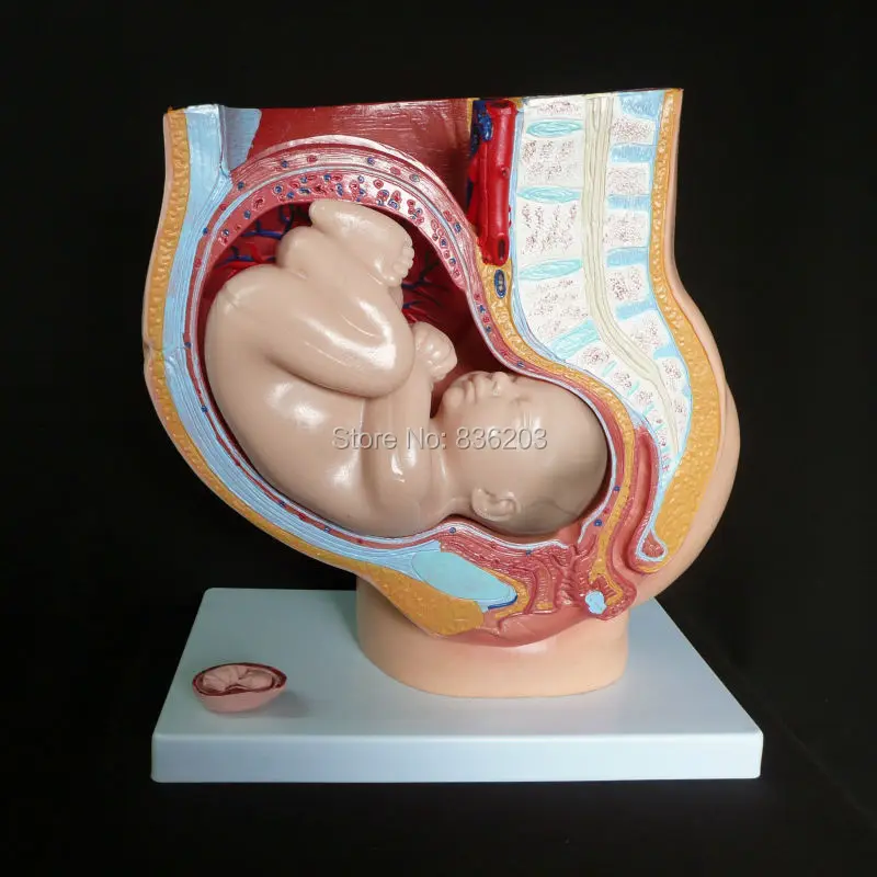 During pregnancy, the network of vessels of the uterus, vagina, and mammary glands decreases sharply. On the external genitalia, in the vagina, lower extremities, there is often an expansion of the veins, sometimes the formation of varicose veins. Heart rate decreases in the second half of pregnancy. It is generally accepted that the rise in blood pressure over 120-130 and a decrease to 100 mm Hg. signal the occurrence of pregnancy complications. But it is important to have data on the initial level of blood pressure. nine0003
And changes in the blood system. During pregnancy, blood formation increases, the number of red blood cells, hemoglobin, plasma and bcc increases. BCC by the end of pregnancy increases by 30-40%, and erythrocytes by 15-20%. Many healthy pregnant women have a slight leukocytosis. ESR during pregnancy increases to 30-40. Changes occur in the coagulation system that contribute to hemostasis and prevent significant blood loss during childbirth or placental abruption and in the early postpartum period.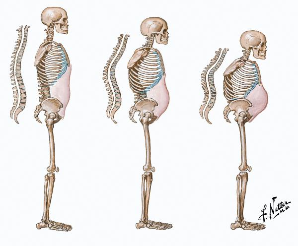 nine0003
Kidneys work hard during pregnancy. They secrete decay products of substances from the body of the mother and fetus (the waste products of the fetus pass through the placenta into the mother's blood).
Changes in the digestive system are represented by increased appetite (in most cases), craving for salty and sour foods. In some cases, there is an aversion to certain foods or dishes that were well tolerated before the onset of pregnancy. Due to the increased tone of the vagus nerve, constipation may occur. nine0003
The most significant changes, however, occur in the genitals of pregnant women. These changes prepare the woman's reproductive system for childbirth and breastfeeding.
The uterus of a pregnant woman increases significantly in size. Its mass increases from 50 g - at the beginning of pregnancy to 1200 g - at the end of pregnancy. The volume of the uterine cavity by the end of pregnancy increases by more than 500 times! The blood supply to the uterus is greatly increased. In the walls of the uterus, the number of muscle fibers increases. The cervix is filled with thick mucus that clogs the cavity of the cervical canal. The fallopian tubes and ovaries also increase in size. In one of the ovaries, there is a "corpus luteum of pregnancy" - a place for the synthesis of hormones that support pregnancy. Walls vaginas will loosen and become more elastic. External genitalia (labia minor and major), also increase in size and become more elastic. The tissues of the perineum are loosened. In addition, there is an increase in mobility in the joints of the pelvis and a divergence of the pubic bones. The changes in the genital tract described above are of extremely important physiological significance for childbirth. Loosening the walls, increasing the mobility and elasticity of the genital tract increases their throughput and facilitates the movement of the fetus through them during childbirth. nine0003
Skin in the genital area and in the midline of the abdomen usually becomes darker in color. Sometimes "stretch marks" form on the skin of the lateral parts of the abdomen, which turn into whitish stripes after childbirth.
Mammary glands increase in size, become more elastic, tense. When pressing on the nipple, colostrum (first milk) is released.
Changes of the bone skeleton and muscular system . An increase in the concentration of the hormones relaxin and progesterone in the blood contributes to the leaching of calcium from the skeletal system. This helps to reduce the rigidity of the joints between the bones of the pelvis and increase the elasticity of the pelvic ring. Increasing the elasticity of the pelvis is of great importance in increasing the diameter of the internal bone ring in the first stage of labor and further reducing the resistance of the birth tract to fetal movement in the second stage of labor. Also, calcium, washed out of the mother's skeletal system, is used to build the skeleton of the fetus. nine0003
It should be noted that calcium compounds are washed out of all bones of the maternal skeleton (including the bones of the foot and spine).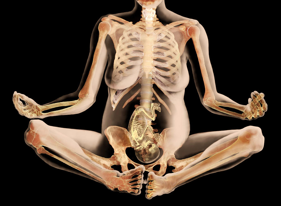 As shown earlier, a woman's weight increases during pregnancy by 10 -12 kg. This additional load against the background of a decrease in bone stiffness can cause foot deformity and the development of flat feet. A shift in the center of gravity of the body of a pregnant woman due to an increase in the weight of the uterus can lead to a change in the curvature of the spine and the appearance of pain in the back and pelvic bones. Therefore, for the prevention of flat feet, pregnant women are advised to wear comfortable shoes with low heels. It is advisable to use insoles that support the arch of the foot. For the prevention of back pain, special physical exercises are recommended that can unload the spine and sacrum, as well as wearing a comfortable bandage. Despite an increase in calcium loss by the bones of the skeleton of a pregnant woman and an increase in their elasticity, structure and bone density (as is the case with osteoporosis in older women). nine0003
Changes in the nervous system .