Prenatal brain development
Fetal brain development: When does the brain develop?
- Community
- Getting Pregnant
- Pregnancy
- Baby names
- Baby
- Toddler
- Child
- Health
- Family
- Courses
- Registry Builder
- Baby Products
Advertisement
Your baby's brain begins developing early in pregnancy, just three weeks after fertilization, and continues throughout your pregnancy. The third trimester is when major developments happen, and your baby's brain triples in weight. Taking folic acid and eating a well-balanced diet that includes fish rich in omega-3s can help support your baby's brain development during pregnancy and beyond.
Photo credit: Jonathan Dimes for BabyCenter
It's probably no surprise that your baby's brain is one of the first major organs to start developing. But you may not know that it will continue to grow until your child is in their early 20s!
Together, the brain and spinal cord make up the central nervous system. The brain is enclosed within the skull, and the spinal cord is encased within a flexible spinal column made up of 33 separate bones (vertebrae). A network of nerves branches off the spinal cord, and these nerves send signals to and receive information from various organs.
Early fetal brain development
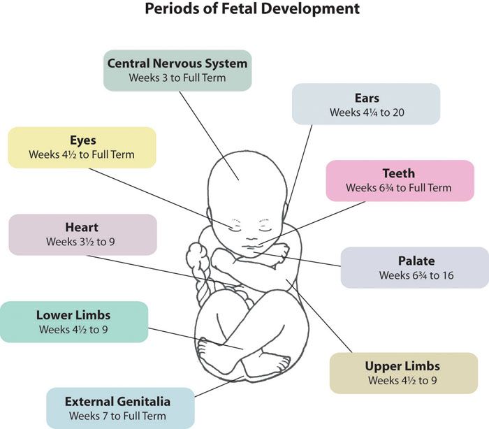
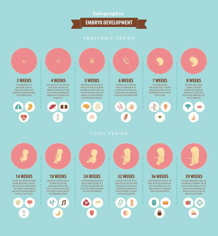
The first part develops just three weeks after fertilization as an oval-shaped disk of tissue called the neural plate. This early (5 weeks pregnant), your baby is an embryo that looks like a tiny tadpole, with the neural plate running down the middle from head to tail.
Over the course of this week, the edges of the plate rise and fold toward each other, forming the neural tube that will become your baby's spine and brain. At this point, the ends of the tube remain open, and the brain starts to take shape at the top of the tube. Near the bottom is the structure that will eventually become the tailbone.
By the time you're 6 weeks pregnant, the neural tube is completely closed at both ends, and at the top of the tube, the brain consists of three areas:
- The forebrain develops into the cerebrum, which controls certain brain functions , like thinking and problem-solving.
- The midbrain is involved in processing visual and auditory information.
- The hindbrain develops into the cerebellum, which manages balance and coordination, as well as the medulla, which is the control center for the body's automatic activities, like blood pressure and heart rate.
How the nervous system develops
Located along the edges of the developing neural tube is the neural crest. This crest, along with the brain and spinal cord, give rise to the millions of nerves that branch out all over the body.
From 8 weeks of pregnancy on, these nerves are making connections not only with each other, but also with muscles and other tissues as well as organs, like the eyes and ears. At 12 weeks, the nerves start sending out simple signals that cause reflex behaviors. For example, your baby's fingers can open and close, and their toes can curl. Your baby can also squint their eye muscles and make sucking movements with their mouth.
By about 28 weeks, nerves connect with their designated organs so your baby's senses of hearing, smell, and taste can begin to function. Some nerve cells develop a sheath of insulating material called myelin that speeds up signaling between nerves. Myelin starts to form in the third trimester and continues after birth and well into adulthood.
Advertisement | page continues below
Although brain development takes place throughout pregnancy, it really kicks into high gear in the last trimester as the brain triples in weight. During these final weeks, the cerebrum also develops deep grooves that provide extra surface area without taking up more room in the skull. This wrinkly outer layer is known as the cerebral cortex.
Your baby's brain at birth
How well does your baby's brain function at birth? Your newborn arrives equipped with all kinds of fascinating abilities that reflect the amazing growth from tiny neural plate to full-fledged nervous system:
- They'll have a wide range of reflexes that you can test yourself. For example, if you stroke your baby's cheek, they'll turn their head toward you (rooting reflex).
 Put your finger in their mouth, and they'll automatically begin to suck (sucking reflex). When you hold your baby upright with feet touching the floor, they'll make little stepping movements (stepping reflex).
 - Your baby can recognize your voice! Starting around the third trimester, your little one can eavesdrop on your conversations, and by the time they're born, they'll show a clear preference for your voice over others. Your little one will even turn their head when they hear you.
- Although you might imagine that your baby would be just as interested in looking at a toy or the TV, research shows that babies are especially attuned to human faces, preferring them to random designs.
And keep in mind that because your baby's brain continues to grow after birth, every day new neural connections form between the different parts of the brain, adding to your child's growing store of knowledge, memory, and experience. Find out how you can help raise a smart baby.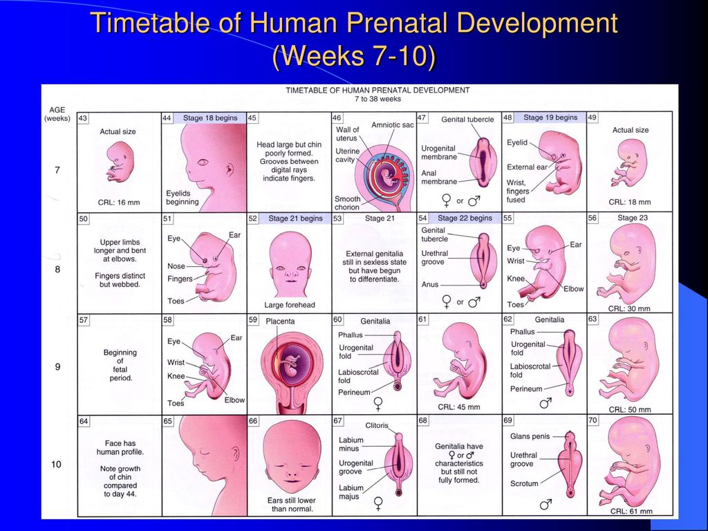
What you can do to support fetal brain development
- Take a folic acid supplement during (and even before) pregnancy. Folic acid is a B vitamin that's crucial to the development of the brain and spinal cord. Getting enough folic acid lowers the risk of neural tube defects like spina bifida and anencephaly. The neural tube develops very early (even before many women even know they're pregnant), so experts recommend that you take 400 micrograms of folic acid daily at least a month before you start trying to get pregnant. You can get it from certain prenatal vitamins or you can take it as a separate supplement.
- Eat cooked fish 2 to 3 times a week. Fish – especially fatty fish like salmon – contains omega-3 fatty acids, which research suggests boosts your baby's brain development during pregnancy and into childhood. See our article on how to choose fish that's rich in omega-3's but low in mercury and other contaminants, which can harm a baby's developing nervous system.
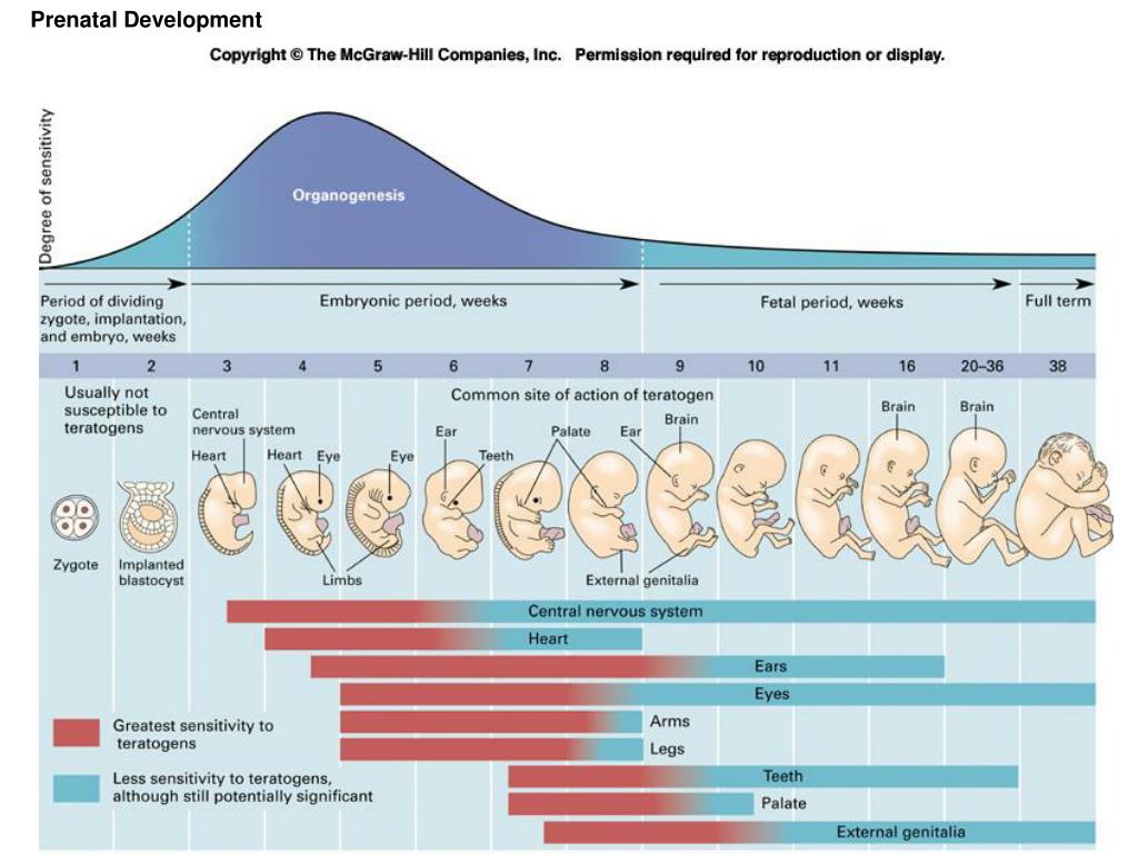
Key milestones in fetal brain development
| Weeks pregnant | Milestone |
|---|---|
| 5 weeks | The neural plate forms. |
| 6 weeks | The neural tube forms and closes. The brain is now made up of three areas (forebrain, midbrain, and hindbrain), and the ventricles have formed. |
| 8 weeks | A network of nerves starts to extend throughout the body. |
| 12 weeks | Fetal reflexes are present. |
| 28 weeks | Senses of hearing, smell, and touch are developed and functional. |
| 28 to 39 weeks | The brain triples in weight, and deep grooves develop in the cerebrum to allow more surface area for brain neurons. Myelin starts to develop along some neural pathways. |
Learn more:
- Fetal development, week by week
- How your baby's bones develop
- How your baby's eyes develop
Was this article helpful?
Yes
No
Kathleen Scogna
Kathleen Scogna is the senior director of education at the Society for Maternal-Fetal Medicine and a former freelance medical writer based in Baltimore.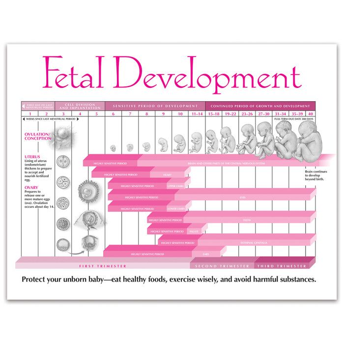
My pregnancy week by week
2
weeks
pregnant
3
weeks
pregnant
4
weeks
pregnant
5
weeks
pregnant
6
weeks
pregnant
7
weeks
pregnant
8
weeks
pregnant
9
weeks
pregnant
10
weeks
pregnant
11
weeks
pregnant
12
weeks
pregnant
13
weeks
pregnant
14
weeks
pregnant
15
weeks
pregnant
16
weeks
pregnant
17
weeks
pregnant
18
weeks
pregnant
19
weeks
pregnant
20
weeks
pregnant
21
weeks
pregnant
22
weeks
pregnant
23
weeks
pregnant
24
weeks
pregnant
25
weeks
pregnant
26
weeks
pregnant
27
weeks
pregnant
28
weeks
pregnant
29
weeks
pregnant
30
weeks
pregnant
31
weeks
pregnant
32
weeks
pregnant
33
weeks
pregnant
34
weeks
pregnant
35
weeks
pregnant
36
weeks
pregnant
37
weeks
pregnant
38
weeks
pregnant
39
weeks
pregnant
40
weeks
pregnant
41
weeks
pregnant
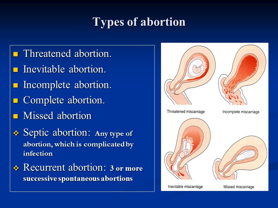
When Does a Fetus Develop a Brain?
Pregnancy is an exciting time full of rapid change and development for both you and your baby.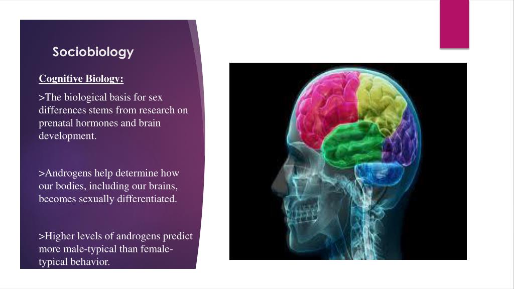 While the growth happening on the outside is clear to everyone (hello, growing belly!), it’s the development we can’t see that is truly fascinating.
Your fetus will begin the process of developing a brain around week 5, but it isn’t until week 6 or 7 when the neural tube closes and the brain separates into three parts, that the real fun begins.
Around week 5, your baby’s brain, spinal cord, and heart begin to develop. Your baby’s brain is part of the central nervous system, which also houses the spinal cord. There are three key components of a baby’s brain to consider. These include:
- Cerebrum: Thinking, remembering, and feeling occurs in this part of the brain.
- Cerebellum: This part of the brain is responsible for motor control, which allows the baby to move their arms and legs, among other things.
- Brain stem: Keeping the body alive is the primary role of the brain stem. This includes breathing, heartbeat, and blood pressure.

The first trimester is a time of rapid development and separation of the various parts of the brain, according to Kecia Gaither, MD, MPH, double board certified in obstetrics and gynecology and maternal-fetal medicine, and director of perinatal services at NYC Health + Hospitals/Lincoln.
Within 4 weeks, the rudimentary structure known as the neural plate develops, which Gaither says is considered the precursor to the nervous system. “This plate elongates and folds on itself forming the neural tube — the cephalad portion of the tube becomes the brain, while the caudal portion elongates to eventually become the spinal cord,” she explains.
The neural tube continues to grow, but around week 6 or 7, Gaither says it closes, and the cephalad portion (aka the rudimentary brain) separates into three distinct parts: front brain, midbrain, and hindbrain.
It’s also during this time that neurons and synapses (connections) begin to develop in the spinal cord. These early connections allow the fetus to make its first movements.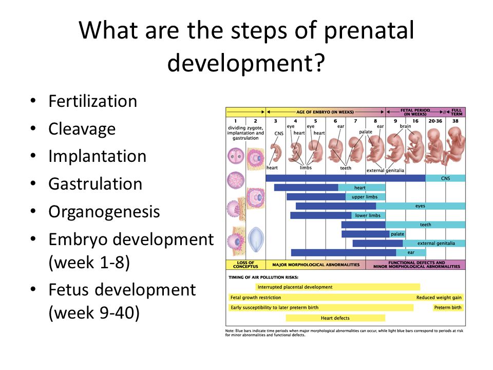
During the second trimester, Gaither says the brain begins to take command of bodily functions. This includes specific movements that come from the hindbrain, and more specifically, the cerebellum.
One of the first notable developments, sucking and swallowing, are detectable around 16 weeks. Fast-forward to 21 weeks, and Gaither says baby can swallow amniotic fluid.
It’s also during the second trimester that breathing movements begin as directed by the developing central nervous system. Experts call this “practice breathing” since the brain (and more specifically, the brain stem) is directing the diaphragm and chest muscles to contract.
And don’t be surprised if you feel some kicking during this trimester. Remember the cerebellum or the part of the brain responsible for motor control? Well, its directing the baby’s movements, including kicking and stretching.
Gaither points out that a fetus can begin to hear during the late second trimester, and a sleep pattern emerges as the brainwaves from the developing hypothalamus become more mature.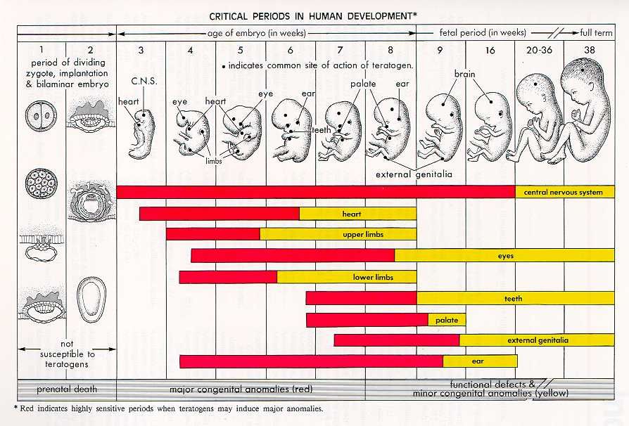
By the end of the second trimester, Gaither says the fetal brain looks structurally much like the adult brain with the brain stem almost entirely developed.
The third trimester is full of rapid growth. In fact, as your baby continues to grow, so does the brain. “All the convoluted surfaces of the brain materialize, and the halves (right brain and left brain) will separate,” explains Gaither.
The most notable part of the brain during this final trimester is the cerebellum — hence, the kicking, punching, wiggling, stretching, and all of the other movements your baby is performing.
Share on PinterestIllustration by Alyssa Kiefer
While it may feel like you have control over nothing for the next 9 months, you do have a say in the foods you eat. Healthy brain development starts before pregnancy.
According to the Centers for Disease Control and Prevention, a healthy diet that includes folic acid, both from foods and dietary supplements, can promote a healthy nervous system.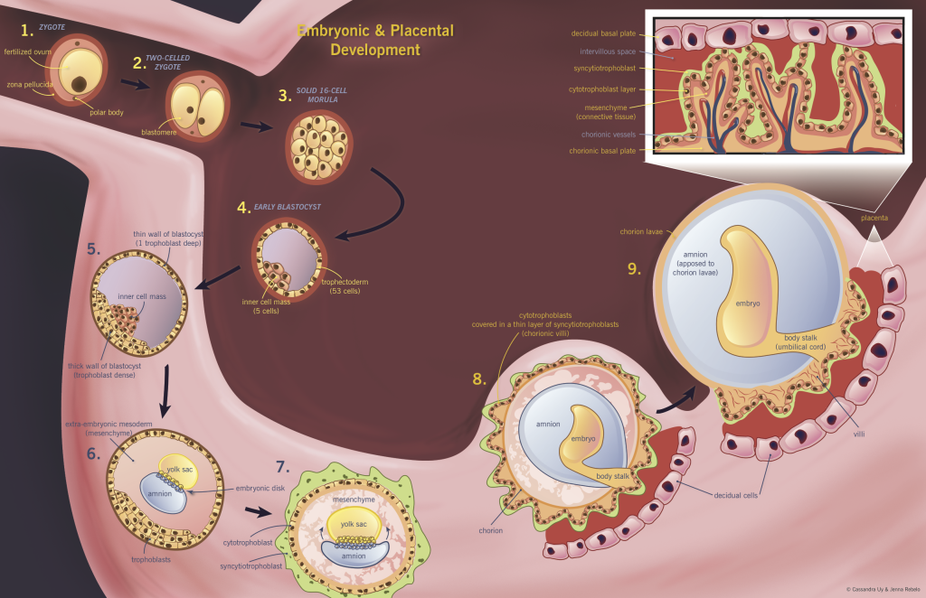
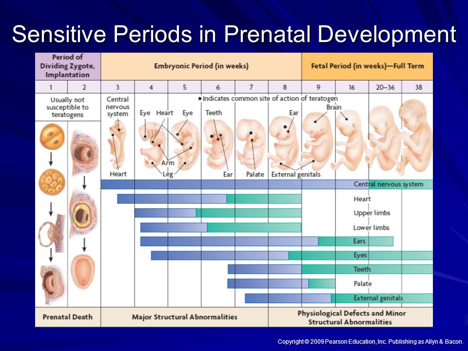
“There are a number of defects along the baby’s brain and spinal cord that can occur when there is an abnormality occurring within the first weeks of brain development,” says Gaither. This may include anencephaly or spina bifida.
Gaither says two supplements in particular are involved with fetal brain development:
Folic acidFolic acid (vitamin B9, specifically) supports fetal brain and spinal development. Not only does it play a role forming the neural tube, but Gaither says it’s also involved in the production of DNA and neurotransmitters, and it’s important for the production of energy and red blood cells.
Gaither recommends taking at least 400 to 600 micrograms of folic acid daily while you’re trying to conceive, and then continue with 400 micrograms daily during pregnancy.
“If you’ve had a child with a neural tube defect, then 4 grams daily in the preconceptual period is advised,” says Gaither.
Foods rich in folate/folic acid include dark green leafy vegetables, flaxseed, and whole grains.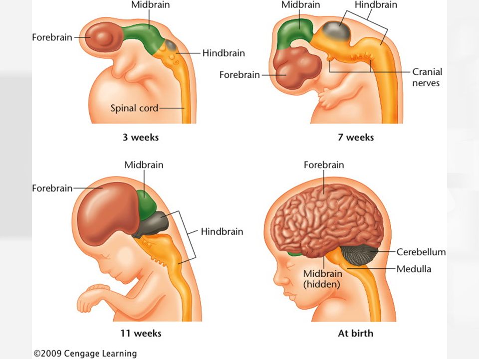
Omega-3 fatty acids
Also important for fetal brain development are omega-3 fatty acids. “The brain has a high fat content, and the omegas are helpful in the deposition of the fat in not only the brain, but the eyes as well,” explains Gaither.
Omegas are also helpful in the neural synapse development or nerve connections to each other.
Foods rich in omega 3-fatty acids include salmon, walnuts, and avocados.
Fetal brain development starts before you may even realize you’re pregnant. That’s why it’s important to start on a prenatal vitamin that contains folic acid right away. If you’re not pregnant, but thinking about having a baby, add a prenatal vitamin to your daily routine.
The brain begins to form early in the first trimester and continues until you give birth. During pregnancy, fetal brain development will be responsible for certain actions like breathing, kicking, and the heartbeat.
Talk to your doctor if you have any questions about your pregnancy, fetal brain development, or how to nurture the baby’s developing brain.
Development of the child's brain in the prenatal period
Author Olga Stepanova Reading 9 min Published
The human nervous system develops from the outer germinal lobe - the ectoderm. From the same part of the embryo, in the process of development, sensory organs, skin and sections of the digestive system are formed. Already on the 17-18th day of intrauterine development (gestation), a layer of nerve cells is released in the structure of the embryo - the neural plate, from which subsequently, by the 27th day of gestation, the neural tube is formed - the anatomical precursor of the central nervous system. The process of neural tube formation is called neurulation. During this period, the edges of the neural plate gradually fold upward, connect, and fuse with each other (Figure 1).
Figure 1. Stages of formation of the neural tube (in section).
Viewed from above, this movement may be associated with zippering (Figure 2).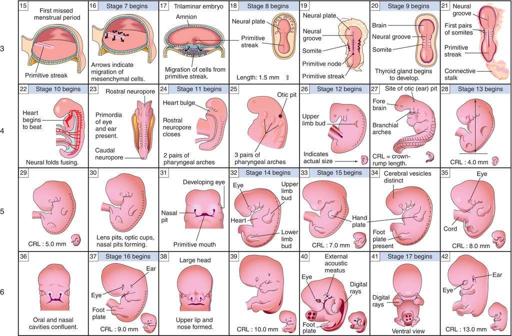
Figure 2. Stages of neural tube formation (top view).
One "zipper" is fastened from the center to the head end of the embryo (rostral wave of neurulation), the other - from the center to the tail end (caudal wave of neurulation). There is also a third "zipper", which ensures the fusion of the lower edges of the neural plate, which "zips" towards the head end and meets the first wave there. All these changes happen very quickly, in just 2 weeks. By the time neurulation is completed (31-32 days of gestation), not all women even know that they will have a child.
However, by this moment, the brain of the future person begins to form, the rudiment of two hemispheres appears. The hemispheres grow rapidly, and by the end of the 32nd day they make up ¼ of the entire brain! Then an attentive researcher will be able to see the rudiment of the cerebellum. During this period, the formation of the sense organs also begins.
Exposure to hazards during this period can lead to various malformations of the nervous system.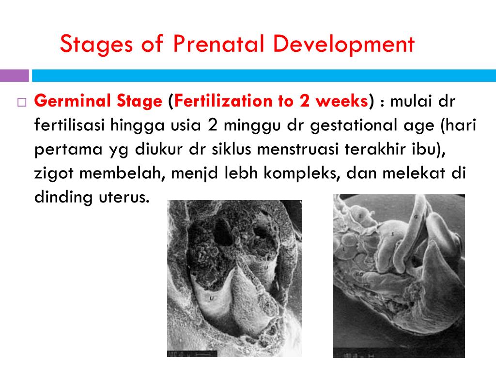 One of the most common defects is a spinal hernia, which is formed as a result of improper “fastening” of the second “zipper” (impaired passage of the caudal wave of neurulation). Even erased, almost imperceptible variants of such spinal hernias sometimes reduce the child's quality of life, leading to various forms of incontinence (urinary and fecal incontinence). If a child has a problem such as enuresis (urinary incontinence) or encopresis (fecal incontinence), it is necessary to check if he has an erased form of spinal hernia. This can be found out by making an MRI of the child's lumbosacral spine. If a spinal hernia is detected, surgical treatment is indicated, which will lead to an improvement in pelvic functions.
In my practice there was a case of a 9-year-old boy who suffered from encopresis. Only on the 6th attempt was it possible to make a high-quality MRI image, which showed the presence of a spinal hernia. Unfortunately, up to this point, the child had already been observed by a psychiatrist and received appropriate treatment, since neurologists disowned him, believing that he had mental problems.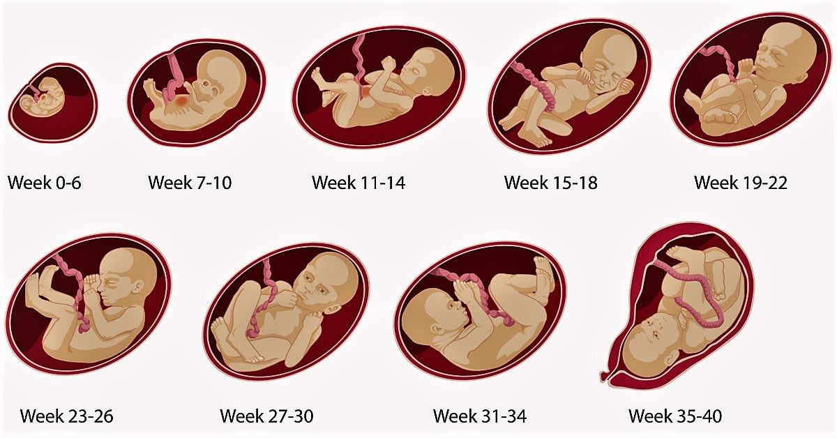 A simple operation allowed the boy to return to a normal lifestyle, to fully control his pelvic functions. Even more revealing was the story of a 16-year-old who suffered from encopresis all his life. Neurologists sent him to gastroenterologists, gastroenterologists to psychiatrists. By the time we met, he had already received psychiatric treatment for ten (!!!) years. No one ever ordered him an MRI scan. Due to the fact that our recommendations for additional examination were carried out, the guy was diagnosed with serious disorders in the lumbar spine, which led to compression of the nerves and a violation of the sensitivity of the pelvic organs. Obviously, psychiatric treatment, as well as psychotherapy or other methods of psychological influence in all these cases, are completely useless and perhaps even harmful.
To prevent malformations such as spina bifida, pregnant women are advised to take folic acid early in pregnancy. Folic acid plays the role of a protector of the cells of the nervous system (neuroprotector), and with its regular intake, the effect of various harmful factors is significantly weakened.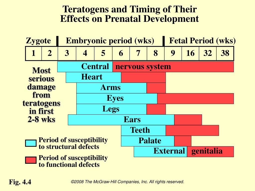
In order to minimize the risk of malformations, the expectant mother must also avoid various adverse effects on the body. Such effects include taking sedatives containing phenobarbital (including Valocordin and Corvalol), hypoxia (oxygen starvation), overheating of the mother's body. Unfortunately, certain anticonvulsant drugs also lead to adverse effects. Therefore, if a woman who is forced to take such drugs plans to become pregnant, she should consult with her doctor.
Throughout the first half of pregnancy, new nerve cells (neurons) are born and develop very actively in the baby's future brain. First of all, the processes of generation of new nerve cells occur in the area surrounding the cerebral ventricles. Another area of the birth of new neurons is the hippocampus - the inner part of the cortex of the temporal regions of the right and left hemispheres. New nerve cells continue to appear after birth, but less intensively than in the prenatal period. Even in adults, young neurons have been found in the hippocampus.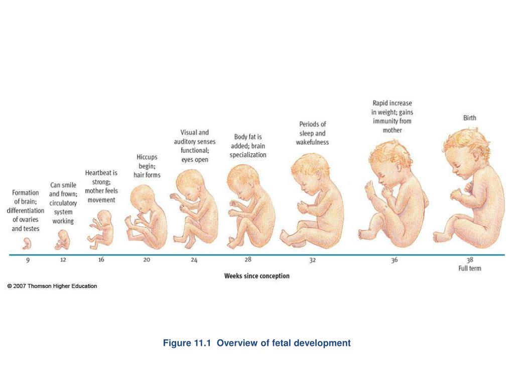 It is believed that this is one of the mechanisms due to which, if necessary, the human brain can plastically rebuild, restore damaged functions.
Newly born neurons do not stay in one place, but "crawl" to the places of their permanent "deployment" in the cortex and deep structures of the brain. This process begins towards the end of the second month of pregnancy and actively continues up to 26-29 weeks of intrauterine development. By the 35th week, the fetal cerebral cortex already has a structure inherent in the adult cortex.
Each neuron has processes through which it interacts with other cells of the body.
Figure 3. Neuron. The long process is the axon. Short branched processes - dendrites.
Neurons that have taken their place in the brain try to establish new relationships with other nerve cells, as well as with cells in other tissues of the body (for example, with muscle cells). The place where one cell connects to another is called a synapse.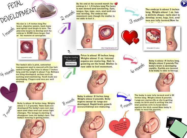 Such connections are very important, because it is thanks to them that the brain forms complex systems in which information can be quickly transmitted from one cell to another. Inside the cell, information is transmitted in the direction from the body to the end in the form of an electrical impulse. This impulse provokes the release of specific chemicals (neurotransmitters) into the synaptic cleft, which are stored at the end of the neuron, and through which information is transmitted from the neuron to the next cell.
Figure 4. Synapse
The first synapses were found in embryos at the age of 5 weeks of intrauterine development. The formation of synaptic contacts between neurons is most active starting from 18 weeks of intrauterine development. New connections between nerve cells are formed almost throughout life. During the period of active formation of synapses, the child's brain is subject to the negative influence of narcotic substances and certain medications that affect the exchange of neurotransmitters.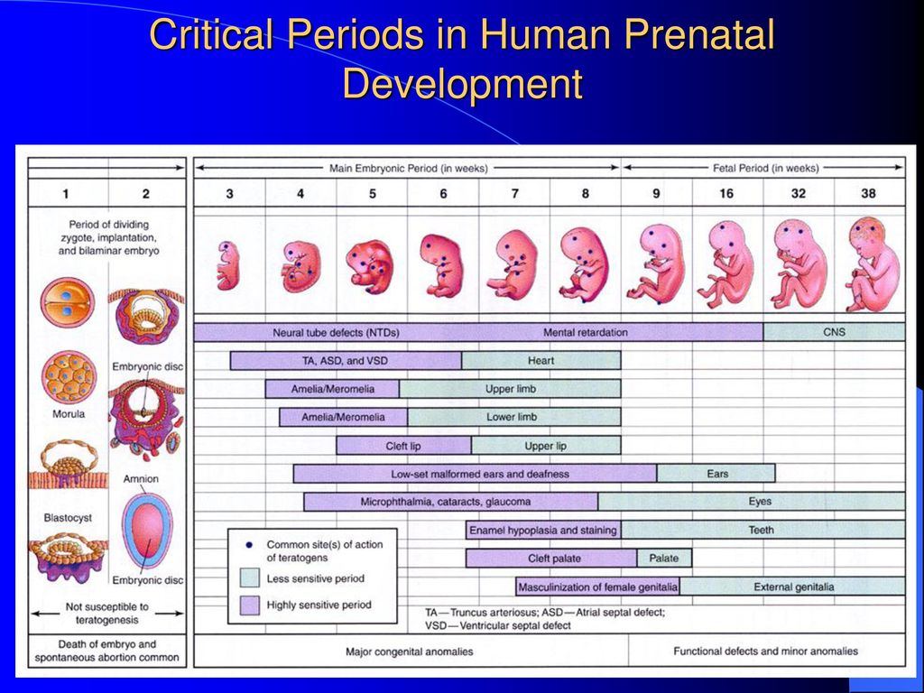 These substances include, in particular, antipsychotics, tranquilizers and antidepressants - drugs that treat mental disorders. If the expectant mother is forced to take such drugs, she should consult with her doctor. And, of course, a pregnant woman should avoid the use of psychoactive substances if she is concerned about the mental development of her child.
Neurotransmitters are specific chemical compounds that transmit information in the nervous system.
A lot in human behavior depends on their correct exchange. Including, his mood, activity, attention, memory. There are factors that can affect their exchange. One such adverse effect is maternal smoking during pregnancy. The impact of nicotine generates several effects at once. The brain recognizes nicotine as an activating agent and begins to develop systems that are sensitive to it. Simply, the number of elements perceiving nicotine in the brain increases, the transmission of information through nicotine improves.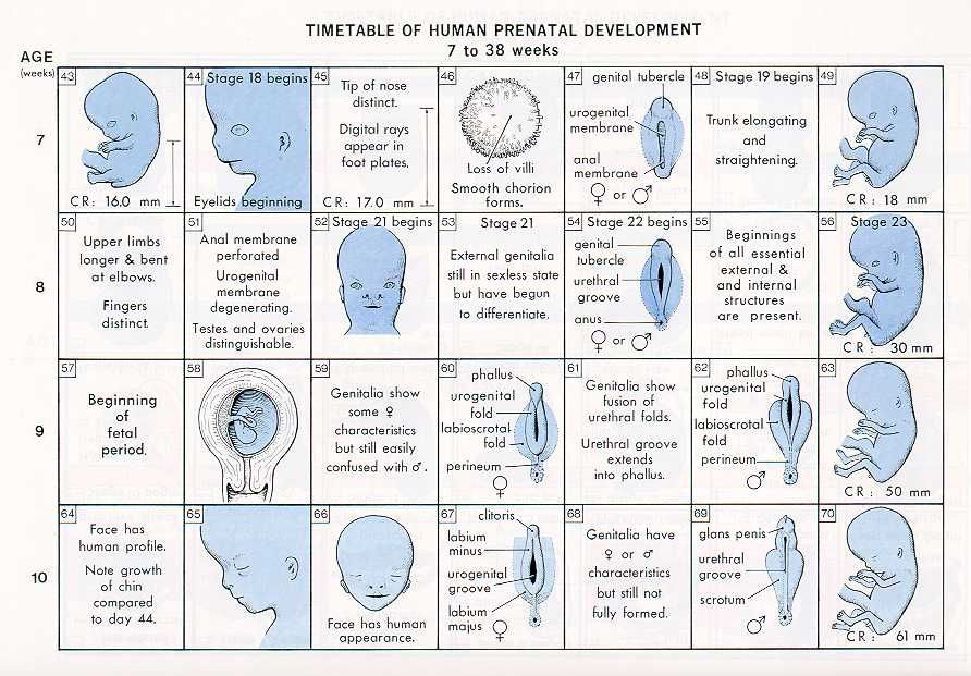 At the same time, there is a negative impact on the exchange of those neurotransmitters that should be produced by the brain itself. First of all, this applies to those substances that are related to the provision of attention and the regulation of emotions. Studies have shown that maternal smoking during pregnancy increases the risk of having a child with attention deficit hyperactivity disorder (ADHD) by several times. The second consequence of intrauterine nicotine use after ADHD is oppositional defiant disorder, which is characterized by such manifestations as irritability, anger, constantly changing, often negative, mood, vindictiveness. Another effect of smoking is a deterioration in the condition of blood vessels, malnutrition of the fetus. Children of smoking mothers are born with low birth weight, and low birth weight itself is a risk factor for the development of subsequent behavioral problems. Due to vasospasm caused by exposure to nicotine, the fetal brain is prone to ischemic strokes - impaired blood supply to certain areas of the brain, their hypoxia, which has a very detrimental effect on all subsequent mental development [Nomura et al.
 , 2010].
One of the most important processes occurring in the developing brain of an unborn child is the covering of the long endings of nerve cells (axons) with myelin (myelination). A myelinated axon is shown in one of the previous drawings (a drawing of a neuron). Myelin is a substance that is somewhat like the insulation that covers wires. Thanks to him, the electrical signal moves from the body of the neuron to the end of the axon very quickly. The first signs of myelination are found in the brain of 20-week-old fetuses. This process is uneven. The axons that form the visual and motor nerve pathways, which are primarily useful to a newborn baby, are the first to be covered with myelin. A little later (almost before birth), the auditory pathways begin to become covered with myelin.
Cells of one of the brain tissues - neuroglia, which produce myelin, are very sensitive to lack of oxygen. Also, the myelination of the fetal brain can be affected by exposure to toxins, narcotic substances, a deficiency of the substances necessary for the brain from food (in particular, B vitamins, iron, copper and iodine), improper metabolism of certain hormones, such as thyroid hormones.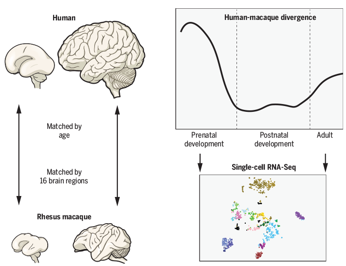
Alcohol is extremely harmful to the normal course of myelination processes. It interferes with myelination and, as a result, can cause severe mental development disorders, accompanied by mental retardation of the child. The impact of alcohol can also have a non-specific effect, leading to a variety of malformations.
About how intensively the brain of a child in the womb develops, at least the fact that in the period from 29 to 41 weeks, the brain increases almost 3 times! In many ways, this is due to myelination.
Relatively little is known about the mental development of a child in the prenatal period. At the same time, there are some interesting facts.
From 10 weeks of fetal development, babies suck their thumb (75% right). It turns out that future right-handers, for the most part, prefer to suck their right thumb, while future left-handers prefer their left [Hepper, 2013].
When exposed to sound on the abdomen of pregnant women (37-41 weeks of pregnancy) through headphones, significant activation was found in the temporal areas in four and in the frontal areas in one fetus - the same areas of the cerebral cortex that will subsequently take part in the processing of speech information .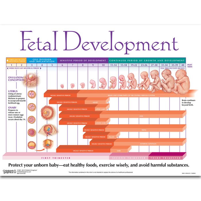 This suggests that the child's brain is actively preparing to exist in the environment that is intended for him.
Development of the child's brain from birth to age 3
The intrauterine period accounts for 70% of the development of the child's brain, 15% during infancy and another 15% during the preschool years. Until the baby is born, as well as in the first months after birth, that is, during the breastfeeding period, its development and health are almost completely dependent on the mother's nutrition. Therefore, it is extremely important that you carefully monitor your diet and remember a number of nutrients that are especially important for the development of a child’s brain.
Important!
In the first year of life, your baby literally grows by leaps and bounds. In a year, his height doubles and his weight triples! But even more incredible speed of development at this time reaches his brain.
The medulla is laid in the fetal cranium already in the first weeks of intrauterine development of the baby. At the tenth week of pregnancy, the baby's brain is divided into three parts. In a child who was born, the brain is almost no different from the brain of an adult. By twelve months, the final formation of the brain structure is completed. The number of neurons remains approximately at the same level until the end of life. And from birth, a lot of reflexes and skills are embedded in the brain: breathing, sucking, grasping...
From birth, the neurons of the brain exist for the most part independently of each other. The task of the brain during the first 3 years is to establish and strengthen the connections between them. At this time, the cells of the child's brain create 2 million new connections - synapses - per second! As the child develops, the synapses become more complex: they grow like a tree with many branches and twigs.
The period from birth to three years is the time of the highest brain activity. By the age of three, a child's brain is already 80% the size of an adult's brain. The increase in brain volume occurs due to special glial cells: they are necessary for the existence of neurons. Starting from the age of three, a sharp slowdown in the rate of brain development begins, and after six years it almost completely slows down and the formation ends. The abilities of the brain of a six-year-old child practically coincide with those of an adult!
For the harmonious development of the baby's brain, an environment full of positive emotions and new impressions is needed. Such an environment will make the brain work more actively, stimulate its development. It is in the first three years that the future foundations of health, thinking, various skills, and adaptability to life are laid in the baby. Therefore, it is very important in these first three years to help the formation of the brain. The child should be surrounded by images, sounds, touches, smells. All these are stimuli that are perceived by the brain and help it form faster.
Adherents of the ideas of "early development" - the intensive development of a child's abilities at an early age (from 0 to 3 years) - pay special attention to this.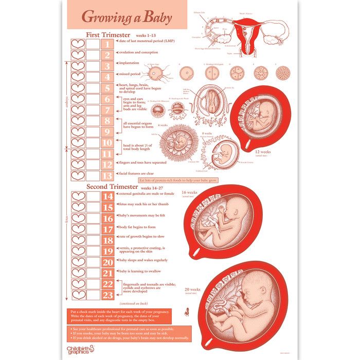 In their opinion, it is necessary to introduce the baby to various activities as early as possible: develop his speech, draw, sculpt, play musical instruments, etc.
Equally important is the baby's nutrition. Of particular importance in the development and proper functioning of the baby's nervous system are long-chain polyunsaturated fatty acids. These include docosahexaenoic and arachidonic acids (DHA and ARA).
The daily diet for the "future genius" should include DHA and APA of breast milk or baby milk in case of supplementary feeding. Breast milk does not have an exact ratio of these fats, as their presence is highly dependent on the diet of the nursing mother and how much she consumes foods containing them. So, for example, the milk of Japanese mothers has a very high amount of DHA due to the high consumption of seafood, while the concentration of DHA in the milk of American mothers is very low. Also, sources of DHA in the mother's diet can be, for example, seafood, various vegetable oils, walnuts.