Pregnant female human body organs
Anatomy of pregnancy and birth - uterus
Anatomy of pregnancy and birth - uterus | Pregnancy Birth and Baby beginning of content5-minute read
Listen
What does the uterus look like?
One of the most recognised changes in a pregnant woman’s body is the appearance of the ‘baby bump’, which forms to accommodate the baby growing in the uterus. The primary function of the uterus during pregnancy is to house and nurture your growing baby, so it is important to understand its structure and function, and what changes you can expect the uterus to undergo during pregnancy.
The uterus (also known as the ‘womb’) has a thick muscular wall and is pear shaped. It is made up of the fundus (at the top of the uterus), the main body (called the corpus), and the cervix (the lower part of the uterus ). Ligaments – which are tough, flexible tissue – hold it in position in the middle of the pelvis, behind the bladder, and in front of the rectum.
The uterus wall is made up of 3 layers. The inside is a thin layer called the endometrium, which responds to hormones – the shedding of this layer causes menstrual bleeding. The middle layer is a muscular wall. The outside layer of the uterus is a thin layer of cells.
Illustration showing the female reproductive system.The size of a non-pregnant woman's uterus can vary. In a woman who has never been pregnant, the average length of the uterus is about 7 centimetres. This increases in size to approximately 9 centimetres in a woman who is not pregnant but has been pregnant before. The size and shape of the uterus can change with the number of pregnancies and with age.
How does the uterus change during pregnancy?
During pregnancy, as the baby grows, the size of a woman’s uterus will dramatically increase. One measure to estimate growth is the fundal height, the distance from the pubic bone to the top of the uterus..png) Your doctor (GP) or obstetrician or midwife will measure your fundal height at each antenatal visit from 24 weeks onwards. If there are concerns about your baby’s growth, your doctor or midwife may recommend using regular ultrasound to monitor the baby.
Your doctor (GP) or obstetrician or midwife will measure your fundal height at each antenatal visit from 24 weeks onwards. If there are concerns about your baby’s growth, your doctor or midwife may recommend using regular ultrasound to monitor the baby.
Fundal height can vary from person to person, and many factors can affect the size of a pregnant woman’s uterus. For instance, the fundal height may be different in women who are carrying more than one baby, who are overweight or obese, or who have certain medical conditions. A full bladder will also affect fundal height measurement, so it’s important to empty your bladder before each measurement. A smaller than expected fundal height could be a sign that the baby is growing slowly or that there is too little amniotic fluid. If so, this will be monitored carefully by your doctor. In contrast, a larger than expected fundal height could mean that the baby is larger than average and this may also need monitoring.
As the uterus grows, it can put pressure on the other organs of the pregnant woman's body.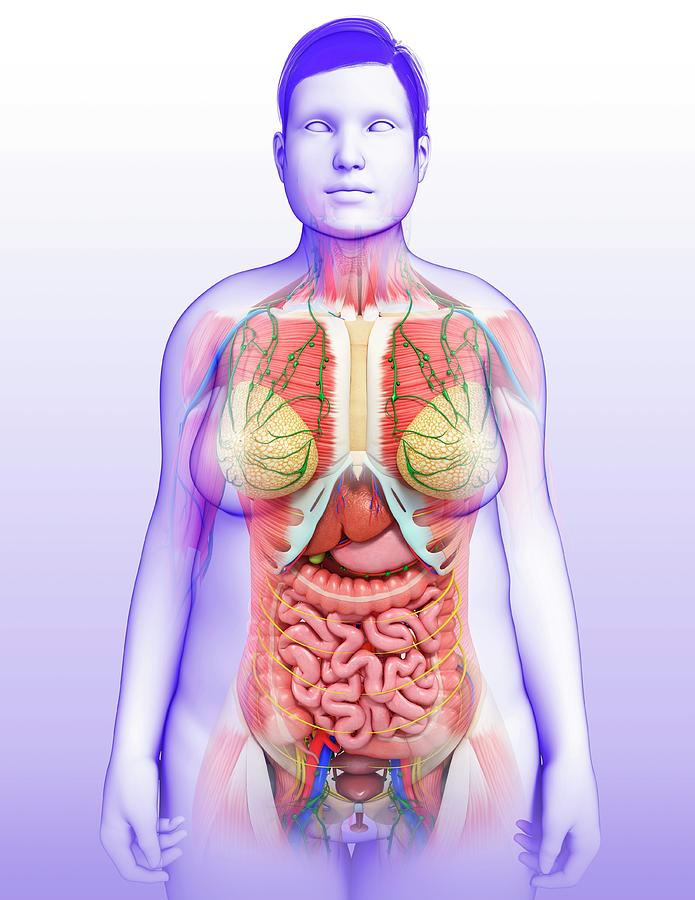 For instance, the uterus can press on the nearby bladder, increasing the need to urinate.
How does the uterus prepare for labour and birth?
Braxton Hicks contractions, also known as 'false labour' or 'practice contractions', prepare your uterus for the birth and may start as early as mid-way through your pregnancy, and continuing right through to the birth. Braxton Hicks contractions tend to be irregular and while they are not generally painful, they can be uncomfortable and get progressively stronger through the pregnancy.
During true labour, the muscles of the uterus contract to help your baby move down into the birth canal. Labour contractions start like a wave and build in intensity, moving from the top of the uterus right down to the cervix. Your uterus will feel tight during the contraction, but between contractions, the pain will ease off and allow you to rest before the next one builds. Unlike Braxton Hicks, labour contractions become stronger, more regular and more frequent in the lead up to the birth.
How does the uterus change after birth?
After the baby is born, the uterus will contract again to allow the placenta, which feeds the baby during pregnancy, to leave the woman’s body. This is sometimes called the ‘after birth’. These contractions are milder than the contractions felt during labour. Once the placenta is delivered, the uterus remains contracted to help prevent heavy bleeding known as ‘postpartum haemorrhage‘.
The uterus will also continue to have contractions after the birth is completed, particularly during breastfeeding. This contracting and tightening of the uterus will feel a little like period cramps and is also known as 'afterbirth pains'.
Read more here about the first few days after giving birth.
Sources:
The Royal Australian and New Zealand College of Obstetricians and Gynaecologists (Labour and birth), StatPearls Publishing (Anatomy, Abdomen and Pelvis), Department of Health (Clinical practice guidelines: Pregnancy care), Better Health Channel Victoria (Pregnancy stages and changes), Mater Mother's Hospital (Labour and birth information), Royal Australian and New Zealand College of Obstetricians and Gynaecologists (The First Few Weeks Following Birth), Queensland Health (Queensland Clinical Guidelines – maternity and neonatal), King Edward Memorial Hospital (Fundal height: Measuring with a tape measure), Royal Hospital for Women (Fetal growth assessment (clinical) in pregnancy), MSD Manual (Female internal genital organs)Learn more here about the development and quality assurance of healthdirect content.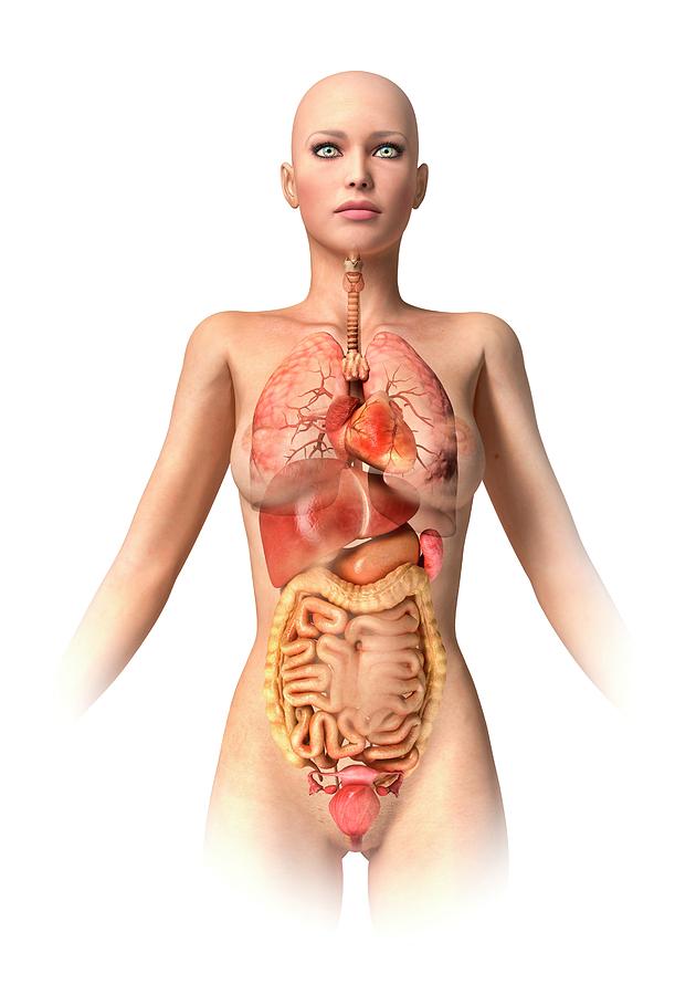
Last reviewed: October 2020
Back To Top
Related pages
- Anatomy of pregnancy and birth
- Anatomy of pregnancy and birth - abdominal muscles
- Anatomy of pregnancy and birth - cervix
- Anatomy of pregnancy and birth - pelvis
- Anatomy of pregnancy and birth - perineum and pelvic floor
Need more information?
Prolapsed uterus - Better Health Channel
The pelvic floor and associated supporting ligaments can be weakened or damaged in many ways, causing uterine prolapse.
Read more on Better Health Channel website
Uterus, cervix & ovaries - fact sheet | Jean Hailes
This fact sheet discusses some of the health conditions that may affect a woman's uterus, cervix and ovaries.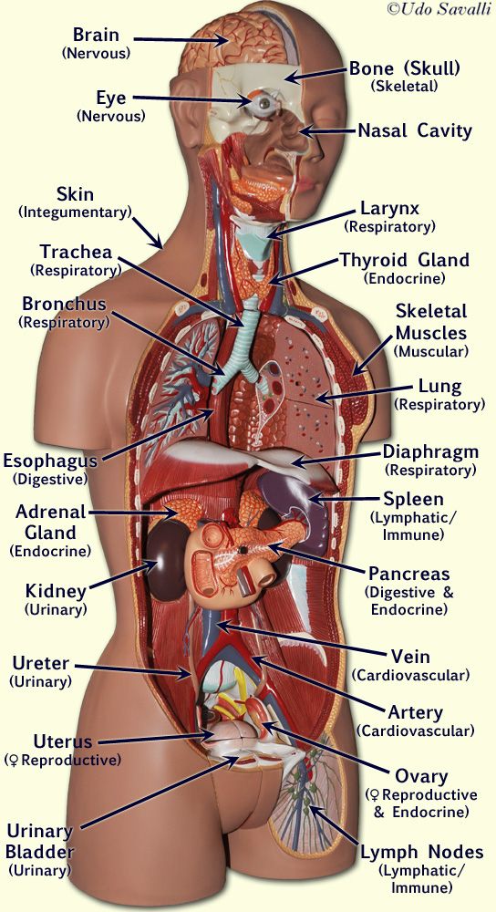
Read more on Jean Hailes for Women's Health website
Dilatation and curettage (D&C)
A D&C is an operation to lightly scrape the inside of the uterus (womb).
Read more on WA Health website
Ectopic pregnancy
An ectopic pregnancy occurs when a fertilised egg implants outside the uterus (womb)
Read more on WA Health website
Hormonal IUD (intrauterine device, Mirena®) | Body Talk
The hormonal IUD is a type of contraception and is placed inside the uterus by a specially trained doctor or nurse. Find out all the facts here.
Read more on Body Talk website
Placental abruption - Better Health Channel
Placental abruption means the placenta has detached from the wall of the uterus, starving the baby of oxygen and nutrients.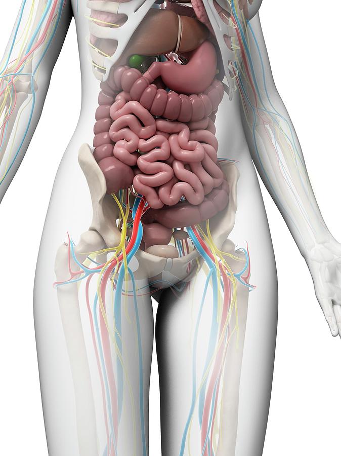
Read more on Better Health Channel website
Copper IUD (intrauterine device) | Body Talk
The copper IUD is a type of contraception and is placed inside the uterus by a specially trained doctor or nurse. Find out all the facts here.
Read more on Body Talk website
Placenta previa - Better Health Channel
Placenta previa means the placenta has implanted at the bottom of the uterus, over the cervix or close by.
Read more on Better Health Channel website
Mirena IUD | Hormonal IUD Mirena | IUD Mirena insertion | IUD Mirena cost | Mirena IUD Melbourne - Sexual Health Victoria
The hormonal intrauterine device (IUD) is a small contraceptive device that is put into the uterus (womb) to prevent pregnancy.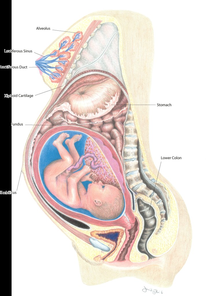
Read more on Sexual Health Victoria website
Contraception - intrauterine devices (IUD) - Better Health Channel
An intrauterine device (IUD) is a small contraceptive device that is put into the uterus (womb) to prevent pregnancy.
Read more on Better Health Channel website
Disclaimer
Pregnancy, Birth and Baby is not responsible for the content and advertising on the external website you are now entering.
OKNeed further advice or guidance from our maternal child health nurses?
1800 882 436
Video call
- Contact us
- About us
- A-Z topics
- Symptom Checker
- Service Finder
- Subscribe to newsletters
- Sign in
- Linking to us
- Information partners
- Terms of use
- Privacy
Pregnancy, Birth and Baby is funded by the Australian Government and operated by Healthdirect Australia.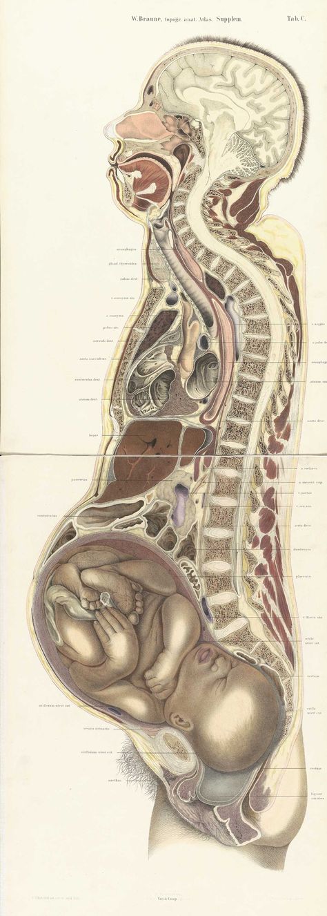
Pregnancy, Birth and Baby’s information and advice are developed and managed within a rigorous clinical governance framework.
This site is protected by reCAPTCHA and the Google Privacy Policy and Terms of Service apply.
Healthdirect Australia acknowledges the Traditional Owners of Country throughout Australia and their continuing connection to land, sea and community. We pay our respects to the Traditional Owners and to Elders both past and present.
This information is for your general information and use only and is not intended to be used as medical advice and should not be used to diagnose, treat, cure or prevent any medical condition, nor should it be used for therapeutic purposes.
The information is not a substitute for independent professional advice and should not be used as an alternative to professional health care.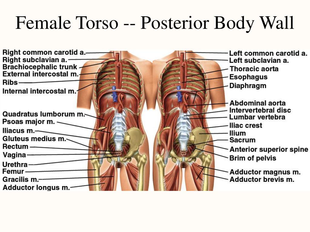 If you have a particular medical problem, please consult a healthcare professional.
Except as permitted under the Copyright Act 1968, this publication or any part of it may not be reproduced, altered, adapted, stored and/or distributed in any form or by any means without the prior written permission of Healthdirect Australia.
Support this browser is being discontinued for Pregnancy, Birth and Baby
Support for this browser is being discontinued for this site
- Internet Explorer 11 and lower
We currently support Microsoft Edge, Chrome, Firefox and Safari. For more information, please visit the links below:
- Chrome by Google
- Firefox by Mozilla
- Microsoft Edge
- Safari by Apple
You are welcome to continue browsing this site with this browser. Some features, tools or interaction may not work correctly.
Female reproductive organ anatomy, parts, and function
The female reproductive organs include several key structures, such as the ovaries, uterus, vagina, and vulva.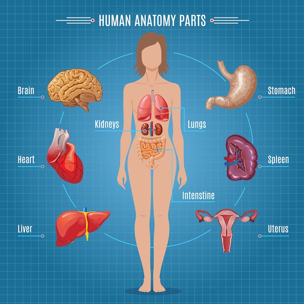 These organs are involved in fertility, conception, pregnancy, and childbirth.
The reproductive organs also have a significant influence on other aspects of health. For example, the ovaries create hormones that impact bone density, cholesterol levels, heart health, and mood.
In this article, we will look at the anatomy of the female reproductive organs in detail, including what they do and how they work.
A note about sex and gender
Sex and gender exist on spectrums. This article will use the terms “male,” “female,” or both to refer to sex assigned at birth. Click here to learn more.
The female reproductive system is a group of organs that work together to enable reproduction, pregnancy, and childbirth. It also produces female sex hormones, including estrogen and progesterone.
The system consists of organs and tissues inside the body and some that are visible outside the body. The internal organs include:
- ovaries
- fallopian tubes
- uterus
- cervix
- vagina
Another organ, the clitoris, extends both inside and outside the body.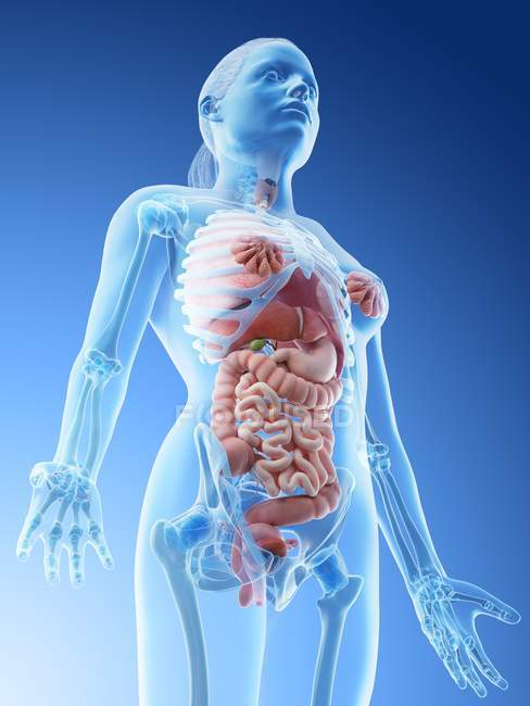 The external area surrounding the vagina is the vulva.
Not everyone who is assigned female at birth has all of these organs. Sometimes, people are born without some parts or with a mixture of female and male characteristics. This is known as intersex.
Some people also undergo procedures to remove some parts of the reproductive system. Some of these procedures take place for medical reasons, while others are the result of harmful cultural practices, such as female genital mutilation.
Most females have two ovaries, one on each side of the uterus. They are about the shape and size of an almond and have two key functions: producing hormones and releasing eggs.
At birth, two ovaries contain approximately 700,000 oocytes, which are immature eggs. When a person reaches puberty, these eggs begin to develop and mature inside the ovary follicles. Around once each month, the ovaries release a mature egg.
This process is known as ovulation, and it is part of the menstrual cycle. It is also what makes pregnancy possible.
The hormones the ovaries produce regulate the menstrual cycle. They also:
- influence the development of female sex traits
- facilitate pregnancy, childbirth, and breast milk production
- contribute to the health of the bones, heart, liver, brain, and other tissues
- influence mood, sleep, and sex drive
The fallopian tubes are passageways that carry eggs toward the uterus. They consist of several parts:
- the infundibulum, which is a funnel-shaped opening near the ovaries
- the fimbriae, which are finger-like projections surrounding the opening
- cilia, which are hair-like structures inside the fallopian tubes
When an ovary releases an egg, fluid and the fimbriae propel it toward the fallopian tube opening. Once inside, the cilia move the egg toward the uterus. This journey takes about 7 days.
During this time, it is possible for sperm to fertilize the egg if a person has sexual intercourse.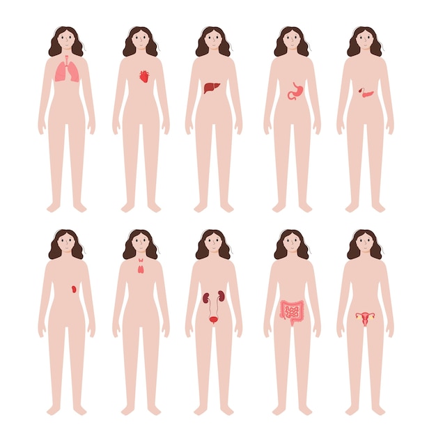 Most fertilization happens in the fallopian tubes.
The uterus is an organ that is about the shape and size of a pear. It is also known as the womb. It consists of muscular walls and a lining (endometrium) that grows and diminishes with each menstrual cycle.
After ovulation, the endometrium gets thicker in preparation for a fertilized egg. If not fertilized, the egg dies, and the lining of the womb sheds after around 2 weeks. The lining breaks down into blood, which then leaves the body through the vagina. This is menstruation, also called a period.
If an egg does become fertilized by sperm, it will implant into the lining of the uterus and begin to develop. The cells divide and grow, becoming an embryo. Over time, it grows into a fetus, which receives oxygen and nutrients from the placenta via the umbilical cord.
When it is time for the fetus to be born, the uterus begins strong muscle contractions that dilate the cervix and push the fetus out.
The cervix is a narrow structure at the bottom of the uterus.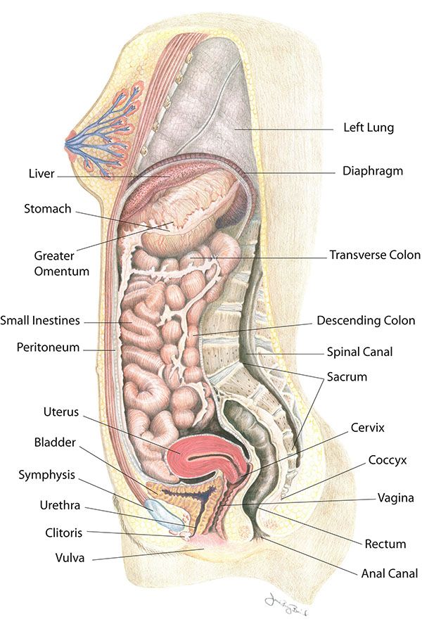 It has several functions:
- Producing mucus: The cervix produces cervical mucus, which stops sperm from entering the uterus when a person is not fertile or when they are pregnant.
- Protecting against bacteria: The mucus also stops bacteria from entering the uterus and keeps the vagina healthy.
- Allowing fluids to drain: At the bottom of the cervix is a small opening that allows fluids, such as menstrual blood, to pass through.
Below the cervix is the vagina, which is a flexible, tubular structure that connects the internal and external reproductive organs. It sits behind the bladder and in front of the digestive tract.
The vagina allows fluids, such as menstrual blood and discharge, to leave the body. It also allows semen, which contains sperm, to enter the body.
This can happen in several ways, such as during penetrative sex with someone who has a penis, or during artificial insemination.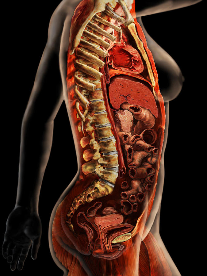 This is a procedure where a doctor inserts semen into the uterus to help someone conceive.
Just inside the body, around the entrance to the vagina, is the clitoris. This organ is most well known for the clitoral glans, which is a small but highly sensitive tissue that sits above the vaginal opening. Most of the clitoris is actually internal.
The clitoral glans is at the top of the clitoris. From there, the clitoris splits into two parts that extend down either side of the vagina. It is around 5 inches (12.7 centimeters) long and consists of spongy tissue that contains thousands of nerve endings.
The clitoris responds to sexual stimulation. When a person experiences arousal, it becomes swollen. It is the main organ responsible for female orgasms.
Share on PinterestDesign by Diego Sabogal
The vulva is the external part of the female reproductive system. It includes the:
- Vestibule: This is the entrance to the vagina. Around the vestibule sit the greater vestibular glands, which produce fluid to keep the area from getting dry.
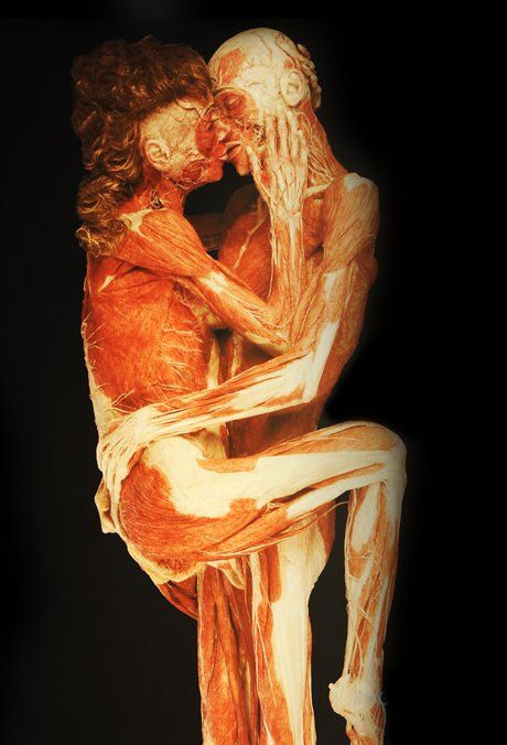 During sexual arousal, these glands produce more fluid to help with lubrication.
 - Hymen: Some people with vulvas also have a hymen. This is a thin, delicate tissue that partially covers the entrance to the vagina. When someone has penetrative sex for the first time, the hymen can stretch or break. But not everyone has a hymen, and it can also stretch for a number of other reasons. Learn more here.
- Urethra: This is where urine comes from. The urethra is part of the urinary system and sits just above the vaginal opening.
- Labia minora: These are smaller lips that surround the entrance to the vagina.
- Clitoral hood and glans: The clitoral hood is a small piece of tissue that protects the external part of the clitoris. It sits at the top of the labia minora.
- Labia majora: These are the larger lips that surround the vulva. After puberty, they typically have pubic hair. At the top of the vulva is also the mons pubis, which is a rounded pad of fat that sits over the pubic bone.
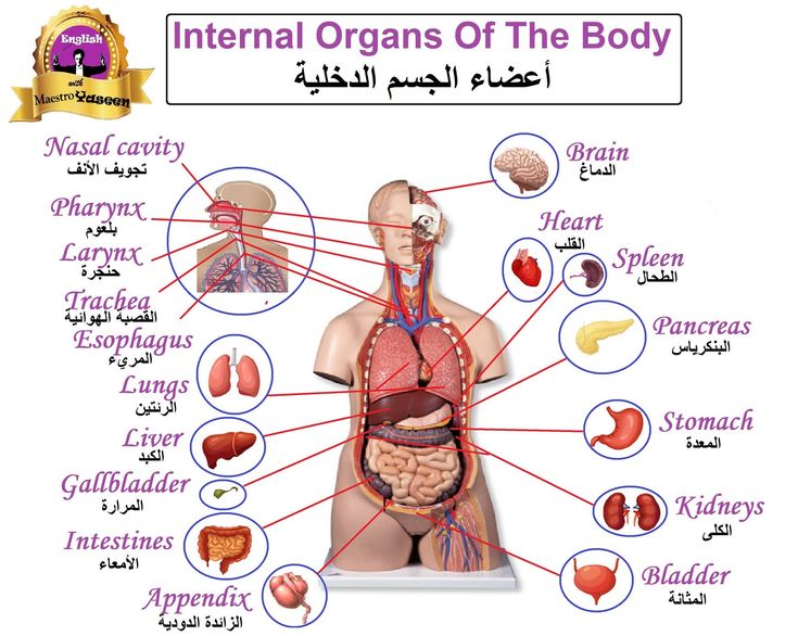
The female reproductive organs include an array of parts that influence health throughout a person’s life. The reproductive system undergoes significant changes during the menstrual cycle, which starts during puberty and ends with menopause. If a person becomes pregnant, it changes further to accommodate a growing fetus.
Female reproductive anatomy also influences sexual well-being, and creates hormones that regulate a wide variety of functions around the body.
Physiological changes in the body during pregnancy
From the very first days of pregnancy, a woman's body undergoes profound transformations. These transformations are the result of the coordinated work of almost all body systems, as well as the result of the interaction of the mother's body with the child's body. During pregnancy, many internal organs undergo significant restructuring. These changes are adaptive in nature, and, in most cases, are short-lived and completely disappear after childbirth. Consider the changes in the basic systems of the vital activity of a woman's body during pregnancy.
The respiratory system during pregnancy works hard. The respiratory rate increases. This is due to an increase in the need of the mother and fetus for oxygen, as well as in the limitation of the respiratory movements of the diaphragm due to an increase in the size of the uterus, which occupies a significant space of the abdominal cavity.
The mother's circulatory system during pregnancy has to pump more blood to ensure an adequate supply of nutrients and oxygen to the fetus. In this regard, during pregnancy, the thickness and strength of the heart muscles increase, the pulse and the amount of blood pumped by the heart in one minute increase. In addition, the volume of circulating blood increases. In some cases, blood pressure increases. The tone of blood vessels during pregnancy decreases, which creates favorable conditions for increased supply of tissues with nutrients and oxygen.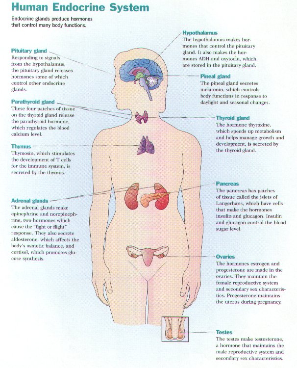 During pregnancy, the network of vessels of the uterus, vagina, and mammary glands decreases sharply. On the external genitalia, in the vagina, lower extremities, there is often an expansion of the veins, sometimes the formation of varicose veins. Heart rate decreases in the second half of pregnancy. It is generally accepted that the rise in blood pressure over 120-130 and a decrease to 100 mm Hg. signal the occurrence of pregnancy complications. But it is important to have data on the initial level of blood pressure.
And changes in the blood system. During pregnancy, blood formation increases, the number of red blood cells, hemoglobin, plasma and bcc increases. BCC by the end of pregnancy increases by 30-40%, and erythrocytes by 15-20%. Many healthy pregnant women have a slight leukocytosis. ESR during pregnancy increases to 30-40. Changes occur in the coagulation system that contribute to hemostasis and prevent significant blood loss during childbirth or placental abruption and in the early postpartum period.
Kidneys work hard during pregnancy. They secrete decay products of substances from the body of the mother and fetus (the waste products of the fetus pass through the placenta into the mother's blood).
Changes in the digestive system are represented by increased appetite (in most cases), cravings for salty and sour foods. In some cases, there is an aversion to certain foods or dishes that were well tolerated before the onset of pregnancy. Due to the increased tone of the vagus nerve, constipation may occur.
The most significant changes, however, occur in the genitals of pregnant women. These changes prepare the woman's reproductive system for childbirth and breastfeeding.
The uterus of a pregnant woman increases significantly in size. Its mass increases from 50 g - at the beginning of pregnancy to 1200 g - at the end of pregnancy. The volume of the uterine cavity by the end of pregnancy increases by more than 500 times! The blood supply to the uterus is greatly increased. In the walls of the uterus, the number of muscle fibers increases. The cervix is filled with thick mucus that clogs the cavity of the cervical canal. The fallopian tubes and ovaries also increase in size. In one of the ovaries, there is a "corpus luteum of pregnancy" - a place for the synthesis of hormones that support pregnancy. Walls vaginas will loosen and become more elastic. External genitalia (labia minor and major), also increase in size and become more elastic. The tissues of the perineum are loosened. In addition, there is an increase in mobility in the joints of the pelvis and a divergence of the pubic bones. The changes in the genital tract described above are of extremely important physiological significance for childbirth. Loosening the walls, increasing the mobility and elasticity of the genital tract increases their throughput and facilitates the movement of the fetus through them during childbirth.
Skin in the genital area and in the midline of the abdomen usually becomes darker in color.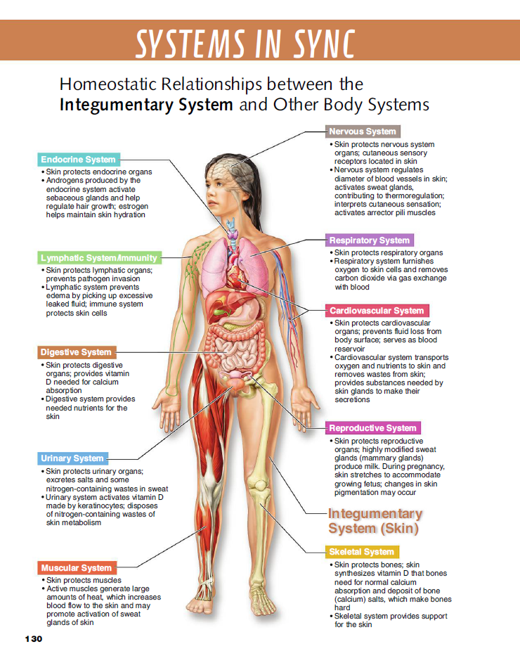 Sometimes "stretch marks" form on the skin of the lateral parts of the abdomen, which turn into whitish stripes after childbirth.
Mammary glands increase in size, become more elastic, tense. When pressing on the nipple, colostrum (first milk) is released.
Changes in the bone skeleton and muscular system . An increase in the concentration of the hormones relaxin and progesterone in the blood contributes to the leaching of calcium from the skeletal system. This helps to reduce the rigidity of the joints between the bones of the pelvis and increase the elasticity of the pelvic ring. Increasing the elasticity of the pelvis is of great importance in increasing the diameter of the internal bone ring in the first stage of labor and further reducing the resistance of the birth tract to fetal movement in the second stage of labor. Also, calcium, washed out of the mother's skeletal system, is used to build the skeleton of the fetus.
It should be noted that calcium compounds are washed out of all bones of the maternal skeleton (including the bones of the foot and spine). As shown earlier, a woman's weight increases during pregnancy by 10 -12 kg. This additional load against the background of a decrease in bone stiffness can cause foot deformity and the development of flat feet. A shift in the center of gravity of the body of a pregnant woman due to an increase in the weight of the uterus can lead to a change in the curvature of the spine and the appearance of pain in the back and pelvic bones. Therefore, for the prevention of flat feet, pregnant women are advised to wear comfortable shoes with low heels. It is advisable to use insoles that support the arch of the foot. For the prevention of back pain, special physical exercises are recommended that can unload the spine and sacrum, as well as wearing a comfortable bandage. Despite an increase in calcium loss by the bones of the skeleton of a pregnant woman and an increase in their elasticity, structure and bone density (as is the case with osteoporosis in older women).
Changes in the nervous system . In the first months of pregnancy and at the end of it, there is a decrease in the excitability of the cerebral cortex, which reaches its greatest degree by the time of the onset of childbirth. By the same period, the excitability of the receptors of the pregnant uterus increases. At the beginning of pregnancy, there is an increase in the tone of the vagus nerve, in connection with which various phenomena often occur: changes in taste and smell, nausea, increased salivation, etc.
Active endocrine glands there are significant changes that contribute to the proper course of pregnancy and childbirth. Changes in body weight. By the end of pregnancy, a woman's weight increases by about 10-12 kg. This value is distributed as follows: fetus, placenta, membranes and amniotic fluid - approximately 4.0 - 4.5 kg, uterus and mammary glands -1.0 kg, blood - 1.5 kg, intercellular (tissue) fluid - 1 kg , an increase in the mass of adipose tissue of the mother's body - 4 kg.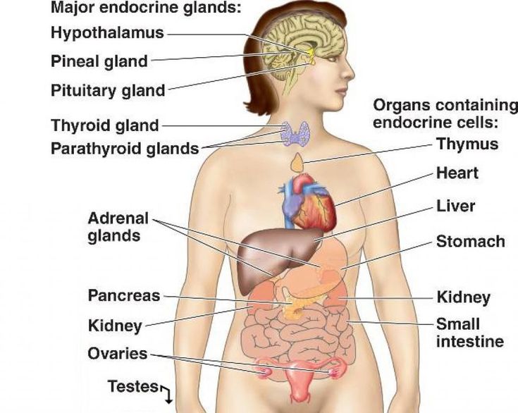
Physiological changes in a woman's body during pregnancy
A woman's pregnancy lasts an average of 280 days. Starting from the very first days, a woman's body is completely rebuilt in order to prepare for childbirth and the birth of a new person into the world. There is not a single tissue and not a single organ that would not take part in this process of transformation. Each system of the body adapts to a new state according to its own scheme.
1) The hormonal system comes into action first. Chorionic gonadotropin, or pregnancy hormone, informs the whole body about the new condition already on the 6-8th day. The number of female hormones that previously regulated the menstrual cycle is reduced. The content of prostaglandin, a hormone that preserves pregnancy, increases. Of course, these changes affect behavior and character. Tearfulness and impressionability of pregnant women, whims, mood swings - all these are consequences of hormonal changes.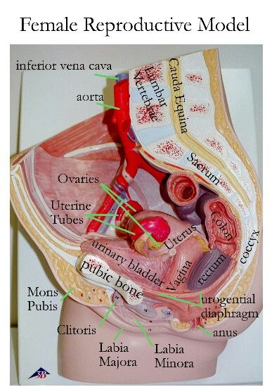
2) Since hormones are produced in the endocrine glands, the glands themselves also change. The pituitary gland, adrenal glands, ovaries are reorganized into a new mode of operation. The substances they secrete now contribute to the growth of the uterus and mammary glands.
3) The nervous system reacts to changes in hormones and also adapts. It is changes in the nervous system that cause nausea and vomiting in the first half of pregnancy. During this period, the processes of excitation prevail over the processes of inhibition. But in the second half of pregnancy, the processes of inhibition gradually take over - this is associated with drowsiness and a weakening of the cognitive functions of the pregnant woman. The body makes this sacrifice to smooth out the shock of labor activity.
4) Changes in the cardiovascular system are associated with the fact that a pregnant woman has a third circle of blood circulation - uteroplacental, through which the fetus is supplied with maternal blood.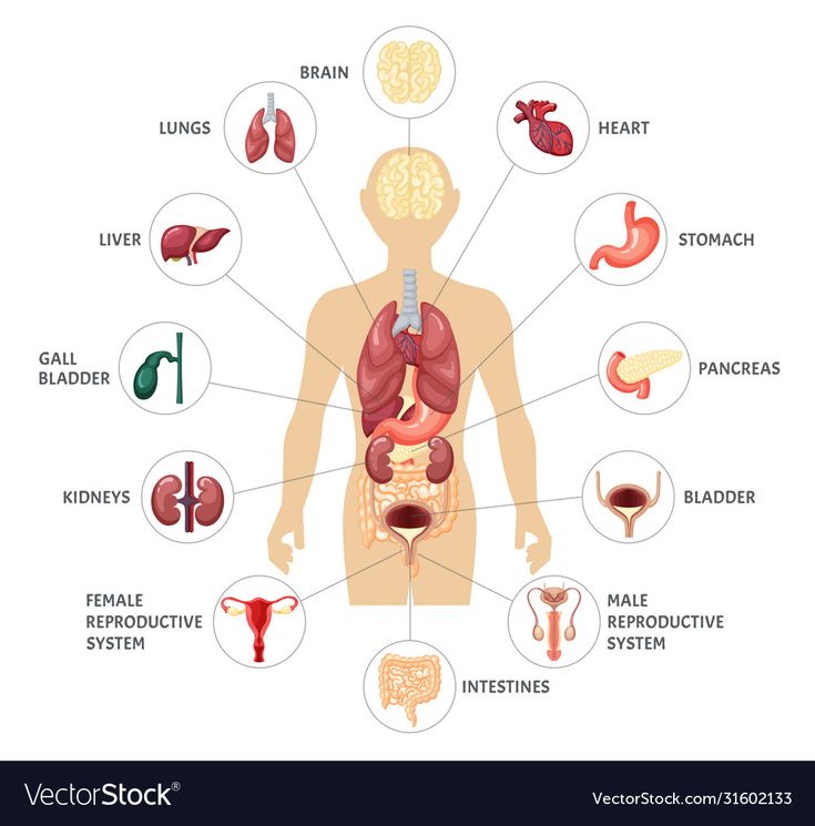 Increases body weight and volume of circulating blood - from 4000 to 5200 ml on average; metabolism is enhanced. This increases cardiac output, that is, the amount of blood the heart ejects during contraction. In a healthy woman, the heart quickly adapts to additional loads, but in some cases complications are possible.
5) An increased load falls on the digestive system. First, the secretion of gastric juice is inhibited, which slows down the digestion process. Secondly, the blood supply to the digestive system does not increase - blood is needed for the baby. Therefore, intestinal motility weakens, because of this, pregnant women often complain of constipation. In addition, the fetus presses on the stomach, due to which part of the food mass can return to the esophagus and cause heartburn. Therefore, pregnant women in the last weeks are recommended fractional meals - frequent meals in small portions.
6) The lungs and other organs of the respiratory system also adapt to increased stress. More oxygen is required, but at the same time, the growing belly props up the chest and presses on the diaphragm. The lungs expand in width, the respiratory rate quickens.
7) The excretory system also works for two, removing toxins from the blood produced by two organisms, mother and child. Therefore, the daily volume of urine may increase. In this case, the bladder is in hypotonia due to the hormone prostaglandin.
8) Musculoskeletal system. Major changes in the spine occur under the weight of the growing belly. The lumbar lordosis becomes deeper, and the woman may suffer from back pain from the unaccustomed bending. Shortly before childbirth, hormones begin to be released in the body that help soften the joints. This is necessary so that the baby's head passes through the mother's pelvis without injury. Because of these hormones, the joints of the limbs can also become slightly loose, and then the woman develops a characteristic duck gait.
9) Metabolism.

