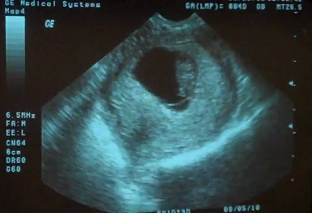
Miscarriage ultrasound 8 weeks
Ultrasound scans - The Miscarriage Association
Ultrasound scans in pregnancy may be routine or they may be offered because of pain or bleeding or because of problems in a previous pregnancy.
There are two ways of doing an ultrasound scan.
In early pregnancy, especially before 11 weeks, it is usual to have a trans-vaginal (internal) scan, where a probe is placed in the vagina. This gives the clearest and most accurate picture in early pregnancy. It may also be offered after 11 or 12 weeks if a trans-abdominal scan doesn’t give a clear enough picture.
From 11 or 12 weeks, including at the routine booking-in scan, it is more common to have a trans-abdominal scan. The person doing the scan spreads a special gel on your lower abdomen (below your belly button and above the line of pubic hair). He or she then moves the scanner over the gel, sometimes pressing down, until the uterus (womb) and pregnancy can be seen.
If you don’t want a trans-vaginal scan, you can ask for a trans-abdominal scan. That may give some information about your pregnancy, but it is less clear than an internal scan and that could possibly delay diagnosis.
Can ultrasound scans harm the baby?There is no evidence that having a vaginal or an abdominal scan will cause a miscarriage or harm your baby. If you bleed after a vaginal scan, it will most likely be because there was already blood pooled higher in the vagina and the probe dislodged it.
When can I have an ultrasound scan? When can you see the baby’s heartbeat?An ultrasound scan may be able to detect a pregnancy and a heartbeat in a normal pregnancy at around 6 weeks, but this varies a great deal and isn’t usually advised. All too often, a scan at 6 weeks shows very little or nothing, even in a perfectly developing pregnancy, whereas waiting a week or 10 days will make the findings much clearer.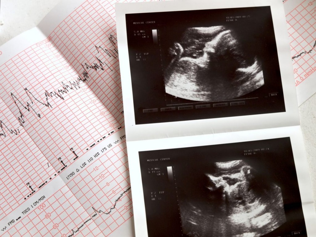
Most pregnant women are referred for their first routine (or booking or dating) ultrasound scan somewhere between 11 and 14 weeks of pregnancy. The purpose of the scan is:
- to confirm that there is a heartbeat
- to assess the baby’s size and growth
- to estimate the delivery date and
- to check whether there is one baby, or twins or more.
Some women may be offered a nuchal scan between 11 and 14 weeks. The purpose of this scan is to try to detect some chromosome abnormalities, such as Downs syndrome.
Most hospitals also offer a further anomaly scan at 20 weeks, making a more detailed check of the baby’s development.
You can find out more about routine, nuchal and anomaly scans at the website of the charity ARC – Antenatal Results and Choices
Sadly…Sadly, sometimes these scans show that the baby has died, possibly some weeks earlier and often without any signs or symptoms such as bleeding or pain. This is often called a “missed”, “silent” or “delayed” miscarriage. This can come as a considerable shock and it may take time before you can take this information in. You can find information about missed miscarriage here.
You may also have to make some difficult decisions about how to manage the miscarriage process. You can read more about this here.
Early scansYou may be referred for an early scan because of vaginal bleeding or spotting, or possibly because you have had problems in a previous pregnancy.
The best time to have a scan is from about 7 weeks’ gestation when it should be possible to see the baby’s heartbeat in a normal pregnancy. But it can be hard to detect a heartbeat in early pregnancy and in those cases it can be hard to know whether the baby has died or not developed at all, or whether it is simply smaller than expected but still developing.
For that reason, you may be asked to return for another scan a week or so later. At that time, the person doing the scan will be looking for a clear difference in the size of the pregnancy sac and for a developing baby and a heartbeat.
Sometimes, it can take several scans before you know for sure what is happening. It can be very stressful dealing with this uncertainty – some women describe it as being “in limbo”. You may need to find some support for yourself if this happens to you.
There’s a heartbeat, but I’m still bleeding…If the scan does pick up a heartbeat and the baby appears to be the right size according to your dates, this can be very reassuring, even if you are still bleeding. Seeing a heartbeat on scan also increased the likelihood of the pregnancy continuing.
Research amongst women with a history of recurrent miscarriage has shown that while those who reached six weeks of pregnancy had a 78% chance of the pregnancy continuing, seeing a heartbeat at 8 weeks increased the chance of a continuing pregnancy to 98% and at 10 weeks that went up to 99.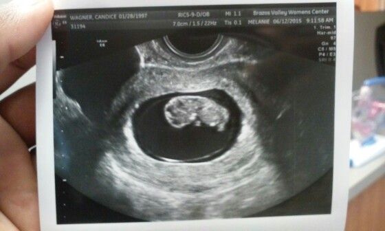 4%.
4%.
The numbers may be even more positive for women without previous miscarriages.
So things could still go wrong and sadly sometimes do, but once a heartbeat is seen, the risk of miscarriage decreases as the weeks go by.
Other investigationsIn some cases, if there is no sign of a pregnancy in the uterus, you may be given a blood test and possibly asked to return two days later for a repeat test.
These blood tests measure the level of the pregnancy hormone ßhCG. In a normally developing pregnancy the hormone levels roughly double about every 48 hours and if the pattern is different, this can help to identify what is happening to the pregnancy.
If there is no sign of a pregnancy in the uterus and you have symptoms that suggest ectopic pregnancy, you are more likely to have both a blood test and an investigation called a laparoscopy, which is done under general anaesthetic. You can read more about this here and in our leaflet Ectopic pregnancy.
The ultrasound scan may show:
- A viable ongoing pregnancy. There is a heartbeat (or heartbeats if it’s a twin or multiple pregnancy) and the pregnancy is the “right size for dates” – that is, the size that would be expected based on the first day of your last period. Those are positive signs, but if you continue to bleed, you may need a further scan in a week or two to check what’s happening.
- An ongoing pregnancy that suggests a problem. The pregnancy may be much smaller than it should be according to dates or the heartbeat might be particularly slow or faint. Perhaps there is something that suggests a problem with the baby’s development. With a twin or multiple pregnancy, the scan may show that one (or more) baby has a heartbeat and one (or more) doesn’t. You may be asked to come back for another scan, possibly in a week or two when things should be clearer.
- A pregnancy of unknown location (PUL).
 The pregnancy is not visible and it’s not clear what is happening. You may be asked to come back for another scan, possibly in a week or two when things should be clearer. Or if the doctor thinks you might have an ectopic pregnancy, you will have blood tests and/or a laparoscopy (keyhole surgery to look inside the abdomen).
- A complete miscarriage. The pregnancy has miscarried. There may still be a small amount of pregnancy tissue or blood in the uterus.
- A non-viable pregnancy. This means a pregnancy that hasn’t survived but hasn’t yet miscarried. You may hear this described in one of the following ways:
- Missed miscarriage (also called silent or delayed miscarriage or early embryonic demise) This is where the baby has died or failed to develop but your body has not miscarried him or her. The scan picture shows a pregnancy sac with a baby (or fetus or embryo) inside, but there is no heartbeat and the pregnancy looks smaller than it should be at this stage.
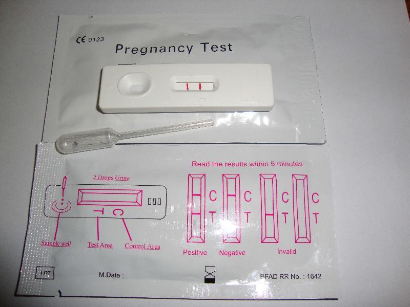
 You may have had little or no sign that anything was wrong and you may still feel pregnant. You might still have a positive pregnancy test.
- Missed miscarriage (also called silent or delayed miscarriage or early embryonic demise) This is where the baby has died or failed to develop but your body has not miscarried him or her. The scan picture shows a pregnancy sac with a baby (or fetus or embryo) inside, but there is no heartbeat and the pregnancy looks smaller than it should be at this stage.
- Blighted ovum or anembryonic pregnancy (which means a pregnancy without an embryo). This is a rather old-fashioned way of describing a missed miscarriage (see above). The scan picture usually shows an empty pregnancy sac.
- Incomplete miscarriage The process of miscarriage has started but there is still pregnancy tissue in the uterus (womb) and you may still have pain and heavy bleeding.
In all of these situations, the pregnancy will fully miscarry with time, but there are several ways of managing the process. You may be offered a choice, or the hospital might make a recommendation. In most cases, you should be able to have time to think about what you can best cope with. You can read more here.
The ultrasound scan might show- An ectopic pregnancy.
 This means a pregnancy that is developing outside the uterus (Ectopic means “out of place”). Ectopic pregnancies usually develop in one of the Fallopian tubes, but they can develop elsewhere inside the abdomen.
- A molar pregnancy. This is a pregnancy where the baby can’t develop but the cells of the placenta grow very quickly. It can’t always be diagnosed on scan so you might find out only after the miscarriage.
Ultrasound is a Critical Tool of Managing Miscarriage
This story originally appeared in Diagnostic Imaging's sister site, Contemporary Ob/Gyn
Introduction
Early pregnancy loss (miscarriage) is defined as a nonviable, intrauterine pregnancy with either an empty gestational sac or a gestational sac containing an embryo or fetus without cardiac activity within the first 12 6/7 weeks of gestation.1
The cessation of development occurs in approximately 10 percent to 20 percent of clinically recognized pregnancies, increasing with advancing parental age. 1-3 With the adoption of early home pregnancy testing, women are commonly referred for sonographic evaluations very early in gestation to determine pregnancy location and viability.
Although we believe ultrasound to be indispensable for the management of suspected early pregnancy failure, if performed too early or without strict adherence to guidelines, it may lead to inconclusive results or an incorrect diagnosis of an early pregnancy loss.4
Ultrasonography (usually transvaginal) in combination with β-hCG and clinical history is instrumental for making the diagnosis of a nonviable pregnancy with certainty.
Historically, these criteria were based on small studies; a CRL of ≥ 5mm and mean sac diameter of≥ 16-17 mm without an embryo were considered diagnostic for an early pregnancy loss.1,5 Over the last decade, the reliability of these thresholds has been called into question.6,7
In 2013, the Society of Radiologists in Ultrasound (SRU) convened a multispecialty panel on early first-trimester diagnosis of miscarriage and exclusion of a viable intrauterine pregnancy (cannot result in the birth of a live baby) and published a more conservative approach to defining a pregnancy as nonviable. 5
5
This change was made with the expectation of a diagnostic specificity of 100 percent (no false positives), while accounting for intra and inter-observer variability in measurements as well as a range in practice conditions and experience.6,7 Inadvertent misclassification of potentially viable pregnancy as nonviable with resultant medical or surgical intervention resulting in the iatrogenic termination of a desired pregnancy has significant consequences to the family and may be an inciting factor in malpractice cases.8,9
This article will focus on the criteria for the sonographic diagnosis of a nonviable intrauterine pregnancy early in gestation, those sonographic findings that are suspicious to result in an early pregnancy loss, and the vital role of ultrasound in these frequent clinical scenarios.
The sonographic findings are only one part of the puzzle in determining if a pregnancy is nonviable.
In order to apply the findings to a clinical situation, the obstetric care provider must take other clinical factors into consideration, such as accounting for the certitude of menstrual dating, the woman’s desire to continue the pregnancy until the definitive diagnosis is made along with logistical challenges such as the possibility of heavy bleeding leading to emergency room visits, unexpected passage of products of conception or unscheduled surgical procedures.
On another note, when diagnosing a complete miscarriage, care must be taken that an intrauterine pregnancy has previously been confirmed. If not, serial β-hCG should be performed so as to not miss the diagnosis of an ectopic pregnancy.
Embryologic development in early pregnancy is quite linear and follows a dependable and fairly tight timetable (Figs. 1A, B, C, D).
Ultrasound can reliably characterize the progression of a normally developing pregnancy from very early in gestation.
The gestational sac is first identified at approximately 5 weeks, the yolk sac is visible at approximately 5.5 weeks and an embryo with cardiac activity located in close proximity to the yolk sac should be evident at approximately 6 weeks gestation.
Embryonic cardiac activity is often seen when the embryo is first identified, measuring as little as 2 mm in length. The mean sac diameter is the average of three orthogonal diameters of the fluid portion of the gestational sac but is not as accurate as crown rump length for gestational dating. 10
10
Nomograms for development of embryonic length, heart rate, gestational sac diameter, and yolk sac diameter are available.11,12
Transvaginal sonographic findings diagnostic of an early pregnancy loss
Given the newer, more conservative guidelines that are now well-accepted, a single transvaginal ultrasound identifying an embryo with a crown rump length of 7 mm or greater without cardiac activity or a gestational sac with a mean sac diameter of 25 mm or greater without an embryo is considered definitive proof that a pregnancy is nonviable5 (Figs. 2 and 3).
These metrics have also been validated in a prospective observational multicenter study to have a specificity of 100 percent.13
Given the expected linear development of normal early pregnancies, the definitive diagnosis of an early pregnancy loss can also be made based on sequential transvaginal ultrasound scans over a specified time interval (Figure 4).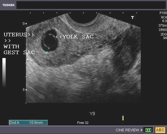
In a normally developing viable pregnancy, an embryo with cardiac activity must be demonstrated ≥11 days after a gestational sac with a yolk sac or ≥14 days in which a gestational sac without a yolk sac was identified by transvaginal sonography.
The lack of cardiac activity after that interval of observation is definitive evidence for an early pregnancy loss. A shorter interval of observation with no embryonic heart motion would raise suspicion for an early pregnancy loss, however, it is not definitive.5,13
Predictably, patients are anxious when the results of an indeterminant status of viability are disclosed, and the rationale behind the recommended time interval for follow up of the sonographic evaluation should be communicated.
Transvaginal sonographic findings suspicious (but not diagnostic) for early pregnancy loss
If an embryo is seen with a crown-rump length of <7 mm and without cardiac activity, it is suspicious for an early pregnancy loss (Figure 5). Similarly, a gestational sac with a mean sac diameter of 16-24 mm and no embryo is suspicious for an early pregnancy loss.
The development of gestational sac landmarks is progressive and therefore the sonographic finding of an amnion with an adjacent yolk sac and without a visualized embryo is suspicious for an early pregnancy loss (Figure 6). It is important not to mistake a second yolk sac for the amnion as may be seen with early monochorionic diamniotic twins. If ascertainment of viability is indeterminant based on a suspicious finding, it is generally appropriate to repeat the ultrasound examination in 7-10 days.5,13
Sonographic signs concerning for an increased risk of early pregnancy loss
In embryos in which cardiac activity is demonstrated, there are additional sonographic features that may signal an increased risk for early pregnancy loss. These include findings such as embryonic bradycardia, a small gestational sac, a large yolk sac, and a subchorionic hematoma.
Embryonic or fetal bradycardia
In normal pregnancies, the embryonic heart rate progressively increases up to 8 weeks’ gestation. It has been 25 years since the seminal paper by Doubilet and Benson in which the rate of first-trimester pregnancy loss was directly correlated with embryonic heart rate 14 (Figure 7).
These investigators evaluated 1,185 early pregnancies and provided pregnancy loss rates stratified by the degree of bradycardia and gestational age. The slower the heart rate, the higher the risk of pregnancy loss (Table 1).
A recent systematic review and diagnostic accuracy meta-analysis evaluating the prediction of miscarriage found that the most predictive factor for early pregnancy loss was embryonic/fetal heart rate.16 This predictive effect was even more pronounced among those with bleeding in early pregnancy.
In their hierarchical summary receiver operating characteristic model curve, the authors found that bradycardia had a sensitivity of 68. 4 percent, specificity of 97.8 percent, positive likelihood ratio of 31.7, and negative likelihood ratio of 0.32 for predicting early pregnancy loss.
For those with bleeding the sensitivity of heart rate to predict miscarriage increased further (sensitivity, 84.2 percent, specificity, 95.7 percent, positive likelihood ratio, 19.51, and negative likelihood ratio, 0.16).
The authors found that the best cut-off for heart rate was <110 bpm for the prediction of miscarriage. A heart rate of >134 bpm at seven weeks’ gestation and a heart rate of >158 bpm at 8 weeks’ gestation were predictive of on-going pregnancies.
These data highlight the importance of assessing embryonic heart rate by M-mode while performing an early ultrasound, as it is the most predictive sign of pregnancy loss. It must be recognized, however, that in an embryo of less than 6 weeks gestation, the very initiation of cardiac pulsations may be reflected in a slower rate,17 which underscores the value of a repeat scan.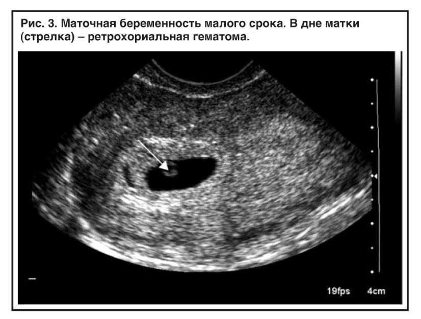
A bradycardic heart rate should prompt a follow-up sonographic examination to assess embryonic viability, whereas a normal heart rate provides considerable reassurance for the patient.
Small gestational sac in relation to crown-rump lengthOn occasion, an embryo with a normal heart rate will appear sonographically ‘crowded’ within the gestational sac (Figure 8), a finding which has been associated with an increased risk of early pregnancy loss.18,19
Objectively, this has been characterized as a mean sac size – crown rump length (MSS-CRL) of < 5 mm.18 Bromley et al. demonstrated that the risk of miscarriage was 94 percent with a small gestational sac in the first trimester despite a normal cardiac rate.
In a larger and more recent study of patients conceiving via IVF, the early pregnancy loss rate among patients with a MSS-CRL of< 5 mm was approximately 44 percent compared with a referent population with a MSS-CRL of 5-9. 9mm in whom the loss rate was 15.8 percent (P < .0001).19
Of note, there is no appreciable increase in the risk of early pregnancy loss between those conceiving using assisted reproductive technology compared with spontaneously conceived pregnancies.20 The sonographic finding of an embryo in a small gestational sac should prompt follow-up sonography to assess persistence of development.
Yolk sac size
The yolk sac is the first structure that is identifiable within the gestational sac by transvaginal ultrasound when the gestational sac reaches 8-10 mm. Abnormalities in size and appearance of the yolk sac have been reported to be associated with an increased risk of pregnancy loss, although not all studies have found this to be a useful metric for predicting pregnancy loss.5,12,21
The sonographic assessment of the yolk sac in asymptomatic patients as a predictor of miscarriage has shown sensitivities ranging from 17 percent to 69 percent and specificities ranging from 79 percent to 99 percent. 16 A yolk sac of greater than 7 mm has been suggested as a concerning threshold for an increased risk of pregnancy loss, although methods of measurement have been inconsistent.5
Recent data suggests yolk sac diameter does not improve the prediction of miscarriage over bradycardic heart rates and limited embryonic/fetal crown-rump length.22
One should be very cautious in the use of this sonographic sign, especially as an isolated finding, for prediction of first-trimester pregnancy loss until stronger guidance from the literature is available.
Crown-rump length
In a well dated pregnancy, a small crown-rump length for gestational age may reflect early growth disturbance and is associated with an increased risk of aneuploidy as well as early pregnancy loss.22 Among an IVF population with clear gestational age determination, those with a crown-rump length of < 10 percent had an increased risk of miscarriage as compared to those normally grown, 17. 2 percent vs. 6.6 percent,P = 0.005, OR = 2.93, 95% CI 1.2,6.7).23
Should a much smaller than expected crown-rump length be encountered in the first trimester, especially those that would lead to a change in pregnancy dating, a follow-up evaluation to assess interval growth in 2 weeks should be considered.
Subchorionic hematoma
Bleeding in early pregnancy is common, and a subchorionic hematoma can be sonographically identified in this setting (Figure 9). Subchorionic hematoma has been associated with an increased rate of pregnancy loss, especially if the hematoma is large, associated with bleeding or the patient is 35 years of age or older.24
The method of assessing the size of the subchorionic hematoma has been controversial, but it appears that in one study, the subjective assessment of hematoma size based on the fraction of the gestational sac size correlates best with first-trimester pregnancy outcome.25 The rate of spontaneous pregnancy loss in the first trimester is reported to be highest for those hematomas diagnosed before 8 weeks (19. 6 percent) compared to those diagnosed after 8 weeks (3.6 percent; P < .001).25
A review and meta-analysis demonstrated that the identification of a subchorionic hematoma was associated with an increased risk for miscarriage, increasing from 8.9 percent to 17.6 percent, pooled OR 2.18, 95% CI 1.29, 3.68).26
A recent retrospective cohort study of 2,446 patients with singletons presenting for ultrasound between 6 weeks and 13+6d weeks at a single center, demonstrated that subchorionic hematoma was associated with an increased risk of pregnancy loss before 20 weeks gestation (7.5 percent vs. 4.9 percent P=.026) on univariate analysis, however, when adjusting for patient age and bleeding, this association was no longer significant.27
Similarly, these authors showed no increased risk of adverse outcome later in gestation.28 Given the possible increased risk for pregnancy loss, follow-up sonography can be considered in these cases.
Chorionic Bump
A chorionic bump is a focal convex protuberance that develops at the choriodecidual surface and protrudes into the gestational sac in the early first trimester, likely reflecting a hematoma or necrotic decidua (Figure 10).
The finding has been associated with an increased risk of early pregnancy loss when identified before 11 weeks gestation. If a chorionic bump is identified and the pregnancy is otherwise normal in appearance with a normal heart rate, the live birth rate has been reported to be approximately 83 percent.29
In addition, there was no significant relationship between the volume of the chorionic bump or bleeding per vagina and the risk of pregnancy loss.29 In the latter part of the first trimester, the presence of a chorionic bump is not thought to be clinically relevant.30
Conclusions
Ultrasound is a powerful tool in the diagnosis and prediction of early pregnancy loss.
Practitioners must be diligent to follow guidelines for diagnosing a pregnancy as non-viable in order to prevent an iatrogenic pregnancy termination. Although some patients will have sonographic findings that definitively allow the diagnosis of a failed pregnancy, many will have findings that are suggestive or inconclusive of miscarriage. A follow-up scan can be very helpful in these cases.
The timing of the sonographic evaluation is important in management, as too early an evaluation is likely to lead to an ultrasound report of a pregnancy of unknown location or an intrauterine pregnancy of uncertain viability.
In a study of asymptomatic women attending an early pregnancy ultrasound unit, the diagnosis of a miscarriage could not be made on initial ultrasound examination until 35 days from LMP and most miscarriages were diagnosed when the first assessment was between 63 and 85 days after the LMP.4
These authors recommended that in order to reduce the number of inconclusive scans, asymptomatic women without a history of ectopic, delay an initial ultrasound until 49 days from LMP.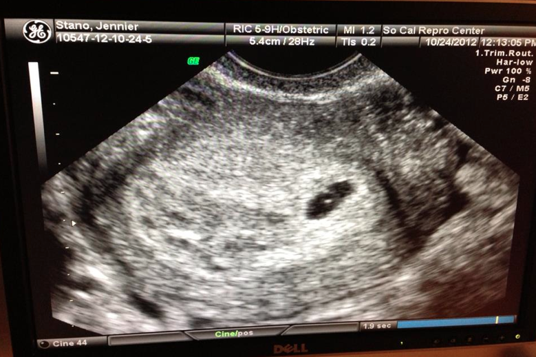 4
4
Healthcare providers taking care of women suspected of having or experiencing an early pregnancy loss should have training in how to compassionately and effectively communicate difficult news as the patient is at risk for posttraumatic stress, anxiety and depression.9
About the Authors
DR. BROMLEY practices in the Department of OB/GYN at Massachusetts General Hospital in Boston and with Diagnostic Ultrasound Associates, PC, in Brookline, Mass. She is a part-time professor at Harvard Medical School.
DR. SHIPP practices in the Department of OB/GYN at Brigham & Women’s Hospital in Boston and with Diagnostic Ultrasound Associates, PC, in Brookline, Mass. He is an associate professor at Harvard Medical School.
References1.ACOG Practice Bulletin No. 200: Early Pregnancy Loss. Obstet Gynecol. 2018;132(5):e197-e207.
2.Wise LA, Wang TR, Willis SK, Wesselink AK, Rothman KJ, Hatch EE. Effect of a Home Pregnancy Test Intervention on Cohort Retention and Pregnancy Detection: A Randomized Trial. Am J Epidemiol. 2020.
3.Ammon Avalos L, Galindo C, Li DK. A systematic review to calculate background miscarriage rates using life table analysis. Birth Defects Res A Clin Mol Teratol. 2012;94(6):417-423.
4.Bottomley C, Van Belle V, Mukri F, et al. The optimal timing of an ultrasound scan to assess the location and viability of an early pregnancy. Hum Reprod. 2009;24(8):1811-1817.
5.Doubilet PM, Benson CB, Bourne T, et al. Diagnostic criteria for nonviable pregnancy early in the first trimester. N Engl J Med. 2013;369(15):1443-1451.
6.Pexsters A, Luts J, Van Schoubroeck D, et al. Clinical implications of intra- and interobserver reproducibility of transvaginal sonographic measurement of gestational sac and crown-rump length at 6-9 weeks' gestation. Ultrasound Obstet Gynecol. 2011;38(5):510-515.
7.Abdallah Y, Daemen A, Kirk E, et al. Limitations of current definitions of miscarriage using mean gestational sac diameter and crown-rump length measurements: a multicenter observational study. Ultrasound Obstet Gynecol. 2011;38(5):497-502.
8.Shwayder JM. Waiting for the tide to change: reducing risk in the turbulent sea of liability. Obstet Gynecol. 2010;116(1):8-15.
9.Farren J, Jalmbrant M, Falconieri N, et al. Posttraumatic stress, anxiety and depression following miscarriage and ectopic pregnancy: a multicenter, prospective, cohort study. Am J Obstet Gynecol. 2020;222(4):367 e361-367 e322.
10.Committee on Obstetric Practice tAIoUiM, the Society for Maternal-Fetal M. Committee Opinion No 700: Methods for Estimating the Due Date. Obstet Gynecol. 2017;129(5):e150-e154.
11.Papaioannou GI, Syngelaki A, Poon LC, Ross JA, Nicolaides KH. Normal ranges of embryonic length, embryonic heart rate, gestational sac diameter and yolk sac diameter at 6-10 weeks.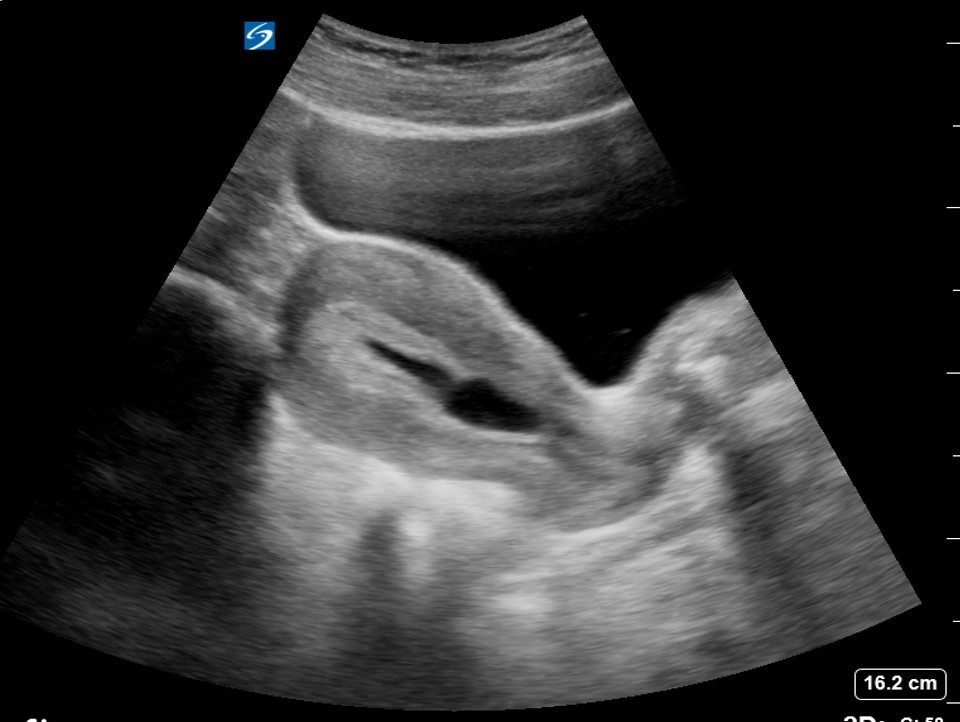 Fetal Diagn Ther. 2010;28(4):207-219.
12.Detti L, Francillon L, Christiansen ME, et al. Early pregnancy ultrasound measurements and prediction of first trimester pregnancy loss: A logistic model. Sci Rep. 2020;10(1):1545.
13.Preisler J, Kopeika J, Ismail L, et al. Defining safe criteria to diagnose miscarriage: prospective observational multicentre study. BMJ. 2015;351:h5579.
14.Bromley B, Doubilet P, Frigoletto FD, Jr., Krauss C, Estroff JA, Benacerraf BR. Is fetal hyperechoic bowel on second-trimester sonogram an indication for amniocentesis? Obstet Gynecol. 1994;83(5 Pt 1):647-651.
15.Doubilet PM, Benson CB. Embryonic heart rate in the early first trimester: what rate is normal? J Ultrasound Med. 1995;14(6):431-434.
16.Pillai RN, Konje JC, Richardson M, Tincello DG, Potdar N. Prediction of miscarriage in women with viable intrauterine pregnancy-A systematic review and diagnostic accuracy meta-analysis. Eur J Obstet Gynecol Reprod Biol. 2018;220:122-131.
17.DuBose TJ. Embryonic heart rates. Fertil Steril. 2009;92(4):e57; author reply e58.
18.Bromley B, Harlow BL, Laboda LA, Benacerraf BR. Small sac size in the first trimester: a predictor of poor fetal outcome. Radiology. 1991;178(2):375-377.
19.Kapfhamer JD, Palaniappan S, Summers K, et al. Difference between mean gestational sac diameter and crown-rump length as a marker of first-trimester pregnancy loss after in vitro fertilization. Fertil Steril. 2018;109(1):130-136.
20.Schieve LA, Tatham L, Peterson HB, Toner J, Jeng G. Spontaneous abortion among pregnancies conceived using assisted reproductive technology in the United States. Obstet Gynecol. 2003;101(5 Pt 1):959-967.
21.Taylor TJ, Quinton AE, de Vries BS, Hyett JA. First-trimester ultrasound features associated with subsequent miscarriage: A prospective study.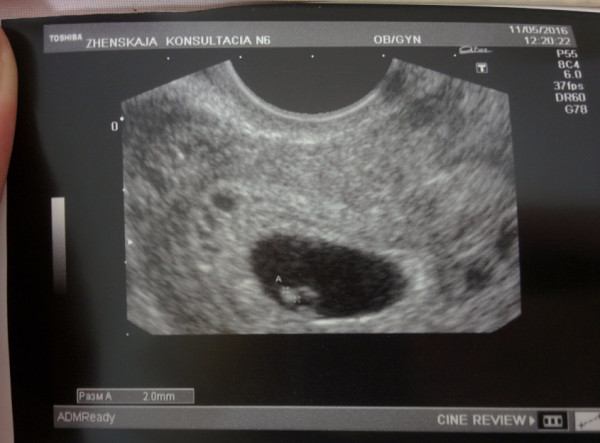 Aust N Z J Obstet Gynaecol. 2019;59(5):641-648.
22.DeVilbiss EA, Mumford SL, Sjaarda LA, et al. Prediction of pregnancy loss by early first trimester ultrasound characteristics. Am J Obstet Gynecol. 2020.
23.Gabbay-Benziv R, Dolev A, Bardin R, Meizner I, Fisch B, Ben-Haroush A. Prediction of fetal loss by first-trimester crown-rump length in IVF pregnancies. Arch Gynecol Obstet. 2017;295(3):771-775.
24.Bennett GL, Bromley B, Lieberman E, Benacerraf BR. Subchorionic hemorrhage in first-trimester pregnancies: prediction of pregnancy outcome with sonography. Radiology. 1996;200(3):803-806.
25.Heller HT, Asch EA, Durfee SM, et al. Subchorionic Hematoma: Correlation of Grading Techniques With First-Trimester Pregnancy Outcome. J Ultrasound Med. 2018;37(7):1725-1732.
26.Tuuli MG, Norman SM, Odibo AO, Macones GA, Cahill AG. Perinatal outcomes in women with subchorionic hematoma: a systematic review and meta-analysis.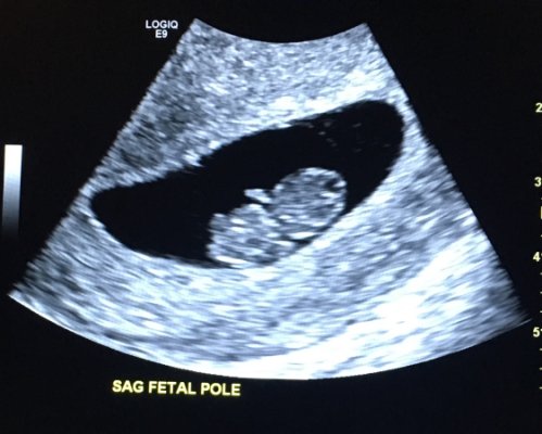 Obstet Gynecol. 2011;117(5):1205-1212.
27.Naert MN, Khadraoui H, Muniz Rodriguez A, Naqvi M, Fox NS. Association Between First-Trimester Subchorionic Hematomas and Pregnancy Loss in Singleton Pregnancies. Obstet Gynecol. 2019;134(2):276-281.
28.Naert MN, Muniz Rodriguez A, Khadraoui H, Naqvi M, Fox NS. Association Between First-Trimester Subchorionic Hematomas and Adverse Pregnancy Outcomes After 20 Weeks of Gestation in Singleton Pregnancies. Obstet Gynecol. 2019;134(4):863-868.
29.Arleo EK, Dunning A, Troiano RN. Chorionic bump in pregnant patients and associated live birth rate: a systematic review and meta-analysis. J Ultrasound Med. 2015;34(4):553-557.
30.Sepulveda W. Chorionic bump at 11 to 13 weeks' gestation: Prevalence and clinical significance. Prenat Diagn. 2019;39(6):471-476.
Spontaneous miscarriage and miscarriage
Over the past 10 years, the number of spontaneous miscarriages has been growing rapidly. The International Histological Classification Organization (FIGO) has declared the situation with an increase in the frequency of miscarriages an epidemic.
Spontaneous miscarriage is the termination of pregnancy before the fetus reaches a viable term (up to 22 weeks of pregnancy and fetal weight 500 g.).
Most miscarriages (about 80%) occur before 12 weeks of gestation. Moreover, in the early stages up to 8 weeks of pregnancy, the cause of miscarriage is chromosomal abnormalities in 50% of cases. It turns out that nature eliminates the defective product of conception. And these causes are difficult to prevent, especially in the presence of hereditary diseases. Fortunately, accidental breakdowns are much more common than genetically determined ones. Therefore, subsequent pregnancies usually end happily. nine0003 But the remaining 50% of miscarriages have completely real and removable causes. They can be easily identified at the stage of preparation for pregnancy by a gynecologist.
What are the reasons?
- chronic diseases: inflammatory diseases of the uterus and appendages, polycystic ovary syndrome, uterine fibroids, endometriosis, malformations of the genital organs.
- infections: toxoplasmosis, listeriosis, genital tuberculosis, sexual infections - chlamydia, mycoplasma, ureaplasma, syphilis. nine0007 - antiphospholipid syndrome.
- endocrine diseases: diabetes, thyroid disease.
- metabolic disorders in the body: obesity, folic acid deficiency, iron deficiency, vitamin D deficiency.
- male factor.
Of course, these causes are identified and eliminated before the planned conception.
There are harmful factors that can affect the development of the fetus in the early stages of pregnancy and lead to miscarriage:
- alcohol consumption. nine0007 - caffeine use (4-5 cups of coffee per day).
- smoking (more than 10 cigarettes a day).
- drug use.
- taking medications with a teratogenic effect (for example: aspirin, nise and others from this group of drugs; antifungals; antidepressants; some antibiotics and a number of other drugs).
- toxins and occupational hazards: ionizing radiation, pesticides, inhalation of anesthetic gases.
What are the signs of possible pregnancy loss? nine0004
These are complaints of pain in the lower abdomen and lower back, bloody discharge from the genital tract. It is necessary to consult a doctor to rule out an ectopic pregnancy and conduct an additional examination (hCG test, blood test for progesterone, ultrasound).
In early pregnancy, with dubious ultrasound data or suspected non-developing (missing) pregnancy, expectant management is chosen with a repeat examination by a gynecologist, ultrasound, tests after 7-10 days. If the diagnosis was made and the fact of uterine pregnancy was confirmed, with a threatened miscarriage, preservation therapy is carried out in an outpatient day hospital. A miscarriage that has begun requires hospitalization in the gynecological department. In the case of a non-developing pregnancy, an abortion is performed. nine0005
In accordance with the clinical treatment protocol approved by the Ministry of Health of the Russian Federation dated 07. 06.2016. Preference is given to drug therapy aimed at terminating pregnancy with prostaglandin analogues (misoprostol) with or without prior use of an antiprogestin (mifepristone). In case of need for surgical treatment (with incomplete miscarriage with infected miscarriage), it is recommended to use aspiration curettage (with an electric vacuum source or a manual vacuum aspirator). What has a significant advantage over curettage of the uterine cavity because it is less traumatic and can be performed on an outpatient basis. nine0005
All women who have had a miscarriage need treatment to prevent complications and prevent recurrent miscarriages. Why is rehabilitation therapy necessary?
According to the decision of the XVIII World Congress of Obstetricians and Gynecologists , the diagnosis of chronic endometritis should be made to absolutely all women who have had an undeveloped pregnancy. Two out of three miscarriages according to Professor V. E. Radzinsky are caused by this disease. When examining the material from the uterine cavity, infectious pathogens were isolated: ureaplasmas, mycoplasmas, streptococci, staphylococci, Escherichia coli, viruses (herpes, HPV). Therefore, it is very important to carry out treatment immediately after the termination of pregnancy. nine0007 If time is lost, it is necessary to carry out additional diagnostics: a pipel biopsy of the endometrium with a histological examination and a study for infections, including tuberculosis. Then, taking into account the results obtained, symptomatic anti-inflammatory therapy is carried out (immunomodulators, antibacterial drugs, physiotherapy, gynecological massage, mud therapy). In parallel, an examination is prescribed to identify other causes of miscarriage (male factor, chronic maternal diseases, genital infections, antiphospholipid syndrome). nine0007 In the medical center "Mifra-Med" at the level of modern requirements of medicine, all the possibilities for a complete adequate examination have been created: all types of tests, ultrasound, hysteroscopy, aspiration biopsy, consultations of narrow specialists (endocrinologist, therapist, neurologist, urologist).
 Our gynecologists of the highest category Melko O.N., Novitskaya E.L., Tikhonova T.N. and urologist of the highest category Kanaev S.A. have sufficient experience in the rehabilitation and preparation of couples for the next pregnancy with a successful outcome. Treatment is carried out in a day hospital with the use of drugs, physiotherapy, gynecological massage, prostate massage. nine0005
WE WILL HELP YOU!
st. Yakovleva, 16 st. Kirova 47 B
tel. 244-744 tel. 46-43-57
Obstetrician-gynecologist of the Reproductive Health Center "SM-Clinic" told about whether it is possible not to notice a miscarriage
Unfortunately, the loss of a child at an early stage of pregnancy is quite common. After the first miscarriage, a woman lives in constant fear and is afraid that the second attempt to become a mother will turn into a tragedy. nine0005
“A miscarriage is the spontaneous termination of a pregnancy before the fetus reaches a viable term. A fetus weighing up to 500 g is considered viable, which corresponds to a period of less than 22 weeks of pregnancy. Many women face this diagnosis. About 80 percent of miscarriages occur before 12 weeks of pregnancy.”
Causes of miscarriage
Approximately half of early miscarriages occur due to genetic pathologies in the development of the fetus, that is, from defects in the number and composition of chromosomes. It is in the first weeks that the formation of the baby's organs begins, which requires 23 normal chromosomes from each of the future parents. When at least one abnormal changes occur, there is a risk of losing a child. nine0005
At 8-11 weeks of gestation, the rate of these miscarriages is 41-50 percent, at 16-19 weeks of gestation, the rate of miscarriages due to chromosomal defects drops to 10-20 percent.
There are other causes of miscarriage. Among them:
- Congenital and acquired disorders of the anatomy of the genital organs If there are fibroids, polyps in the uterus, this can cause abnormal development of the embryo. The threat of miscarriage may be in women with an abnormal development of the uterus. nine0078 Infectious causes Numerous studies have shown that the risk of miscarriage increases in the presence of sexually transmitted infections. Dangerous for a pregnant woman are measles, rubella, cytomegalovirus, as well as diseases that occur with an increase in body temperature. Intoxication of the body often leads to the loss of a child.
- Endocrine causes Problems with gestation occur with diabetes, thyroid diseases, disorders of the adrenal glands. nine0078 Unfavorable ecology, exposure
- Blood clotting disorder (thrombosis, antiphospholipid syndrome) APS (antiphospholipid syndrome) is a disease in which the human body produces a lot of antibodies to phospholipids, the chemical structures that make up parts of cells.
 The body mistakenly perceives its own phospholipids as foreign and begins to defend itself against them: it produces antibodies to them that damage blood components. Blood clotting increases, microthrombi appear in small vessels that feed the fetal egg and placenta. Blood circulation in the fetal egg is disturbed. As a result, the pregnancy stops or the growth of the fetus slows down. Both of these lead to miscarriage. All this is due to the hormonal background that has changed during pregnancy. nine0078 Lifestyle and bad habits Nicotine addiction, alcohol use, obesity.
 Is it possible not to notice a miscarriage
Sometimes women mistake a miscarriage for normal menstruation. This occurs during the so-called biochemical pregnancy, when there is a violation of the implantation of the embryo at a very early stage and menstruation begins. But before the appearance of spotting, the test will show two strips.
The classic variant is when a miscarriage is manifested by bleeding against the background of a long delay in menstruation, which rarely stops on its own.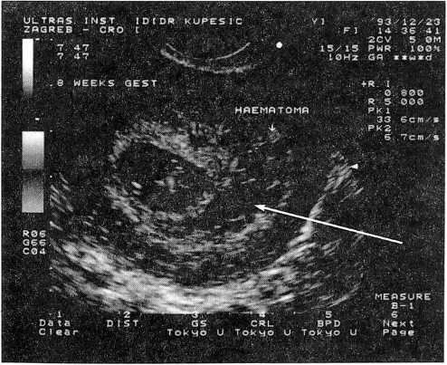 Therefore, even if a woman does not follow the menstrual cycle, the signs of an interrupted pregnancy will be immediately noticed by the doctor during examination and ultrasound. nine0007
Alarm
The symptoms of a miscarriage can be completely different, and depending on them, as a rule, it is possible to predict the likelihood of maintaining and successfully continuing this pregnancy.
For the threat of miscarriage is characterized by pulling pains in the lower abdomen and lumbar region, scanty bloody discharge from the genital tract. Ultrasound signs: the tone of the uterus is increased, the cervix is not shortened and closed, the body of the uterus corresponds to the gestational age, the fetal heartbeat is recorded. nine0005
Started miscarriage - pain and discharge from the genital tract are more pronounced, the cervix is ajar.
Miscarriage in progress - cramping pains in the lower abdomen, copious bloody discharge from the genital tract. On examination, as a rule, the uterus does not correspond to the gestational age, the cervix is open, the elements of the fetal egg are in the cervix or in the vagina.
Incomplete miscarriage - the pregnancy was interrupted, but there are delayed elements of the fetal egg in the uterine cavity. This is manifested by ongoing bleeding due to the lack of a full contraction of the uterus. nine0005
Non-progressive pregnancy - the death of an embryo (up to 9 weeks) or a fetus up to 22 weeks of gestation in the absence of any signs of termination of pregnancy.
Important!
Severe abdominal pain and spotting at any stage of pregnancy is a reason for an urgent appeal to an obstetrician-gynecologist with a solution to the issue of hospitalization in a gynecological hospital.
Is it possible to avoid miscarriage
“Today, there are no methods for preventing miscarriages,” the doctor says.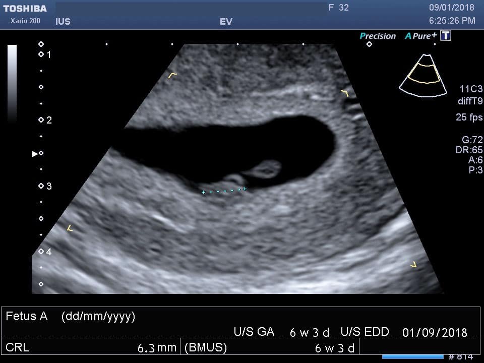 “Therefore, it is very important to comprehensively prepare for pregnancy before it occurs by visiting an obstetrician-gynecologist and following all the necessary recommendations for examination and taking the necessary drugs.”
But if, nevertheless, the pregnancy could not be maintained, then the birth of a child can be planned again no earlier than 3-6 months after the miscarriage. This time is needed to figure out, together with the attending physician, what are the causes of miscarriage and whether it is possible to avoid them in the future. nine0005
By the way, a common misconception for both women and men is that only the woman is to blame for the loss of pregnancy, but this is far from being the case.
“A man is also responsible, which is why future dads are required to perform a study - a spermogram and be screened for genital infections, since with a pathology of spermatozoa, the likelihood of miscarriage due to genetic abnormalities increases many times,” emphasizes our expert.