How big does your uterus get during pregnancy
Sizes And How It Works
The uterus creates the placenta for fetal development and blood vessels to nurture it.
Research-backed
MomJunction believes in providing reliable, research-backed information to you. As per our strong editorial policy requirements, we base our health articles on references (citations) taken from authority sites, international journals, and research studies. However, if you find any incongruencies, feel free to write to us.
When you think of pregnancy, the first thing you may envision is a growing belly. While this is a visible change of pregnancy that is hard to ignore, myriad transformations occur inside a pregnant woman’s body. The most important change is the size of the uterus during pregnancy.
The uterus plays a crucial role in pregnancy as the abode of the infant, and it constantly expands during your gestation journey to hold the developing fetus.
This post discusses interesting details about the uterus, including its size during and before pregnancy, functions, and positions. It also mentions how to maintain a healthy uterus.
Uterus During Pregnancy
The uterus is a distensible organ the size of a closed fist. It grows and changes to become large enough to accommodate a full-term baby. It is held in its position by ligaments, which stretch as the uterus grows.
Dr. Alan Lindemann, MD, a former clinical associate professor at the University of North Dakota, says, “When you get 10 or 12 weeks along, about the end of the third month, your uterus could be appreciably enlarged and feel heavy. You also have more blood supply, so your arteries and veins to and from your uterus might be perceived as heavy.”
Read on as we tell you about what exactly the uterus does, how much its size changes, and how you can measure the organ and keep it healthy.
Size Of Uterus During Pregnancy
As you know, the uterus keeps changing in shape and size as your pregnancy progresses. The uterus expands between 500 and 1,000 times its normal size (1).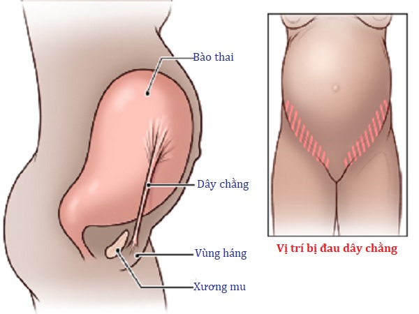 Let’s see how the organ changes during each trimester.
Source: www.newhealthadvisor.com
The first trimester (0 to 12 weeks)
- At 12 weeks of pregnancy, the size of the uterus remains as small as the size of a grapefruit.
- As your pregnancy progresses, the uterus grows, putting pressure on the bladder. This is when the frequency of urination increases (2).
- In the case of twins or multiple pregnancies, the stretching of the uterus will be faster compared to that of a single baby.
Related: First Trimester Of Pregnancy: Changes, Symptoms & Baby Growth
The second trimester (14-28 weeks)
- By the second trimester, the uterus grows to the size of a papaya. The uterus grows upward and develops outside the pelvic area.
- The uterus expands between the naval area and the breasts and starts putting thrust on the other organs, pushing them away from their original positions (3). This may lead to some tensions in the ligaments and the surrounding muscles, leading to body aches and pains.

- In some cases, the naval may pop out because of the pressure put by the uterus on other organs.
The third trimester (28-40 weeks)
- By the third trimester, your uterus will get enlarged to the size of a watermelon. It will move from your pubic brim to the lower bottom of the rib cage.
- Once you approach labor, your baby will descend into the pelvis.
Related: Third Trimester: When It Starts And What Changes Happen
After childbirth
After childbirth, the uterus shrinks back to its normal position and size. This process is known as involution, which will take about six to eight weeks (4).
Apart from changing in size to accommodate the growing fetus, the uterus also plays other roles during pregnancy.
Functions Of The Uterus During Pregnancy
During pregnancy, the uterus:
- Accepts the fertilized ovum that passes through the fallopian tube.
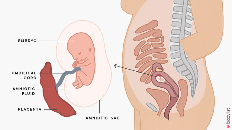
- Creates the placenta for the development of the fetus.
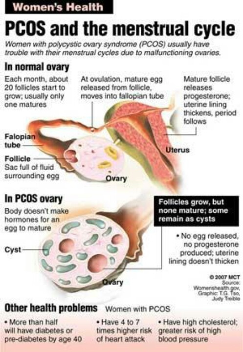
- Nurtures the fetus with nutrients by developing blood vessels exclusively for this purpose.
- Contracts to facilitate the exit of the baby and the placenta through the vagina during childbirth.
- Post-delivery, it shrinks back and starts preparing for the next menstrual cycle.
- It helps the blood flow into the ovaries. It supports other organs such as the vagina, bladder, and rectum. It plays an important role in sexual functions like triggering orgasm (5).
Interesting how important this inconspicuous organ is, right? Read on, and we will give more information about the uterus and its functions.
How Big Is a Uterus? Normal Size Of A Uterus In Women
The size of the uterus varies from woman to woman. It weighs approximately 70 to 125gm (6). However, the size of the uterus is based on factors such as age and hormonal conditions of a woman.
Size of the uterus:
- Before attaining puberty, the length of the uterus is about 3.
 5cm and thickness is approximately 1.4cm (7).
- After attaining puberty, the length is between 5 and 8cm, width is 3.5cm, and thickness is between 1.5 and 3cm.
- During pregnancy at term, the uterus measures 38cm in length and 24 to 26cm in width.
Just like the size of the uterus, the position of the uterus also varies from woman to woman. It can be in an anteverted, anteflexed, or any other position.
What Is The Uterus Made Of?
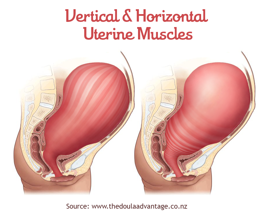
The uterus is an inverted, pear-shaped, and hollow, muscular reproductive organ (8) located in the pelvic region of a woman’s body.
Image: iStock
It is made of smooth muscles lined with various glands. These muscles contract during orgasm, menstruation, and labor, whereas the glands grow thicker with the stimulation of ovarian hormones every month. If you do not become pregnant during the ovulation period, the glands will cast off through the monthly menstrual cycle.
The uterus extends into the vagina through the cervix, which is made up of fibromuscular tissues, and it controls the flow of material into and out of the uterus.
Did you know?
Some women may have a short cervix, which increases the risk of premature labor. Medical or surgical intervention can help prevent complications (15).
Related: Cervical Cerclage: Why And How It Is Done During Pregnancy
Positions Of The Uterus
One of the factors that influence the position of the uterus is the degree of dilation of the bladder. The uterus normally lies right above the bladder and in front of the (anterior) rectum. The normal uterine position is straight and vertical.
Common uterus positions
The uterus position can also be anteverted or anteflexed, both of which are common.
Image: Shutterstock
- In the anteverted position, the uterus is bent forward towards the bladder/ pubic bone and towards the front of the body.
- In anteflexed position, the uterus is rotated towards the pubic bone with a concave surface.
Uncommon uterus positions
Certain positions are tilted back towards the rectum.
- A retroverted uterus is tipped backward toward the rectum. Around 25% of women have this type of uterus.
- A retroflexed uterus is the opposite of the anteflexed uterus, as it takes a concave shape towards the rectum (9).
A retro uterus is not known to create any fertility issues. However, you might have pain during sexual intercourse.
A tilted uterus does not usually create a problem during pregnancy, as the changing uterus will retain the forward-tipped position (10).
Research finds
An incarcerated uterus or a trapped uterus is an extremely rare complication of a retroverted uterus (16). A portion of the uterus gets entrapped and expands within the confined areas of the pelvis, which might cause uterine rupture (17).
Related: Where Does Sperm Go During Pregnancy? Is It Safe?
Measuring The Uterus During Pregnancy
Your doctor might measure the size of your uterus, also called the fundal height (fundus is the domed region at the top of the uterus), to understand the fetal growth and development. The fundal height is the measurement of the top of the pubic bone to the top of the uterus, which determines the gestational age.
Note: It should be noted that the size of the uterus varies from woman to woman and depends on height, weight, and age (11).
You, too, can measure the size of your uterus. Before we go into the various methods of measuring it, let’s see the initial steps:
- Lie down on your back on the bed or the floor. While lying down, you should not feel any pain or dizziness.
- Locate your uterus by touching your tummy. If you are over 20 weeks pregnant, you can feel the uterus just above the navel. The uterus is hard, round.
- Now, move your fingers upward to feel the top of the uterus, which is also referred to as fundus.
- Proceed to locate your pubic bone. It is positioned just above your pubic hair line. The bone tip that you can feel is the pubic bone.
Once you locate the fundus and the pubic bone, you can measure the fundal height using various methods.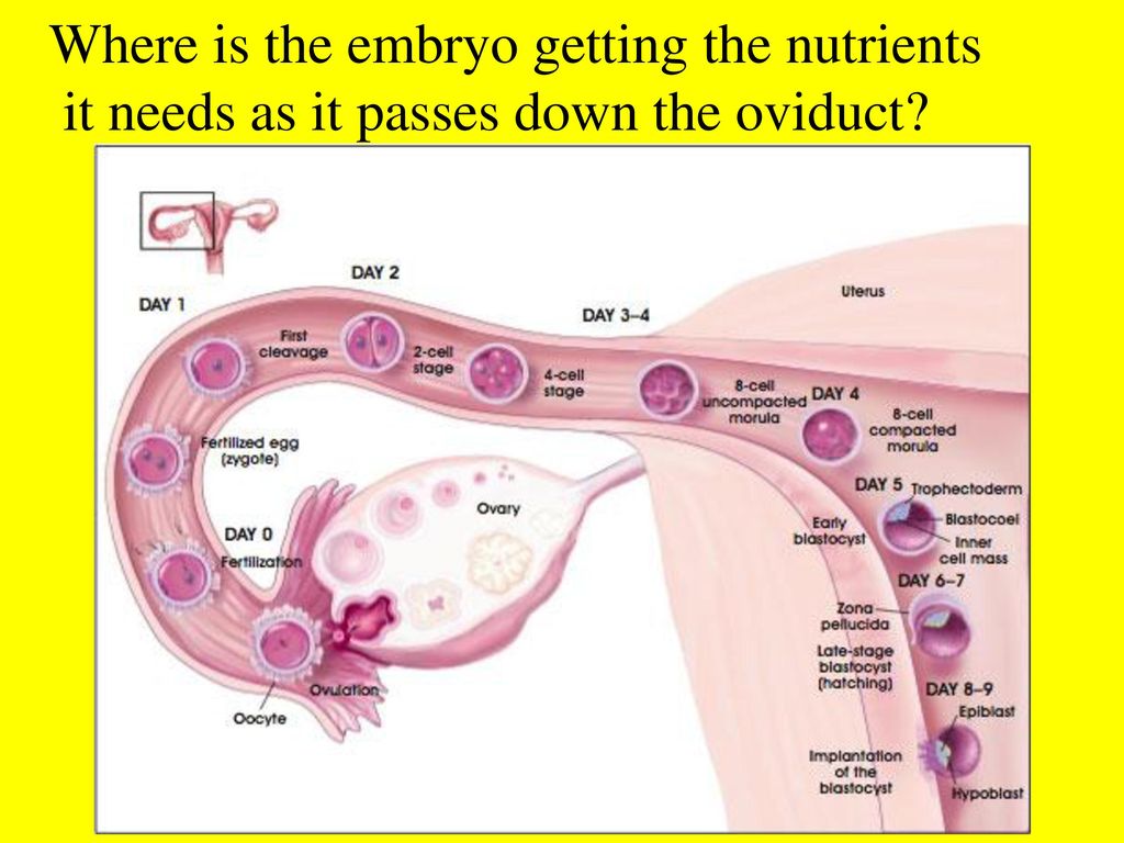
- Measure the distance between your fundus and the pubic bone in centimeters using a tape.
- For example, if you are 24 weeks pregnant, then your uterus would usually measure 22-26cm. The uterus usually grows 1cm in a week or about 4cm in a month.
You can use this method to track your pregnancy growth week by week. However, studies say that more research needs to be done to find out the reliability of this method.
2. Measuring the fundal height using the finger method- If your uterus is below or above the belly button, then measure how many fingers below or above is the uterus from the bellybutton.
- The assumption is the fundal height increases by two finger-widths every month.
- For example, if you are 4 two-finger-widths above the belly button, then it indicates seven months gestation (see the above figure).
This method helps determine the gestational age in months (12).
If you find the fundal height different from the gestational week that you think you are in, talk to your doctor, and get your doubts cleared.
The size of the uterus during pregnancy plays an important role in the well-being of your baby growing inside. Next, we look at how you can maintain the health of your uterus during pregnancy.
How To Keep Your Uterus Healthy?
The best way to keep your uterus healthy is to keep yourself healthy. Here is how you can do it:
- Make sure to eat right and engage in some forms of exercise to maintain.
- Eat omega-3 fatty acid-rich food.
- Quit smoking as it has harmful effects on the uterus.
- Do not hold your urine.
1. When does the mouth of the uterus open in pregnancy?
The opening of the mouth of the uterus is called cervical dilation. The cervix begins to dilate towards the end of pregnancy.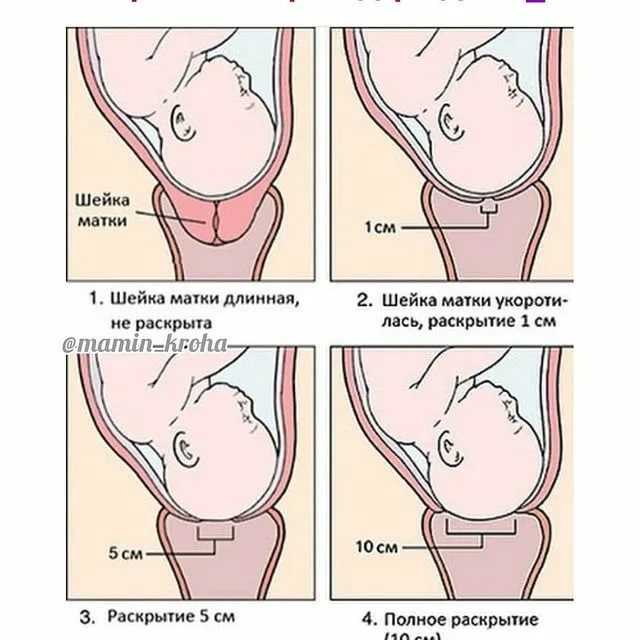 It becomes thinner and dilates during the first stage of labor, and is completely dilated by about 10cm during the last stage of labor (13).
2. How does a weak uterus affect pregnancy?
An incompetent or weak cervix, a kind of womb abnormality, may lead to preterm birth in some women (14).
3. Does a swollen uterus mean I am pregnant?
“A swollen uterus could mean pregnancy. Generally speaking, by the time you can appreciate the swelling in your uterus, you will be about three months along. However, there are other reasons for swelling. The most common reason for swelling is fibroids, which are benign smooth muscle tumors,” observes Dr. Lindemann.
The uterus expands during pregnancy, and its growth indicates the size of the fetus. After birth, the uterus again conforms to its previous size. It is beautiful how a small organ grows over time into the size of a watermelon to accommodate a growing fetus. Understanding how your uterus changes throughout pregnancy will help you appreciate the first three trimesters. Because this organ is so important to your health, you must look after it before, during, and after the pregnancy. This entails eating a nutritious diet, exercising regularly, and seeing a gynecologist for regular examinations. Also, notify your gynecologist if you detect any unusual menstrual indications so that the underlying problem can be treated promptly.
References:
MomJunction's articles are written after analyzing the research works of expert authors and institutions. Our references consist of resources established by authorities in their respective fields. You can learn more about the authenticity of the information we present in our editorial policy.
1. The Developing Baby; Parents Centre
2. The First Trimester; John Hopkins Medicine
3. Pregnancy: Starting Out; Women’s Specialists of New Mexico, Ltd
4. Ms. Rajdawinder Kaur1, et al.; A Quasi- Experimental Study to Assess the Effectiveness of Early Ambulation on Involution of Uterus among Postnatal Mothers Admitted At SGRD Hospital, Vallah, Sri Amritsar, Punjab; International Journal of Health Sciences and Research
5. Role of the uterus in fertility, pregnancy, and developmental programming; American Society of Reproductive Medicine
6. Determining the Route and Method of Hysterectomy; Ethicon Endo-Surgery, Inc
7. Wellington de Paula Martins, et al.; Pelvic ultrasonography in children and teenagers; Scielo
8. Anatomy of Female Pelvic Area; John Hopkins Medicine
9. Dr Anne Marie Coady; The uterus and the endometrium Common and unusual pathologies; The British Medical Ultrasound Society
10. Retroverted Uterus; Better Health; Victoria State Government
11. Dr. Ajay M. Parmar, et al.; Sonographic Measurements Of Uterus And Its Correlation With Different Parameters In Parous And Nulliparous Women; International Journal of Medical Science and Education
12. Antenatal Care Module: 10. Estimating Gestational Age from Fundal Height Measurement; The Open University
13. Cervical Ripening; Cleveland Clinic
14. Uterine abnormality – problems with the womb; Tommy’s
15. Cervical insufficiency and short cervix; March of Dimes
16. Incarcerated uterus; Radiopaedia
17. Richard Slama, et al.; (2015)Uterine Incarceration: Rare Cause of Urinary Retention in Healthy Pregnant Patients; National Library of Medicine
The following two tabs change content below.
- Reviewer
- Author
- Expert
28 Weeks Pregnant: Symptoms, Baby Development And Changes
28 Weeks Pregnant: Symptoms, Baby Development And Changes
27th Week Pregnancy: Symptoms, Baby Development And Tips
27th Week Pregnancy: Symptoms, Baby Development And Tips
25 Weeks Pregnant: Symptoms, Baby Development And Growth
25 Weeks Pregnant: Symptoms, Baby Development And Growth
35th Week Pregnancy: Symptoms, Baby Development And Tips
35th Week Pregnancy: Symptoms, Baby Development And Tips
36th Week Pregnancy: Symptoms And Baby Development
36th Week Pregnancy: Symptoms And Baby Development
19 Weeks Pregnant: Signs, Symptoms, Baby Development & Tips
19 Weeks Pregnant: Signs, Symptoms, Baby Development & Tips
40th Week Pregnancy: Symptoms, Baby Development And Tips
40th Week Pregnancy: Symptoms, Baby Development And Tips
38th Week Pregnancy: Symptoms, Baby Development, And Tips
38th Week Pregnancy: Symptoms, Baby Development, And Tips
20 Weeks Pregnancy: Symptoms, Tips And Baby Development
20 Weeks Pregnancy: Symptoms, Tips And Baby Development
Anatomy of pregnancy and birth - uterus
Anatomy of pregnancy and birth - uterus | Pregnancy Birth and Baby beginning of content5-minute read
Listen
What does the uterus look like?
One of the most recognised changes in a pregnant woman’s body is the appearance of the ‘baby bump’, which forms to accommodate the baby growing in the uterus.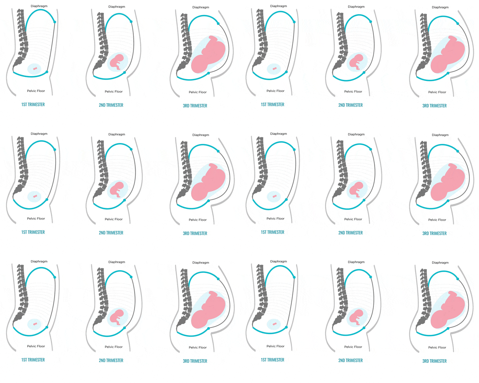 The primary function of the uterus during pregnancy is to house and nurture your growing baby, so it is important to understand its structure and function, and what changes you can expect the uterus to undergo during pregnancy.
The uterus (also known as the ‘womb’) has a thick muscular wall and is pear shaped. It is made up of the fundus (at the top of the uterus), the main body (called the corpus), and the cervix (the lower part of the uterus ). Ligaments – which are tough, flexible tissue – hold it in position in the middle of the pelvis, behind the bladder, and in front of the rectum.
The uterus wall is made up of 3 layers. The inside is a thin layer called the endometrium, which responds to hormones – the shedding of this layer causes menstrual bleeding. The middle layer is a muscular wall. The outside layer of the uterus is a thin layer of cells.
Illustration showing the female reproductive system.The size of a non-pregnant woman's uterus can vary. In a woman who has never been pregnant, the average length of the uterus is about 7 centimetres. This increases in size to approximately 9 centimetres in a woman who is not pregnant but has been pregnant before. The size and shape of the uterus can change with the number of pregnancies and with age.
How does the uterus change during pregnancy?
During pregnancy, as the baby grows, the size of a woman’s uterus will dramatically increase. One measure to estimate growth is the fundal height, the distance from the pubic bone to the top of the uterus. Your doctor (GP) or obstetrician or midwife will measure your fundal height at each antenatal visit from 24 weeks onwards. If there are concerns about your baby’s growth, your doctor or midwife may recommend using regular ultrasound to monitor the baby.
Fundal height can vary from person to person, and many factors can affect the size of a pregnant woman’s uterus. For instance, the fundal height may be different in women who are carrying more than one baby, who are overweight or obese, or who have certain medical conditions. A full bladder will also affect fundal height measurement, so it’s important to empty your bladder before each measurement. A smaller than expected fundal height could be a sign that the baby is growing slowly or that there is too little amniotic fluid. If so, this will be monitored carefully by your doctor. In contrast, a larger than expected fundal height could mean that the baby is larger than average and this may also need monitoring.
As the uterus grows, it can put pressure on the other organs of the pregnant woman's body. For instance, the uterus can press on the nearby bladder, increasing the need to urinate.
How does the uterus prepare for labour and birth?
Braxton Hicks contractions, also known as 'false labour' or 'practice contractions', prepare your uterus for the birth and may start as early as mid-way through your pregnancy, and continuing right through to the birth. Braxton Hicks contractions tend to be irregular and while they are not generally painful, they can be uncomfortable and get progressively stronger through the pregnancy.
During true labour, the muscles of the uterus contract to help your baby move down into the birth canal. Labour contractions start like a wave and build in intensity, moving from the top of the uterus right down to the cervix. Your uterus will feel tight during the contraction, but between contractions, the pain will ease off and allow you to rest before the next one builds. Unlike Braxton Hicks, labour contractions become stronger, more regular and more frequent in the lead up to the birth.
How does the uterus change after birth?
After the baby is born, the uterus will contract again to allow the placenta, which feeds the baby during pregnancy, to leave the woman’s body. This is sometimes called the ‘after birth’. These contractions are milder than the contractions felt during labour. Once the placenta is delivered, the uterus remains contracted to help prevent heavy bleeding known as ‘postpartum haemorrhage‘.
The uterus will also continue to have contractions after the birth is completed, particularly during breastfeeding. This contracting and tightening of the uterus will feel a little like period cramps and is also known as 'afterbirth pains'.
Read more here about the first few days after giving birth.
Sources:
The Royal Australian and New Zealand College of Obstetricians and Gynaecologists (Labour and birth), StatPearls Publishing (Anatomy, Abdomen and Pelvis), Department of Health (Clinical practice guidelines: Pregnancy care), Better Health Channel Victoria (Pregnancy stages and changes), Mater Mother's Hospital (Labour and birth information), Royal Australian and New Zealand College of Obstetricians and Gynaecologists (The First Few Weeks Following Birth), Queensland Health (Queensland Clinical Guidelines – maternity and neonatal), King Edward Memorial Hospital (Fundal height: Measuring with a tape measure), Royal Hospital for Women (Fetal growth assessment (clinical) in pregnancy), MSD Manual (Female internal genital organs)Learn more here about the development and quality assurance of healthdirect content.
Last reviewed: October 2020
Back To Top
Related pages
- Anatomy of pregnancy and birth - perineum and pelvic floor
- Anatomy of pregnancy and birth - pelvis
- Anatomy of pregnancy and birth - cervix
- Anatomy of pregnancy and birth - abdominal muscles
- Anatomy of pregnancy and birth
Need more information?
Prolapsed uterus - Better Health Channel
The pelvic floor and associated supporting ligaments can be weakened or damaged in many ways, causing uterine prolapse.
Read more on Better Health Channel website
Uterus, cervix & ovaries - fact sheet | Jean Hailes
This fact sheet discusses some of the health conditions that may affect a woman's uterus, cervix and ovaries.
Read more on Jean Hailes for Women's Health website
Dilatation and curettage (D&C)
A D&C is an operation to lightly scrape the inside of the uterus (womb).
Read more on WA Health website
Ectopic pregnancy
An ectopic pregnancy occurs when a fertilised egg implants outside the uterus (womb)
Read more on WA Health website
Hormonal IUD (intrauterine device, Mirena®) | Body Talk
The hormonal IUD is a type of contraception and is placed inside the uterus by a specially trained doctor or nurse. Find out all the facts here.
Read more on Body Talk website
Placental abruption - Better Health Channel
Placental abruption means the placenta has detached from the wall of the uterus, starving the baby of oxygen and nutrients.
Read more on Better Health Channel website
Copper IUD (intrauterine device) | Body Talk
The copper IUD is a type of contraception and is placed inside the uterus by a specially trained doctor or nurse. Find out all the facts here.
Read more on Body Talk website
Placenta previa - Better Health Channel
Placenta previa means the placenta has implanted at the bottom of the uterus, over the cervix or close by.
Read more on Better Health Channel website
Mirena IUD | Hormonal IUD Mirena | IUD Mirena insertion | IUD Mirena cost | Mirena IUD Melbourne - Sexual Health Victoria
The hormonal intrauterine device (IUD) is a small contraceptive device that is put into the uterus (womb) to prevent pregnancy.
Read more on Sexual Health Victoria website
Contraception - intrauterine devices (IUD) - Better Health Channel
An intrauterine device (IUD) is a small contraceptive device that is put into the uterus (womb) to prevent pregnancy.
Read more on Better Health Channel website
Disclaimer
Pregnancy, Birth and Baby is not responsible for the content and advertising on the external website you are now entering.
OKNeed further advice or guidance from our maternal child health nurses?
1800 882 436
Video call
- Contact us
- About us
- A-Z topics
- Symptom Checker
- Service Finder
- Linking to us
- Information partners
- Terms of use
- Privacy
Pregnancy, Birth and Baby is funded by the Australian Government and operated by Healthdirect Australia.
Pregnancy, Birth and Baby is provided on behalf of the Department of Health
Pregnancy, Birth and Baby’s information and advice are developed and managed within a rigorous clinical governance framework. This website is certified by the Health On The Net (HON) foundation, the standard for trustworthy health information.
This site is protected by reCAPTCHA and the Google Privacy Policy and Terms of Service apply.
This information is for your general information and use only and is not intended to be used as medical advice and should not be used to diagnose, treat, cure or prevent any medical condition, nor should it be used for therapeutic purposes.
The information is not a substitute for independent professional advice and should not be used as an alternative to professional health care. If you have a particular medical problem, please consult a healthcare professional.
Except as permitted under the Copyright Act 1968, this publication or any part of it may not be reproduced, altered, adapted, stored and/or distributed in any form or by any means without the prior written permission of Healthdirect Australia.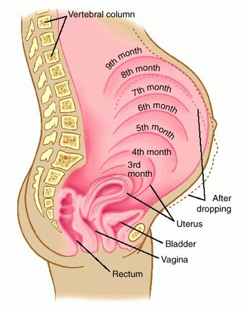
Support this browser is being discontinued for Pregnancy, Birth and Baby
Support for this browser is being discontinued for this site
- Internet Explorer 11 and lower
We currently support Microsoft Edge, Chrome, Firefox and Safari. For more information, please visit the links below:
- Chrome by Google
- Firefox by Mozilla
- Microsoft Edge
- Safari by Apple
You are welcome to continue browsing this site with this browser. Some features, tools or interaction may not work correctly.
How the belly grows - articles from the specialists of the clinic "Mother and Child"
Bagriy Alexander Tarasovich
Anesthesiologist-resuscitator
Clinical Hospital "AVICENNA" GC "Mother and Child"
Let's say right away that how the belly looks and grows during pregnancy depends on many reasons: the physique of a woman, the structure of the pelvis, the condition of the muscles, height uterus and baby, the amount of amniotic fluid.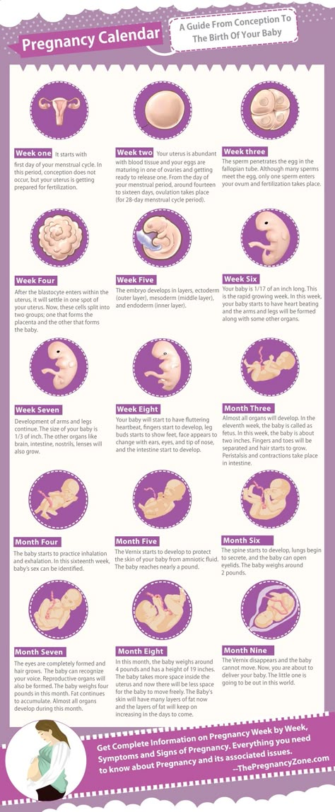 Therefore, for some, the belly grows faster, for some it is slower, for some mothers it is large, for others it can be almost invisible. But still, there are some general patterns of growth and size of the abdomen during pregnancy. nine0003
Growth rate
1st trimester
As a rule, in the early stages of pregnancy, the belly does not increase in size or increases very slightly. This is due to the fact that the uterus is still very small and takes up little space in the pelvis. So, for example, by the end of 4th week the uterus reaches only the size of a chicken egg, by the 8th week increases to the size of a goose, but most importantly, at this time it still does not reach the pubic articulation (located in the lower abdomen ). That is why in the early stages no increase in the abdomen is visible. And only after 12th week (end of the first trimester of pregnancy) the fundus of the uterus begins to rise above the womb.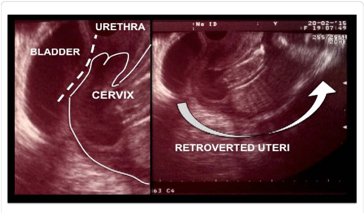
II trimester
At this time, the child is rapidly gaining height and weight, and the uterus is also rapidly growing. That is why, at a period of 12-16 weeks, an attentive mother will see that the stomach has already become noticeable. True, others will pay attention to the new position of a woman at about 20-q weeks, especially if she wears tight-fitting things.
III trimester
By the beginning of the third trimester, no one doubts pregnancy. The abdomen is clearly visible even if the woman is wearing loose clothing.
Sharp, round, various
Approximately from the second trimester of pregnancy, during each examination, the obstetrician-gynecologist determines the height of the uterine fundus and measures the circumference of the abdomen at the level of the navel. Why does the doctor so carefully monitor the increase in mother's tummy? The fact is that this is the easiest way to control the growth and development of the unborn baby. nine0003
One of the formulas for estimating fetal weight by fundal height (FHH): the shape of the abdomen often tried to determine the sex of the child. It was believed that a round belly portends a girl, and an elongated, oblong, “sharp” one portends a boy. However, these predictions did not always come true, since the shape and size of the abdomen do not depend at all on the sex of the child. nine0003
 Because of this, the shape of the abdomen can also be "stretched". nine0056
Feeling the belly
The belly of the expectant mother changes not only externally. From the 20th week of pregnancy, the mother begins to feel movements of the baby . At first, they look like light flutters, over time, the movements become more intense, because by the end of pregnancy, the mass and size of the child increase, and now it is not as spacious in the uterus as before. The number of movements gradually decreases, but their strength grows.
The movements of the crumbs, especially intense ones, can cause unpleasant sensations in a woman, especially in the right or left hypochondrium .
This is explained by the fact that during cephalic presentation (the baby is located in the uterus head down), the blows of the baby's legs are projected into the region of the mother's internal organs: the liver, stomach, intestines and spleen. Such sensations and even pains are natural and do not require treatment.
And sometimes pains appear in the lateral sections of the abdomen.
They occur due to the fact that during pregnancy the ligaments that support the uterus and ovaries are stretched. nine0012
In addition, changes occur in the fallopian tubes (they thicken, blood circulation increases in them), in the ovaries (they increase slightly in size, cyclic processes stop in them, and the position of the ovaries changes due to an increase in the size of the uterus).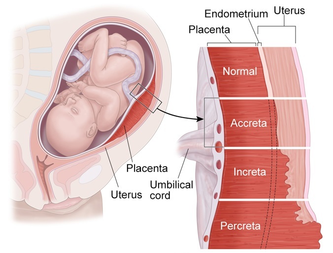 Slight pain in the lower abdomen may occur several times during the day, but, as a rule, they quickly disappear if the woman takes a position that is comfortable for her. Sometimes periodic discomfort in the lower lateral parts of the abdomen appear with constipation, which is also common in pregnant women: during pregnancy, hormones are produced that relax the uterus, they have a similar effect on the intestines: its peristalsis is disturbed, resulting in constipation. nine0003
The belly can be large or small, bulging forward or as if swollen, low or high - everything will depend on the individual characteristics of the pregnancy.
How the belly grows and looks during pregnancy depends on many factors: the physique of the woman, the structure of the pelvis, the condition of the muscles, the growth of the uterus and the baby, the amount of amniotic fluid.
Most often, the belly begins to grow after the 12th week of pregnancy, and others will be able to notice the woman's interesting position only from the 20th week. However, everything is strictly individual, there is absolutely no exact definition of the timing of the appearance of the abdomen, it is simply impossible to predict. nine0003
Don't be upset if the shape or size of your belly does not match the average, because everyone is different. Focus on your constitution, the work of your body and the development of the baby.
REFERENCE
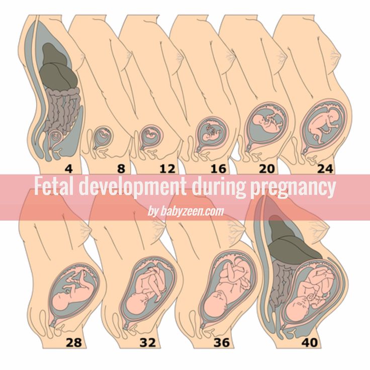
At 4 weeks, the uterus reaches the size of a hen's egg.
Goose egg at 8 weeks.
At 12 weeks, the uterus reaches the upper edge of the pubic bone. The belly is not visible yet.
At 16 weeks, the abdomen is rounded, the uterus is in the middle of the distance between the pubis and the navel. nine0003
At 20 weeks, the abdomen is visible to others, the fundus of the uterus is 4 cm below the navel.
At 24 weeks, the fundus of the uterus is at the level of the navel.
At 28 weeks, the uterus is already above the navel.
At 32 weeks, the navel begins to smooth out. Abdominal circumference - 80-85 cm.
At 40 weeks, the navel protrudes noticeably. Abdominal circumference 96-98 cm
Make an appointment
to the doctor - Bagriy Alexander Tarasovich
Clinical Hospital "AVICENNA" Group of Companies "Mother and Child"
Anesthesiology and resuscitation
By clicking on the send button, I consent to the processing of personal data
Attention! Prices for services in different clinics may vary. To clarify the current cost, select the clinic
The administration of the clinic takes all measures to update the prices for programs in a timely manner, however, in order to avoid possible misunderstandings, we recommend that you check the cost of services by phone / with the managers of the clinic
nine0002 Clinical Hospital "AVICENNA" GK "Mother and Child" Novosibirsk Center for Reproductive MedicineAll directionsGynecological proceduresSpecialist consultations (adults)Specialist consultations (children)Laboratory of molecular geneticsGeneral clinical examinationsProcedural roomTherapeutic examinationsUltrasound examinations for adults
01.
Gynecological procedures
02.
Consultations of specialists (adults)
03.
Consultations of specialists (children's)
04.
Laboratory of molecular genetics
05.
General studies
06. 9000
Therapeutic research
08.
Adult ultrasound
Nothing found
The administration of the clinic takes all measures to timely update the price list posted on the website, however, in order to avoid possible misunderstandings, we advise you to clarify the cost of services and the timing of the tests by calling
Changes in the cervix during pregnancy
Pregnancy is always pleasant, but sometimes not planned. And not all women have time to prepare for it, to be fully examined before its onset. And the detection of diseases of the cervix already during pregnancy can be an unpleasant discovery.
The cervix is the lower segment of the uterus in the form of a cylinder or cone. In the center is the cervical canal, one end of which opens into the uterine cavity, and the other into the vagina. On average, the length of the cervix is 3–4 cm, the diameter is about 2.5 cm, and the cervical canal is closed. The cervix has two parts: lower and upper. The lower part is called the vaginal, because it protrudes into the vaginal cavity, and the upper part is supravaginal, because it is located above the vagina. The cervix is connected to the vagina through the vaginal fornices. There is an anterior arch - short, posterior - deeper and two lateral ones. Inside the cervix passes the cervical canal, which opens into the uterine cavity with an internal pharynx, and is clogged with mucus from the side of the vagina. Mucus is normally impervious to infections and microbes, or to spermatozoa. But in the middle of the menstrual cycle, the mucus thins and becomes permeable to sperm. nine0003
Outside, the surface of the cervix has a pinkish tint, it is smooth and shiny, durable, and from the inside it is bright pink, velvety and loose.
The cervix during pregnancy is an important organ, both in anatomical and functional terms. It must be remembered that it promotes the process of fertilization, prevents infection from entering the uterine cavity and appendages, helps to "endure" the baby and participates in childbirth. That is why regular monitoring of the condition of the cervix during pregnancy is simply necessary. nine0003
During pregnancy, a number of physiological changes occur in this organ. For example, a short time after fertilization, its color changes: it becomes cyanotic. The reason for this is the extensive vascular network and its blood supply. Due to the action of estriol and progesterone, the tissue of the cervix becomes soft. During pregnancy, the cervical glands expand and become more branched.
Screening examination of the cervix during pregnancy includes: cytological examination, smears for flora and detection of infections. Cytological examination is often the first key step in the examination of the cervix, since it allows to detect very early pathological changes that occur at the cellular level, including in the absence of visible changes in the cervical epithelium. The examination is carried out to identify the pathology of the cervix and the selection of pregnant women who need a more in-depth examination and appropriate treatment in the postpartum period. When conducting a screening examination, in addition to a doctor's examination, a colposcopy may be recommended. As you know, the cervix is covered with two types of epithelium: squamous stratified from the side of the vagina and single-layer cylindrical from the side of the cervical canal. Epithelial cells are constantly desquamated and end up in the lumen of the cervical canal and in the vagina. Their structural characteristics make it possible, when examined under a microscope, to distinguish healthy cells from atypical ones, including cancerous ones. nine0003
During pregnancy, in addition to physiological changes in the cervix, some borderline and pathological processes may occur.
Under the influence of hormonal changes that occur in a woman's body during the menstrual cycle, cyclic changes also occur in the cells of the epithelium of the cervical canal. During the period of ovulation, the secretion of mucus by the glands of the cervical canal increases, and its qualitative characteristics change. With injuries or inflammatory lesions, sometimes the glands of the cervix can become clogged, a secret accumulates in them and cysts form - Naboth follicles or Naboth gland cysts that have been asymptomatic for many years. Small cysts do not require any treatment. And pregnancy, as a rule, is not affected. Only large cysts that strongly deform the cervix and continue to grow may require opening and evacuation of the contents. However, this is very rare and usually requires monitoring during pregnancy.
Quite often, in pregnant women, during a mirror examination of the vaginal part, polyps cervix. The occurrence of polyps is most often associated with a chronic inflammatory process. As a result, a focal proliferation of the mucosa is formed, sometimes with the involvement of muscle tissue and the formation of a pedicle. They are mostly asymptomatic. Sometimes they are a source of blood discharge from the genital tract, more often of contact origin (after sexual intercourse or defecation). The size of the polyp is different - from millet grain rarely to the size of a walnut, their shape also varies. Polyps are single and multiple, their stalk is located either at the edge of the external pharynx, or goes deep into the cervical canal. Sometimes during pregnancy there is an increase in the size of the polyp, in some cases quite fast. Rarely, polyps first appear during pregnancy. The presence of a polyp is always a potential threat of miscarriage, primarily because it creates favorable conditions for ascending infection. Therefore, as a rule, more frequent monitoring of the cervix follows. The tendency to trauma, bleeding, the presence of signs of tissue necrosis and decay, as well as questionable secretions require special attention and control. Treatment of cervical polyps is only surgical and during pregnancy, in most cases, treatment is postponed until the postpartum period, since even large polyps do not interfere with childbirth.
nine0003
The most common pathology of the cervix in women is erosion . Erosion is a defect in the mucous membrane. True erosion is not very common. The most common pseudo-erosion (ectopia) is a pathological lesion of the cervical mucosa, in which the usual flat stratified epithelium of the outer part of the cervix is replaced by cylindrical cells from the cervical canal. Often this happens as a result of mechanical action: with frequent and rough sexual intercourse, desquamation of the stratified squamous epithelium occurs. Erosion is a multifactorial disease. The reasons may be: nine0003
- genital infections, vaginal dysbacteriosis and inflammatory diseases of the female genital area;
- is an early onset of sexual activity and a frequent change of sexual partners. The mucous membrane of the female genital organs finally matures by the age of 20–23. If an infection interferes with this delicate process, erosion is practically unavoidable;
- is a cervical injury.
The main cause of such injuries is, of course, childbirth and abortion;
- hormonal disorders; nine0056
- , cervical pathology may also occur with a decrease in the protective functions of immunity.
The presence of erosion does not affect pregnancy in any way, as well as pregnancy on erosion. Treatment during pregnancy consists in the use of general and local anti-inflammatory drugs for inflammatory diseases of the vagina and cervix. And in most cases, just dynamic observation is enough. Surgical treatment is not carried out throughout the entire pregnancy, since the excess of risks and benefits is significant, and after treatment during childbirth, there may be problems with opening the cervix. nine0003
Almost all women with various diseases of the cervix safely bear and happily give birth to beautiful babies!
Attention! Prices for services in different clinics may vary. To clarify the current cost, select the clinic
The administration of the clinic takes all measures to update the prices for programs in a timely manner, however, in order to avoid possible misunderstandings, we recommend that you check the cost of services by phone / with the managers of the clinic
nine0002 Clinical Hospital IDKMother and Child Clinic Enthusiastov SamaraAll directionsSpecialist consultations (adults)Specialist consultations (children)Molecular genetics laboratoryGeneral clinical examinationsProcedural roomOther gynecological operationsTelemedicine for adultsTherapeutic examinationsAdult ultrasound examinations
01.


