Gestation for baby in weeks
Pregnancy - week by week
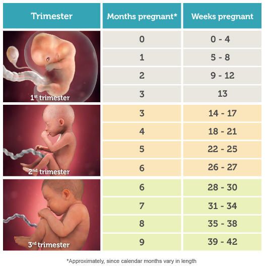
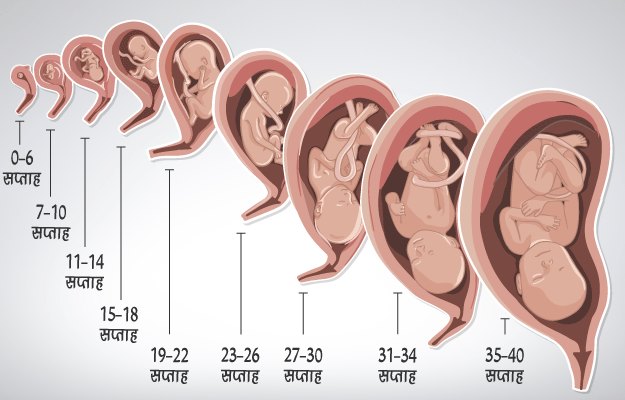
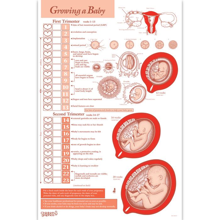
The unborn baby spends around 38 weeks in the womb, but the average length of pregnancy (gestation) is counted as 40 weeks. This is because pregnancy is counted from the first day of the woman’s last period, not the date of conception, which generally occurs two weeks later.
Pregnancy is divided into three trimesters:
- First trimester – conception to 12 weeks
- Second trimester – 12 to 24 weeks
- Third trimester – 24 to 40 weeks.
Conception
The moment of conception is when the woman’s ovum (egg) is fertilised by the man’s sperm. The gender and inherited characteristics are decided in that instant.
Week 1
This first week is actually your menstrual period. Because your expected birth date (EDD or EDB) is calculated from the first day of your last period, this week counts as part of your 40-week pregnancy, even though your baby hasn’t been conceived yet.
Week 2
Fertilisation of your egg by the sperm will take place near the end of this week.
Week 3
Thirty hours after conception, the cell splits into two. Three days later, the cell (zygote) has divided into 16 cells. After two more days, the zygote has migrated from the fallopian tube to the uterus (womb). Seven days after conception, the zygote burrows itself into the plump uterine lining (endometrium). The zygote is now known as a blastocyst.
Week 4
The developing baby is tinier than a grain of rice. The rapidly dividing cells are in the process of forming the various body systems, including the digestive system.
Week 5
The evolving neural tube will eventually become the central nervous system (brain and spinal cord).
Week 6
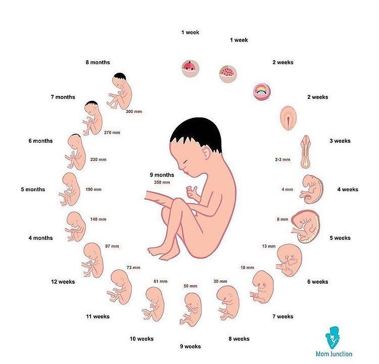
The baby is now known as an embryo.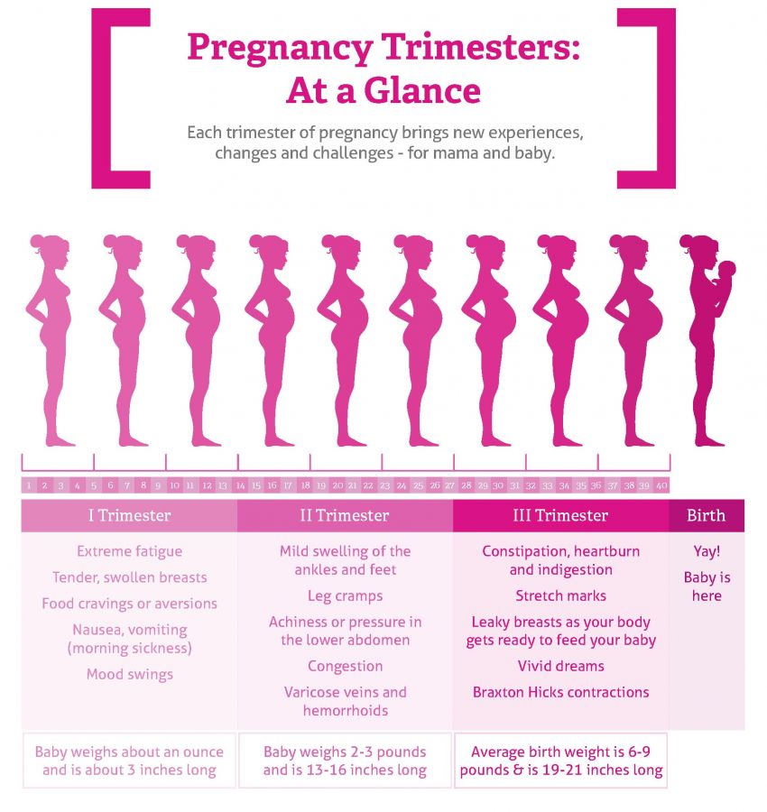 It is around 3 mm in length. By this stage, it is secreting special hormones that prevent the mother from having a menstrual period.
Week 7
The heart is beating. The embryo has developed its placenta and amniotic sac. The placenta is burrowing into the uterine wall to access oxygen and nutrients from the mother’s bloodstream.
Week 8
The embryo is now around 1.3 cm in length. The rapidly growing spinal cord looks like a tail. The head is disproportionately large.
Week 9
The eyes, mouth and tongue are forming. The tiny muscles allow the embryo to start moving about. Blood cells are being made by the embryo’s liver.
Week 10
The embryo is now known as a fetus and is about 2.5 cm in length. All of the bodily organs are formed. The hands and feet, which previously looked like nubs or paddles, are now evolving fingers and toes. The brain is active and has brain waves.
Week 11
Teeth are budding inside the gums. The tiny heart is developing further.
Week 12
The fingers and toes are recognisable, but still stuck together with webs of skin. The first trimester combined screening test (maternal blood test + ultrasound of baby) can be done around this time. This test checks for trisomy 18 (Edward syndrome) and trisomy 21 (Down syndrome).
Week 13
The fetus can swim about quite vigorously. It is now more than 7 cm in length.
Week 14
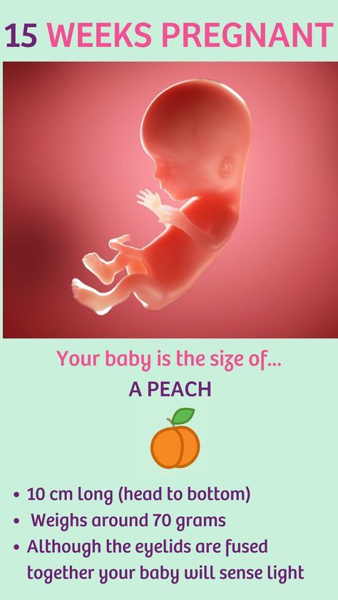
The eyelids are fused over the fully developed eyes. The baby can now mutely cry, since it has vocal cords. It may even start sucking its thumb. The fingers and toes are growing nails.
Week 16
The fetus is around 14 cm in length. Eyelashes and eyebrows have appeared, and the tongue has tastebuds. The second trimester maternal serum screening will be offered at this time if the first trimester test was not done (see week 12).
Week 18-20
An ultrasound will be offered. This fetal morphology scan is to check for structural abnormalities, position of placenta and multiple pregnancies. Interestingly, hiccoughs in the fetus can often be observed.
Week 20
The fetus is around 21 cm in length. The ears are fully functioning and can hear muffled sounds from the outside world. The fingertips have prints. The genitals can now be distinguished with an ultrasound scan.
Week 24
The fetus is around 33 cm in length. The fused eyelids now separate into upper and lower lids, enabling the baby to open and shut its eyes. The skin is covered in fine hair (lanugo) and protected by a layer of waxy secretion (vernix). The baby makes breathing movements with its lungs.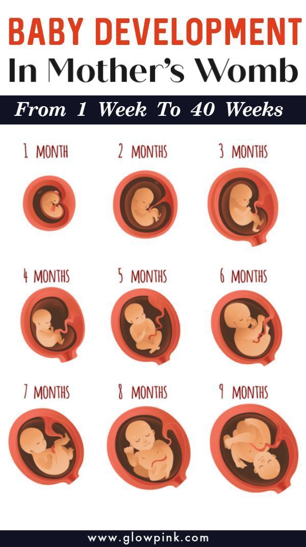
Week 28
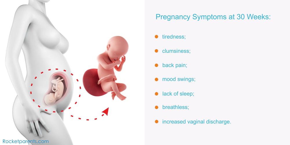
Your baby now weighs about 1 kg (1,000 g) or 2 lb 2oz (two pounds, two ounces) and measures about 25 cm (10 inches) from crown to rump. The crown-to-toe length is around 37 cm. The growing body has caught up with the large head and the baby now seems more in proportion.
Week 32
The baby spends most of its time asleep. Its movements are strong and coordinated. It has probably assumed the ‘head down’ position by now, in preparation for birth.
Week 36
The baby is around 46 cm in length. It has probably nestled its head into its mother’s pelvis, ready for birth. If it is born now, its chances for survival are excellent. Development of the lungs is rapid over the next few weeks.
Week 40
The baby is around 51 cm in length and ready to be born. It is unknown exactly what causes the onset of labour. It is most likely a combination of physical, hormonal and emotional factors between the mother and baby.
Where to get help
- Your doctor
- Obstetrician
- Midwife
Things to remember
- Pregnancy is counted as 40 weeks, starting from the first day of the mother’s last menstrual period. Your estimated date to birth is only to give you a guide. Babies come when they are ready and you need to be patient.
- The gender and inherited characteristics of the baby are decided at the moment of conception.
Baby due date - Better Health Channel
Summary
Read the full fact sheet- The unborn baby spends around 38 weeks in the uterus, but the average length of pregnancy, or gestation, is counted at 40 weeks.
- Pregnancy is counted from the first day of the woman’s last period, not the date of conception which generally occurs two weeks later.
- Since some women are unsure of the date of their last menstruation (perhaps due to period irregularities), a baby is considered full term if its birth falls between 37 to 42 weeks of the estimated last menstruation date.
The unborn baby spends around 37 weeks in the uterus (womb), but the average length of pregnancy, or gestation, is calculated as 40 weeks. This is because pregnancy is counted from the first day of the woman’s last period, not the date of conception which generally occurs two weeks later, followed by five to seven days before it settles in the uterus. Since some women are unsure of the date of their last menstruation (perhaps due to period irregularities), a pregnancy is considered full term if birth falls between 37 to 42 weeks of the estimated last menstruation date.
A baby born prior to week 37 is considered premature, while a baby that still hasn’t been born by week 42 is said to be overdue. In many cases, labour will be induced in the case of an overdue baby.
The average length of human gestation is 280 days, or 40 weeks, from the first day of the woman’s last menstrual period. The medical term for the due date is estimated date of confinement (EDC). However, only about four per cent of women actually give birth on their EDC.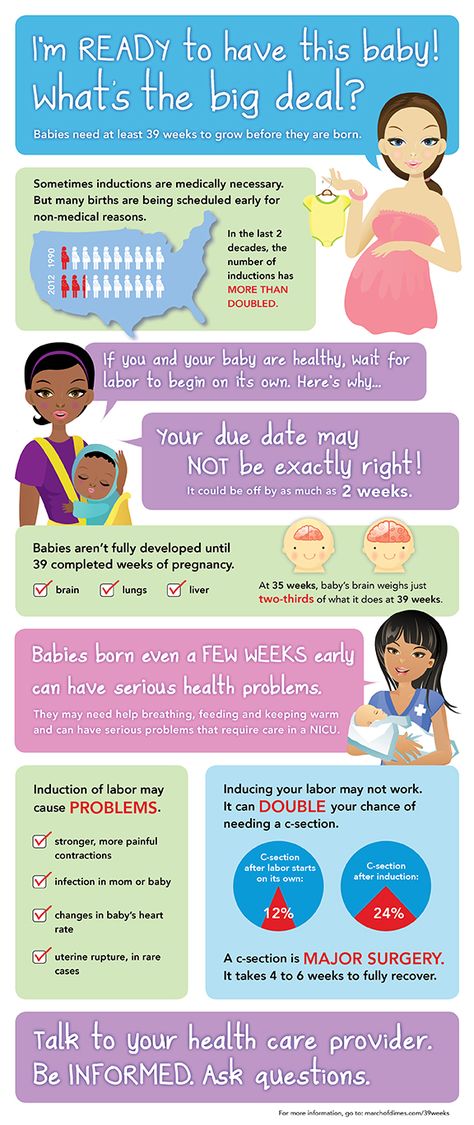 There are many online pregnancy calculators (see Baby due date calculator that can tell you when your baby is due, if you type in the date of the first day of your last period.
A simple method to calculate the due date is to add seven days to the date of the first day of your last period, then add nine months. For example, if the first day of your last period was 1 February, add seven days (8 February) then add nine months, for a due date of 8 November.
Determining baby due date
Irregular menstrual cycles can mean that some women aren’t sure of when they conceived. Some clues to the length of gestation include:
- Ultrasound examination (especially when performed between six and 12 weeks)
- Size of uterus on vaginal or abdominal examination
- The time fetal movements are first felt (an approximate guide only).
Pregnancy ultrasound
A pregnancy ultrasound is a non-invasive test that scans the unborn baby and the mother’s reproductive organs using high frequency sound waves. The general procedure for a pregnancy ultrasound includes:
- The woman lies on a table.
- A small amount of a clear, conductive jelly is smeared on the woman’s abdomen.
- The operator places the small hand-held instrument called a transducer onto the woman’s abdomen.
- The transducer is moved across the abdomen. The sound waves bounce off internal structures (including the baby) and are transmitted back to the transducer. The sound waves are then translated into a two-dimensional picture on a monitor. The mother doesn’t feel or hear the transmission of the sound waves.
- By measuring the baby’s body parts, such as head circumference and the length of long bones, the operator can estimate its gestational age.
The diagnostic uses of pregnancy ultrasound
Apart from helping to pinpoint the unborn baby’s due date, pregnancy ultrasounds are used to diagnose a number of conditions including:
- Multiple fetuses
- Health problems with the baby
- Ectopic pregnancy (the embryo lodges in the fallopian tube instead of the uterus)
- Abnormalities of the placenta such as placenta praevia, where the placenta is positioned over the neck of the womb (cervix)
- The health of the mother’s reproductive organs.
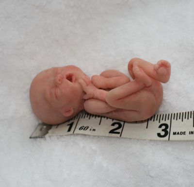
Premature babies
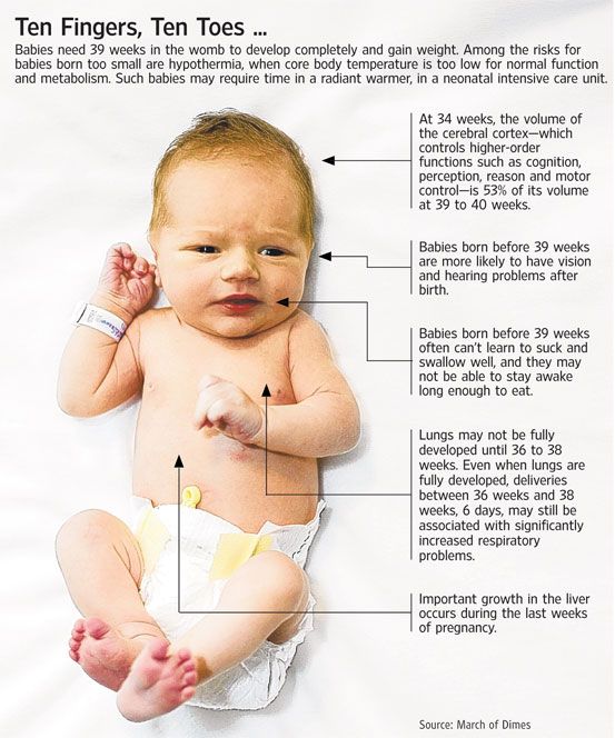
A baby born prior to week 37 is considered premature. The odds of survival depend on the baby’s degree of prematurity. The closer to term (estimated date of confinement, or EDC) the baby is born, the higher its chances of survival - after 34 weeks gestation with good paediatric care almost all babies will survive.
Premature babies are often afflicted by various health problems, caused by immature internal organs. Respiratory difficulties and an increased susceptibility to infection are common.
Often there is no known cause for a premature labour; however, some of the maternal risk factors may include:
- Drinking alcohol or smoking during pregnancy
- Low body weight prior to pregnancy
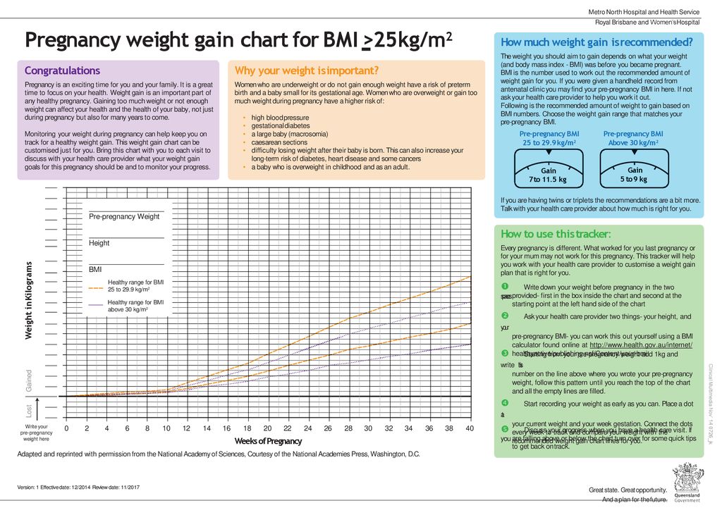
- Inadequate weight gain during pregnancy
- No prenatal care
- Emotional stress
- Placenta problems such as placenta praevia
- Various diseases such as diabetes and congestive heart failure
- Infections such as syphilis.
Overdue babies
Around five out of every 100 babies will be overdue, or more than 42 weeks gestation. If you have gone one week past your due date without any signs of impending labour, your doctor will want to closely monitor your condition. Tests include:
- Monitoring the fetal heart rate
- Using a cardiotocograph machine
- Performing ultrasound scans.
The placenta starts to deteriorate after 38 weeks or so, which means an overdue baby may not get enough oxygen. An overdue baby could also grow too large for vaginal delivery. Generally, an overdue baby will be induced once it is two weeks past its expected date. Some of the methods of induction include:
- Vaginal prostaglandin gel - to help dilate the cervix
- Amniotomy - breaking the waters, sometimes called an artificial rupture of membranes (ARM)
- Oxytocin - a synthetic form of this hormone is given intravenously to stimulate uterine contractions.

Where to get help
- Your doctor
- Your obstetrician
- Midwife or childbirth educator
Things to remember
- The unborn baby spends around 38 weeks in the uterus, but the average length of pregnancy, or gestation, is counted at 40 weeks.
- Pregnancy is counted from the first day of the woman’s last period, not the date of conception which generally occurs two weeks later.
- Since some women are unsure of the date of their last menstruation (perhaps due to period irregularities), a baby is considered full term if its birth falls between 37 to 42 weeks of its estimated due date.
- Common concerns and discomforts: overdue baby, Mother’s Bliss, UK.
- Going Overdue, 2001, Centre for Reproduction and Minimally Invasive Surgery.
- Premmie-L FAQ and advice sheets, Parents of Premature Babies Inc.(Preemie-L). More information here.
- Pregnancy: what to expect when it’s past your due date, Family Doctor, USA.

- Ultrasound, Women Health Information, Royal Women’s Hospital, Melbourne. More information here.
This page has been produced in consultation with and approved by:
Child development by week | Regional Perinatal Center
Expectant mothers are always curious about how the fetus develops at a time when it is awaited with such impatience. Let's talk and look at the photos and pictures of how the fetus grows and develops week by week.
What does the puffer do for 9 whole months in mom's tummy? What does he feel, see and hear?
Let's start the story about the development of the fetus by weeks from the very beginning - from the moment of fertilization. A fetus up to 8 weeks old is called embryo , this occurs before the formation of all organ systems.
Embryo development: 1st week
The egg is fertilized and begins to actively split. The ovum travels to the uterus, getting rid of the membrane along the way.
On the 6th-8th days, implantation of eggs is carried out - implantation into the uterus. The egg settles on the surface of the uterine mucosa and, using the chorionic villi, attaches to the uterine mucosa.
Embryo development: 2-3 weeks
Picture of embryo development at 3 weeks.
The embryo is actively developing, starting to separate from the membranes. At this stage, the beginnings of the muscular, skeletal and nervous systems are formed. Therefore, this period of pregnancy is considered important.
Embryo development: 4–7 weeks
Fetal development by week in pictures: week 4
Fetal development by week photo: week 4
Photo of an embryo before the 6th week of pregnancy.
The heart, head, arms, legs and tail are formed in the embryo :) . Gill slit is defined. The length of the embryo at the fifth week reaches 6 mm.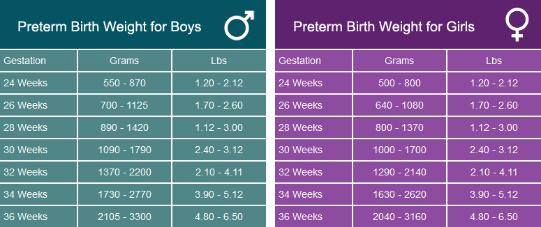
Fetal development by week photo: week 5
At the 7th week, the rudiments of the eyes, stomach and chest are determined, and fingers appear on the handles. The baby already has a sense organ - the vestibular apparatus. The length of the embryo is up to 12 mm.
Fetal development: 8th week
Fetal development by week photo: week 7-8
The face of the fetus can be identified, the mouth, nose, and auricles can be distinguished. The head of the embryo is large and its length corresponds to the length of the body; the fetal body is formed. All significant, but not yet fully formed, elements of the baby's body already exist. The nervous system, muscles, skeleton continue to improve.
Fetal development in the photo already sensitive arms and legs: week 8
The fetus developed skin sensitivity in the mouth (preparation for the sucking reflex), and later in the face and palms.
At this stage of pregnancy, the genitals are already visible. Gill slits die. The fruit reaches 20 mm in length.
Fetal development: 9–10 weeks
Fetal development by week photo: week 9
Fingers and toes already with nails. The fetus begins to move in the pregnant woman's stomach, but the mother does not feel it yet. With a special stethoscope, you can hear the baby's heartbeat. Muscles continue to develop.
Weekly development of the fetus photo: week 10
The entire surface of the fetal body is sensitive and the baby develops tactile sensations with pleasure, touching his own body, the walls of the fetal bladder and the umbilical cord. It is very curious to observe this on ultrasound. By the way, the baby first moves away from the ultrasound sensor (of course, because it is cold and unusual!), And then puts his hands and heels trying to touch the sensor.
It's amazing when a mother puts her hand to her stomach, the baby tries to master the world and tries to touch with his pen "from the back".
The development of the fetus: 11–14 weeks
Development of the fetus in the photo of the legs: weeks 11
The baby, legs and eyelids are formed, and the genitals become distinguishable (you can find out the gender (you can find out the gender child). The fetus begins to swallow, and if something is not to its taste, for example, if something bitter got into the amniotic fluid (mother ate something), then the baby will begin to frown and stick out his tongue, making less swallowing movements.
Fruit skin appears translucent.
Fruit development: Week 12
Photo of the fetus 12 weeks per 3D Uzi
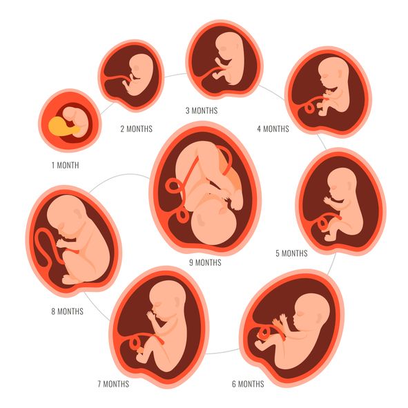
buds are responsible for production for production urine. Blood forms inside the bones. And hairs begin to grow on the head. The skin turns pink, the ears and other parts of the body, including the face, are already visible. Imagine, a child can already open his mouth and blink, as well as make grasping movements. The fetus begins to actively push in the mother's tummy. The sex of the fetus can be determined by ultrasound. Baby sucks his thumb, becomes more energetic. Pseudo-feces are formed in the intestines of the fetus - meconium , kidneys begin to work. During this period, the brain develops very actively. The auditory ossicles become stiff and now they are able to conduct sounds, the baby hears his mother - heartbeat, breathing, voice. The lungs at this stage of fetal development are so developed that the baby can survive in the artificial conditions of the intensive care unit. Lungs continue to develop. Now the baby is already falling asleep and waking up. Downy hairs appear on the skin, the skin becomes wrinkled and covered with grease. The cartilage of the ears and nose is still soft. Lips and mouth become more sensitive. The eyes develop, open slightly and can perceive light and squint from direct sunlight. In girls, the labia majora do not yet cover the small ones, and in boys, the testicles have not yet descended into the scrotum. Fetal weight reaches 900–1200 g, and the length is 350 mm. 9 out of 10 children born at this term survive. The lungs are now adapted to breathe normal air. Breathing is rhythmic and body temperature is controlled by the CNS. The baby can cry and responds to external sounds. Child opens eyes while awake and closes during sleep. The skin becomes thicker, smoother and pinkish. Starting from this period, the fetus will actively gain weight and grow rapidly. Almost all babies born prematurely at this time are viable. The weight of the fetus reaches 2500 g, and the length is 450 mm. The fetus reacts to a light source. Muscle tone increases and the baby can turn and raise his head. On which, the hairs become silky. The child develops a grasping reflex. The lungs are fully developed. The fetus is quite developed, prepared for birth and considered mature. The baby perfectly mastered the movements of his mother , knows when she is calm, excited, upset and reacts to this with her movements. During the intrauterine period, the fetus gets used to moving in space, which is why babies love it so much when they are carried in their arms or rolled in a stroller. For a baby, this is a completely natural state, so he will calm down and fall asleep when he is rocked. The nails protrude beyond the tips of the fingers, the cartilages of the ears and nose are elastic. In boys, the testicles have descended into the scrotum, and in girls, the large labia cover the small ones. The weight of the fetus reaches 3200-3600 g, and the length is 480-520 mm. After the birth, the baby longs for touching his body, because at first he cannot feel himself - the arms and legs do not obey the child as confidently as it was in the amniotic fluid. And one more thing, the baby remembers the rhythm and sound of your heart very well . Therefore, you can comfort the baby in this way - take him in your arms, put him on the left side and your miracle will calm down, stop crying and fall asleep. And for you, finally, the time of bliss will come :) . From a tiny fetus to a small person, a child's body develops in just 9 months. What changes are happening to the expectant mother and what changes are observed inside her during this difficult and joyful period of life? Each new life begins with the union of the egg and sperm. Conception is the process by which a sperm enters an egg and fertilizes it. It should be noted that the embryonic and obstetric terms are different. The thing is that among specialists it is customary to consider the period from the first day of the last menstruation, that is, the obstetric period includes the period of preparation for pregnancy. Until the moment of the meeting, the spermatozoon and the ovum lived for a certain time, being in the stage of development and maturation. The development of the future fetus significantly depends on the quality of these processes. Growth and maturation of the egg starts from the first day of the cycle. A mature egg contains 23 chromosomes as the genetic material for the future embryo, and also contains all the nutrients necessary to start its development. It contains reserves of carbohydrates, proteins and fats, designed to support the embryo during the first days after its occurrence. A certain number of eggs are laid in each ovary of a girl before she is born. During the childbearing period, they only grow and develop, the process of their formation does not occur. The ovary each month gives the opportunity to develop most often one egg, the maturation of which occurs inside a vesicle with a liquid called a follicle. From the first day of the cycle, the uterine mucosa begins to prepare for a possible pregnancy. For implantation, i.e., the introduction of the resulting embryo into the wall of the uterus, an optimal environment is created. To do this, due to the influence of hormones, the endometrium thickens, it becomes covered with a network of vessels and accumulates the nutrients necessary for the future embryo. Male reproductive cells are formed in the gonads - in the testicles or testes. The maturation of spermatozoa occurs in the epididymis, into which they move after formation. The number of spermatozoa is quite large: tens of millions in one milliliter. Despite such a significant number, only one of them will be able to fertilize the egg. In spermatozoa, there is exclusively genetic material - 23 chromosomes, which are necessary for the appearance of the embryo. Spermatozoa are characterized by high motility. Once in the female genital tract, they begin their movement towards the egg. Only half an hour or an hour passes from the moment of ejaculation, when sperm enter the uterine cavity. It takes one and a half to two hours for spermatozoa to penetrate into the widest part, which is called the ampulla. Most spermatozoa die on the way to the egg, meeting the folds of the endometrium, getting into the vaginal environment, cervical mucus. In the middle of the cycle, the egg fully matures and leaves the ovary. It enters the abdominal cavity. This process is called ovulation. With a regular cycle of 30 days, ovulation occurs on the fifteenth. The egg is unable to move on its own. When she leaves the follicle, the fimbriae of the fallopian tube ensure her penetration inside. The fallopian tubes are characterized by longitudinal folding, they are filled with mucus. The muscular movements of the tubes have a wave-like character, which, with a significant number of cilia, creates optimal conditions for transporting the egg. Through the tubes, the egg enters their widest part, which is called the ampulla. This is where fertilization takes place. If there is no meeting with the sperm, the egg dies, and the female body receives the appropriate signal to start a new cycle. There is a rejection of the mucous membrane, which was created by the uterus. The manifestation of such rejection is bloody discharge, which is called menstruation. The waiting period for fertilization by the egg is short. On average, it takes no more than a day. Fertilization is likely on the day of ovulation and maximum on the next. Sperm have a longer lifespan, averaging three to five days, in some cases seven. Accordingly, if a spermatozoon enters the female genital tract before ovulation, there is a possibility that it will be able to wait for the appearance of an egg. When the egg is in a state of waiting for fertilization, certain substances are released that are designed to detect it. If spermatozoa find an egg, they begin to secrete special enzymes that can loosen its shell. As soon as one of the spermatozoa penetrates the egg, the others can no longer do this due to the restoration of the density of its shell. Thus, one egg can only be fertilized by one sperm. After fertilization, the chromosome sets of the parents merge - 23 chromosomes from each. As a result, one cell is formed from two different cells, which is called a zygote. As soon as a day has passed after the formation of the embryo, he will need to make his first journey. The movements of the cilia and the contraction of the muscles of the tube direct it into the uterine cavity. During this process, inside the egg, fragmentation into identical cells occurs. After four days, the appearance of the egg changes: it loses its round shape and becomes vine-shaped. It takes 5-7 days for the embryo to reach the uterus. When implantation occurs in its mucous membrane, the number of cells reaches one hundred. The term implantation refers to the process of implantation of the embryo into the endometrial layer. After fertilization on the seventh or eighth day, implantation takes place. The first critical period of pregnancy is this stage, since the embryo will have to demonstrate its viability for the first time. During implantation, the outer cells of the embryo actively divide, and the process itself takes about forty hours. The embryo at this stage of life is called a blastocyst. It contacts with the endometrium, melts the cells of the endometrium with its activity, creates a path for itself to the deeper layers. The blood vessels of the embryo intertwine with the mother's body, which allows it to immediately begin to extract useful and necessary substances for development. This is vital, because by this time the stock that the mature egg carried in itself is exhausted. Next, the production of the trophoblast cells, i.e., the outer cells of human chorionic gonadotropin, the hCG hormone, begins. After fertilization and before the start of hCG, it usually takes eight or nine days. Therefore, already from the tenth day after fertilization, it becomes possible to determine this hormone in the mother's blood. Such an analysis is the most reliable confirmation of the onset of pregnancy. The tests that are offered today to determine pregnancy are based on the detection of this hormone in the urine of a woman. After the first day of delayed menstruation with its regular cycle, it is already possible to determine pregnancy using a test on your own. If a woman is planning a pregnancy, 21-24 days, subject to a regular cycle, should be important for her. This is a period of possible implantation, when you should pay special attention to your own lifestyle. The woman does not feel anything at this stage, because implantation has no external signs. If you adjust your lifestyle in accordance with the simple rules listed above, you will be able to create optimal conditions for successful implantation. In the fourth obstetric week or the second week of the fetus's life, its body consists of two layers. Endoblast - cells of the inner layer - will become the beginning of the digestive and respiratory systems, ectoblast - cells of the outer layer - will start the development of the nervous system and skin. The size of the embryo at this stage is 1.5 mm. The flat arrangement of the cells determined the name of the embryo of this age - the disc. The fourth week is characterized by intensive development of extra-embryonic organs. Such organs should surround the embryo and create the most favorable conditions for its development.
Development of the fetus for weeks: Week 14 9000 9000  Moves more coordinated.
 Fetal development: 15-18 weeks
Fetal development by weeks photo: week 15 Fetal development: 19-23 weeks
Fetal development by week photo: week 19
Fetal development by weeks photo: week 20  The fetus intensively gains weight, fat deposits are formed. The weight of the fetus reaches 650 g, and the length is 300 mm.
 Fetal development: 24-27 weeks
Fetal development by week photo: week 27 
Fetal development: 28-32 weeks
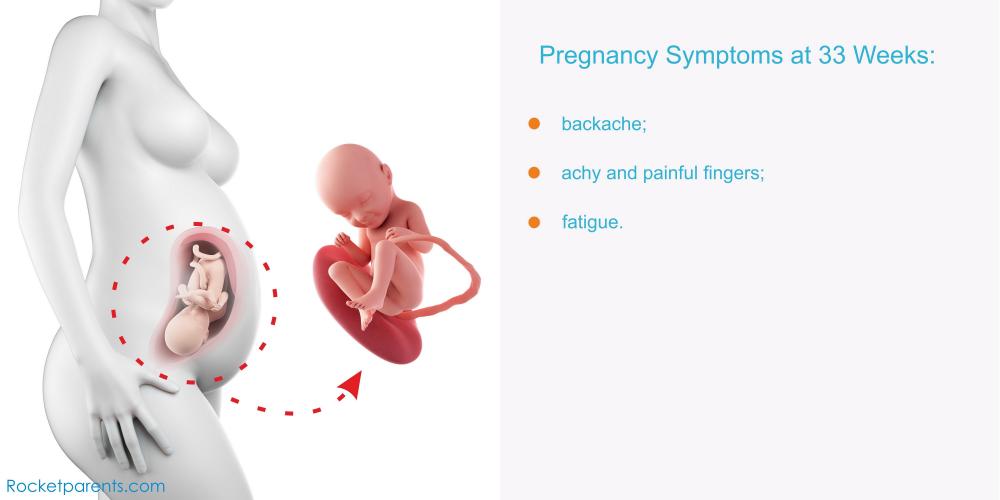
Fetal development: 33-37 weeks
Fetal development by week photo: week 36 Fetal development: 38-42 weeks
 The baby has mastered over 70 different reflex movements. Due to the subcutaneous fatty tissue, the baby's skin is pale pink. The head is covered with hairs up to 3 cm.
Fetal development by weeks photo: week 40 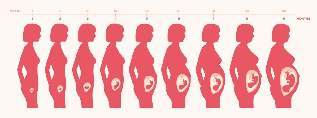 Therefore, so that your baby does not feel lonely, it is advisable to carry him in your arms, press him to you while stroking his body.
 1-4 weeks of pregnancy
 So it turns out that the embryo has just appeared, and the gestation period is already two weeks. It is the obstetric period that is indicated in all the documents of a woman and is the only reporting period for specialists.
 First week
 By the time a girl is born, the number of cells from which eggs can develop in the future reaches a million, but this number decreases significantly over the course of life. So, by the time of puberty, there are several hundred thousand of them, and by maturity - about 500.
  The liquid structure of semen is formed due to the secretion of the seminal vesicles and the prostate gland. A liquid medium is necessary for storing mature spermatozoa and creating favorable conditions for their life.
Second week
 The sex of the unborn child depends on which of the chromosomes, X or Y, was in the sperm. Eggs contain only X chromosomes. When XX is combined, girls are born. If the spermatozoon contains a Y chromosome, that is, with a combination of XY, boys are born. As soon as a zygote is formed in the body, a mechanism is launched in it aimed at maintaining pregnancy. There are changes in the hormonal background, biochemical reactions, immune mechanisms, and the receipt of nerve signals. The female body creates all the necessary conditions for the safe development of the fetus.
 Third week
 This stage is called morula, embryogenesis begins - an important stage in the development of the embryo, during which the formation of the rudiments of organs and tissues occurs. Cleavage of cells continues for several days, on the fifth day their complexes are formed, which have different functions. The central cluster forms directly the embryo, the outer one, called the trophoblast, is designed to melt the endometrium - the inner layer of the uterus.
  The number of cells outside the embryo increases dramatically, they stretch, they penetrate into the uterine mucosa, and the thinnest blood vessels are formed inside, which are necessary for the supply of nutrients to the embryo. Time will pass, and these vessels will be transformed first into the chorion, and subsequently into the placenta, which will be able to supply the fetus with everything necessary until the baby is born.
  The distribution of this hormone throughout the body notifies it of the onset of pregnancy, which causes the launch of active hormonal changes and the beginning of corresponding changes in the body.
 What happens to a woman in the third week of pregnancy
 During this period, thermal effects and excessive physical exertion are undesirable, and the influence of various kinds of radiation should also be prevented.
 Fourth week
