Female anatomy bone
Anatomy, Function of Bones, Muscles, Ligaments
What is the female pelvis?
The pelvis is the lower part of the torso. It’s located between the abdomen and the legs. This area provides support for the intestines and also contains the bladder and reproductive organs.
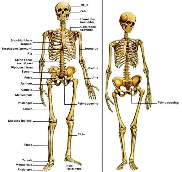
There are some structural differences between the female and the male pelvis. Most of these differences involve providing enough space for a baby to develop and pass through the birth canal of the female pelvis. As a result, the female pelvis is generally broader and wider than the male pelvis.
Below, learn more about the bones, muscles, and organs of the female pelvis.
Female pelvis anatomy and function
Female pelvis bones
Hip bones
There are two hip bones, one on the left side of the body and the other on the right. Together, they form the part of the pelvis called the pelvic girdle.
The hip bones join to the upper part of the skeleton through attachment at the sacrum. Each hip bone is made of three smaller bones that fuse together during adolescence:
- Ilium. The largest part of the hip bone, the ilium, is broad and fan-shaped. You can feel the arches of these bones when you put your hands on your hips.
- Pubis. The pubis bone of each hip bone connects to the other at a joint called the pubis symphysis.
- Ischium. When you sit down, most of your body weight falls on these bones. This is why they’re sometimes called sit bones.
The ilium, pubis, and ischium of each hip bone come together to form the acetabulum, where the head of the thigh bone (femur) attaches.
Sacrum
The sacrum is connected to the lower part of the vertebrae. It’s actually made up of five vertebrae that have fused together. The sacrum is quite thick and helps to support body weight.
Coccyx
The coccyx is sometimes called the tailbone. It’s connected to the bottom of the sacrum supported by several ligaments.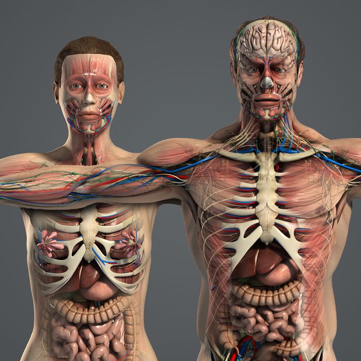
The coccyx is made up of four vertebrae that have fused into a triangle-like shape.
Female pelvis muscles
Levator ani muscles
The levator ani muscles are the largest group of muscles in the pelvis. They have several functions, including helping to support the pelvic organs.
The levator ani muscles consist of three separate muscles:
- Puborectalis. This muscle is responsible for holding in urine and feces. It relaxes when you urinate or have a bowel movement.
- Pubococcygeus. This muscle makes up most of the levator ani muscles. It originates at the pubis bone and connects to the coccyx.
- Iliococcygeus. The iliococcygeus has thinner fibers and serves to lift the pelvic floor as well as the anal canal.
Coccygeus
This small pelvic floor muscle originates at the ischium and connects to the sacrum and coccyx.
Female pelvis organs
Uterus
The uterus is a thick-walled, hollow organ where a baby develops during pregnancy.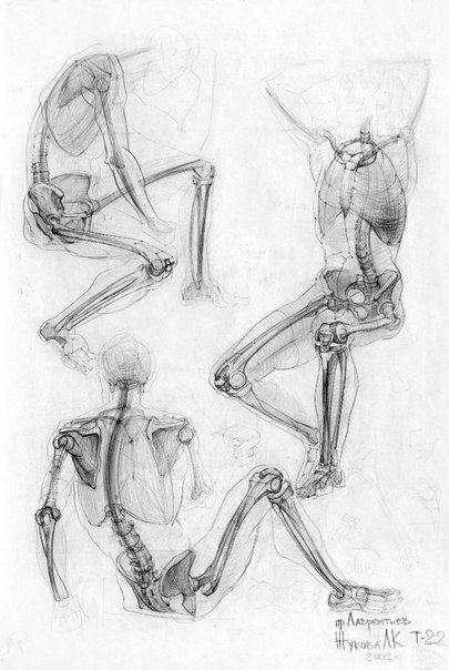
During the reproductive years, the lining of the uterus sheds every month during menstruation if you don’t become pregnant.
Ovaries
There are two ovaries located on either side of the uterus. The ovaries produce eggs and also release hormones, such estrogen and progesterone.
Fallopian tubes
The fallopian tubes connect each ovary to the uterus. Specialized cells in the fallopian tubes use hair-like structures called cilia to help direct eggs from the ovaries toward the uterus.
Cervix
The cervix connects the uterus to the vagina. It’s able to widen, allowing sperm to pass into the uterus.
In addition, thick mucus produced in the cervix can help to prevent bacteria from reaching the uterus.
Vagina
The vagina connects the cervix to the exterior female genitalia. It’s also called the birth canal, as the baby passes through the vagina during delivery.
Rectum
The rectum is the lowest part of the large intestine.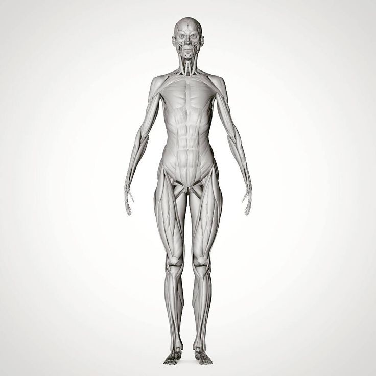 Feces collects here until exiting through the anus.
Bladder
The bladder is the organ that collects and stores urine until it’s released. Urine reaches the bladder through tubes called ureters that connect to the kidneys.
Urethra
The urethra is the tube that urine travels through to exit the body from the bladder. The female urethra is much shorter than the male urethra.
Female pelvis ligaments
Broad ligament
The broad ligament supports the uterus, fallopian tubes, and ovaries. It extends to both sides of the pelvic wall.
The broad ligament can be further divided into three components that are linked to different parts of the female reproductive organs:
- mesometrium, which supports the uterus
- mesovarium, which supports the ovaries
- mesosalpinx, which supports the fallopian tubes
Uterine ligaments
Uterine ligaments provide additional support for the uterus.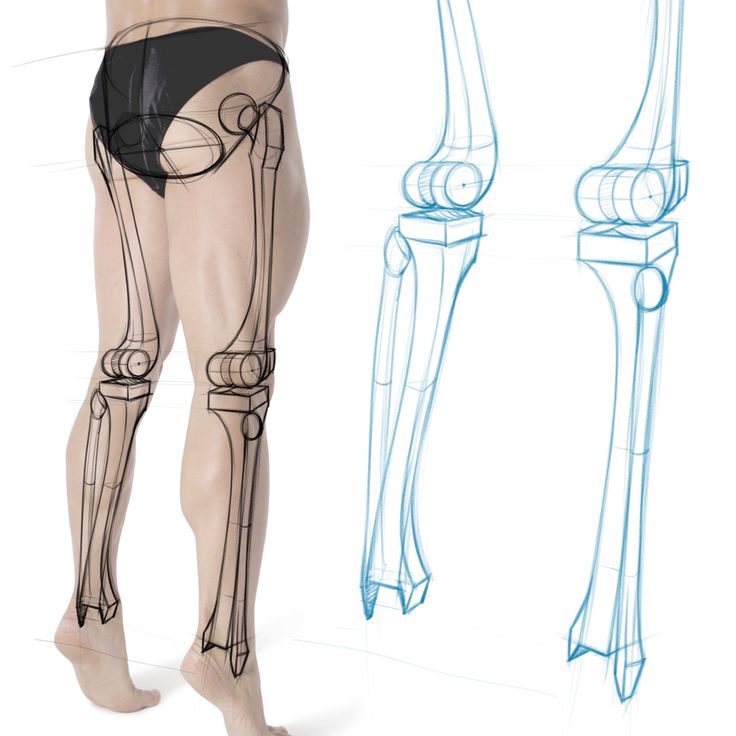 Some of the main uterine ligaments include:
- the round ligament
- cardinal ligaments
- pubocervical ligaments
- uterosacral ligaments
Ovarian ligaments
The ovarian ligaments support the ovaries. There are two main ovarian ligaments:
- the ovarian ligament
- the suspensory ligament of the ovary
Female pelvis diagram
Explore this interactive 3-D diagram to learn more about the female pelvis:
Female pelvis conditions
The pelvis contains a large number of organs, bones, muscles, and ligaments, so many conditions can affect the entire pelvis or parts within it.
Some conditions that can affect the female pelvis as a whole include:
- Pelvic inflammatory disease (PID). PID is an infection that occurs in the female reproductive system. While it’s often caused by a sexually transmitted infection, other infections can also cause PID. Untreated PID can lead to complications, such as infertility or ectopic pregnancy.
- Pelvic organ prolapse. Pelvic organ prolapse occurs when the muscles in the pelvis can no longer support its organs, such as the bladder, uterus, or rectum. This can cause one or more of these organs to press down on the vagina. In some cases, this can cause a bulge to form outside of the vagina.
- Endometriosis. Endometriosis occurs when the tissue that lines the inside walls of the uterus (endometrium) begins to grow outside of the uterus. The ovaries, fallopian tubes, and other tissues in the pelvis are typically affected by the condition. Endometriosis can lead to complications, including infertility or ovarian cancer.
Symptoms of a pelvic condition
Some common symptoms of a pelvic condition can include:
- pain in the lower abdomen or pelvis
- a feeling of pressure or fullness in the pelvis
- unusual or foul-smelling vaginal discharge
- pain during sex
- bleeding in between periods
- painful cramping during or before periods
- pain during bowel movements or when urinating
- a burning feeling when urinating
Tips for a healthy pelvis
The female pelvis is a complex, important part of the body. Follow these tips to keep it in good health:
Stay on top of your reproductive health
See a gynecologist for a yearly health screen. Things like pelvic exams and Pap smears can aid in identifying pelvic conditions or infections early.
You can get a free or low-cost pelvic exam at your local Planned Parenthood clinic.
Practice safe sex
Use barriers — such as condoms or dental dams — during sexual activity, especially with a new partner, to avoid infections that could lead to PID.
Try pelvic floor exercises
These types of exercises can help to strengthen the muscles in the pelvis, including those around the bladder and vagina.
Stronger pelvic floor muscles can aid in preventing things like incontinence or organ prolapse. Here’s how to get started.
Never ignore unusual symptoms
If you’re experiencing anything unusual in your pelvic area, such as bleeding between periods or unexplained pelvic pain, make an appointment with your doctor. Left untreated, some pelvic conditions can have lasting impacts on your health and fertility.
Anatomy, Function of Bones, Muscles, Ligaments
What is the female pelvis?
The pelvis is the lower part of the torso. It’s located between the abdomen and the legs. This area provides support for the intestines and also contains the bladder and reproductive organs.
There are some structural differences between the female and the male pelvis. Most of these differences involve providing enough space for a baby to develop and pass through the birth canal of the female pelvis. As a result, the female pelvis is generally broader and wider than the male pelvis.
Below, learn more about the bones, muscles, and organs of the female pelvis.
Female pelvis anatomy and function
Female pelvis bones
Hip bones
There are two hip bones, one on the left side of the body and the other on the right. Together, they form the part of the pelvis called the pelvic girdle.
The hip bones join to the upper part of the skeleton through attachment at the sacrum. Each hip bone is made of three smaller bones that fuse together during adolescence:
- Ilium. The largest part of the hip bone, the ilium, is broad and fan-shaped. You can feel the arches of these bones when you put your hands on your hips.
- Pubis. The pubis bone of each hip bone connects to the other at a joint called the pubis symphysis.
- Ischium. When you sit down, most of your body weight falls on these bones. This is why they’re sometimes called sit bones.
The ilium, pubis, and ischium of each hip bone come together to form the acetabulum, where the head of the thigh bone (femur) attaches.
Sacrum
The sacrum is connected to the lower part of the vertebrae. It’s actually made up of five vertebrae that have fused together. The sacrum is quite thick and helps to support body weight.
Coccyx
The coccyx is sometimes called the tailbone.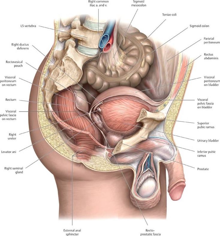 It’s connected to the bottom of the sacrum supported by several ligaments.
The coccyx is made up of four vertebrae that have fused into a triangle-like shape.
Female pelvis muscles
Levator ani muscles
The levator ani muscles are the largest group of muscles in the pelvis. They have several functions, including helping to support the pelvic organs.
The levator ani muscles consist of three separate muscles:
- Puborectalis. This muscle is responsible for holding in urine and feces. It relaxes when you urinate or have a bowel movement.
- Pubococcygeus. This muscle makes up most of the levator ani muscles. It originates at the pubis bone and connects to the coccyx.
- Iliococcygeus. The iliococcygeus has thinner fibers and serves to lift the pelvic floor as well as the anal canal.
Coccygeus
This small pelvic floor muscle originates at the ischium and connects to the sacrum and coccyx.
Female pelvis organs
Uterus
The uterus is a thick-walled, hollow organ where a baby develops during pregnancy.
During the reproductive years, the lining of the uterus sheds every month during menstruation if you don’t become pregnant.
Ovaries
There are two ovaries located on either side of the uterus. The ovaries produce eggs and also release hormones, such estrogen and progesterone.
Fallopian tubes
The fallopian tubes connect each ovary to the uterus. Specialized cells in the fallopian tubes use hair-like structures called cilia to help direct eggs from the ovaries toward the uterus.
Cervix
The cervix connects the uterus to the vagina. It’s able to widen, allowing sperm to pass into the uterus.
In addition, thick mucus produced in the cervix can help to prevent bacteria from reaching the uterus.
Vagina
The vagina connects the cervix to the exterior female genitalia.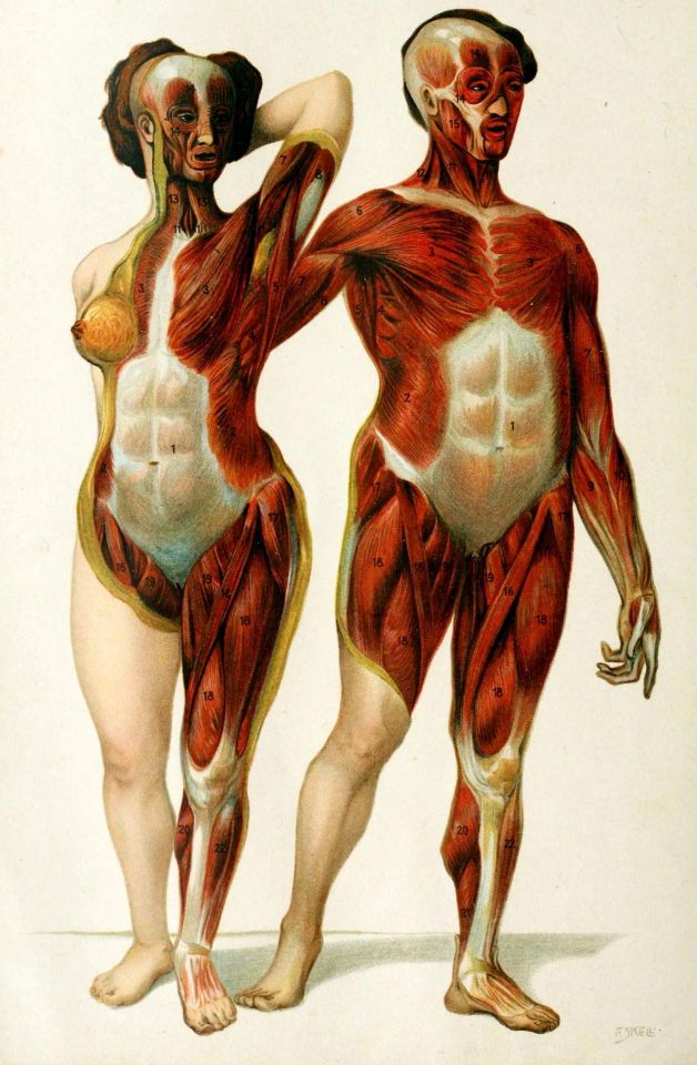 It’s also called the birth canal, as the baby passes through the vagina during delivery.
Rectum
The rectum is the lowest part of the large intestine. Feces collects here until exiting through the anus.
Bladder
The bladder is the organ that collects and stores urine until it’s released. Urine reaches the bladder through tubes called ureters that connect to the kidneys.
Urethra
The urethra is the tube that urine travels through to exit the body from the bladder. The female urethra is much shorter than the male urethra.
Female pelvis ligaments
Broad ligament
The broad ligament supports the uterus, fallopian tubes, and ovaries. It extends to both sides of the pelvic wall.
The broad ligament can be further divided into three components that are linked to different parts of the female reproductive organs:
- mesometrium, which supports the uterus
- mesovarium, which supports the ovaries
- mesosalpinx, which supports the fallopian tubes
Uterine ligaments
Uterine ligaments provide additional support for the uterus.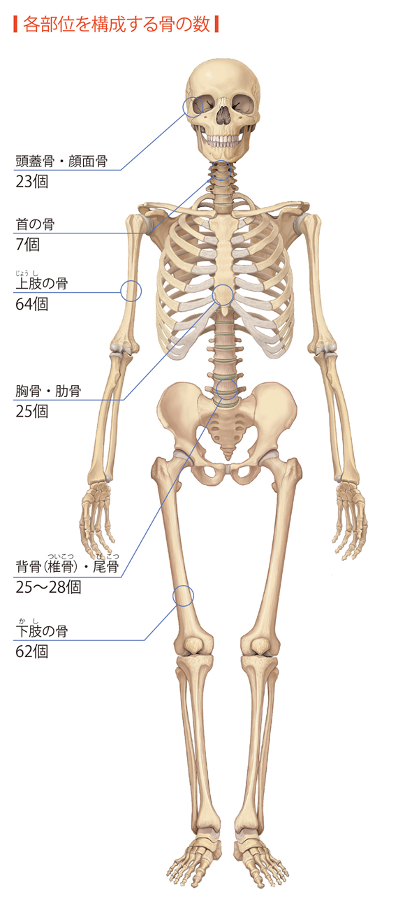 Some of the main uterine ligaments include:
- the round ligament
- cardinal ligaments
- pubocervical ligaments
- uterosacral ligaments
Ovarian ligaments
The ovarian ligaments support the ovaries. There are two main ovarian ligaments:
- the ovarian ligament
- the suspensory ligament of the ovary
Female pelvis diagram
Explore this interactive 3-D diagram to learn more about the female pelvis:
Female pelvis conditions
The pelvis contains a large number of organs, bones, muscles, and ligaments, so many conditions can affect the entire pelvis or parts within it.
Some conditions that can affect the female pelvis as a whole include:
- Pelvic inflammatory disease (PID). PID is an infection that occurs in the female reproductive system. While it’s often caused by a sexually transmitted infection, other infections can also cause PID. Untreated PID can lead to complications, such as infertility or ectopic pregnancy.
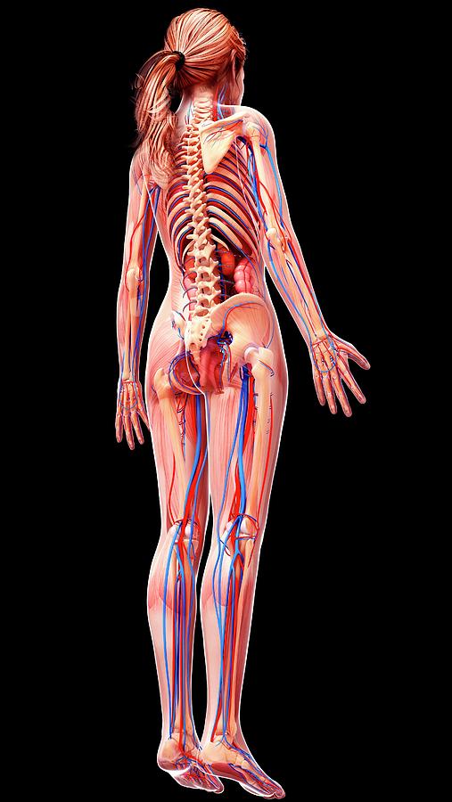
- Pelvic organ prolapse. Pelvic organ prolapse occurs when the muscles in the pelvis can no longer support its organs, such as the bladder, uterus, or rectum. This can cause one or more of these organs to press down on the vagina. In some cases, this can cause a bulge to form outside of the vagina.
- Endometriosis. Endometriosis occurs when the tissue that lines the inside walls of the uterus (endometrium) begins to grow outside of the uterus. The ovaries, fallopian tubes, and other tissues in the pelvis are typically affected by the condition. Endometriosis can lead to complications, including infertility or ovarian cancer.
Symptoms of a pelvic condition
Some common symptoms of a pelvic condition can include:
- pain in the lower abdomen or pelvis
- a feeling of pressure or fullness in the pelvis
- unusual or foul-smelling vaginal discharge
- pain during sex
- bleeding in between periods
- painful cramping during or before periods
- pain during bowel movements or when urinating
- a burning feeling when urinating
Tips for a healthy pelvis
The female pelvis is a complex, important part of the body. Follow these tips to keep it in good health:
Stay on top of your reproductive health
See a gynecologist for a yearly health screen. Things like pelvic exams and Pap smears can aid in identifying pelvic conditions or infections early.
You can get a free or low-cost pelvic exam at your local Planned Parenthood clinic.
Practice safe sex
Use barriers — such as condoms or dental dams — during sexual activity, especially with a new partner, to avoid infections that could lead to PID.
Try pelvic floor exercises
These types of exercises can help to strengthen the muscles in the pelvis, including those around the bladder and vagina.
Stronger pelvic floor muscles can aid in preventing things like incontinence or organ prolapse. Here’s how to get started.
Never ignore unusual symptoms
If you’re experiencing anything unusual in your pelvic area, such as bleeding between periods or unexplained pelvic pain, make an appointment with your doctor. Left untreated, some pelvic conditions can have lasting impacts on your health and fertility.
Diagram of the female pelvis: anatomy, functions of bones, muscles, ligaments
contents
What is the female pelvis?
The pelvis is the lower part of the body. It is located between the abdomen and legs. This area provides support for the intestines and also contains the bladder and reproductive organs.
There are some structural differences between the female and male pelvis. Most of these differences involve providing enough space for the baby to develop and pass through the woman's pelvic birth canal. As a result, the female pelvis is usually wider and wider than the male pelvis. nine0003
Learn more about the bones, muscles and organs of a woman's pelvis below.
Pelvic Anatomy and Function
Female Hip Bones
Pelvic Bones
There are two hip bones, one on the left side of the body and the other on the right side. Together they form a part of the pelvis called the pelvic girdle.
The femurs are connected to the upper part of the skeleton, attaching it to the cruciate ligament. Each hip bone is made up of three smaller bones that fuse during adolescence:
- Ilion. Most of the pelvic bone, the ilium, is broad and rich. You can feel the curves of these bones when you put your hands on your hips.
- Pub. The pubic bone of each pelvic bone joins with the other at a joint called pubis syndrome.
- Sit down. When you sit down, most of your body weight rests on these bones. That is why they are sometimes called tiny bones.
The ilium, pubis, and ischium of each pelvic bone are joined together to form the acetabulum, to which the femoral head is attached. nine0003
sacrum
The sacrum is connected to the lower part of the vertebrae. In fact, it consists of five fused vertebrae. The sacrum is fairly thick and helps support body weight.
Tailbone
A mole is sometimes called a tailbone.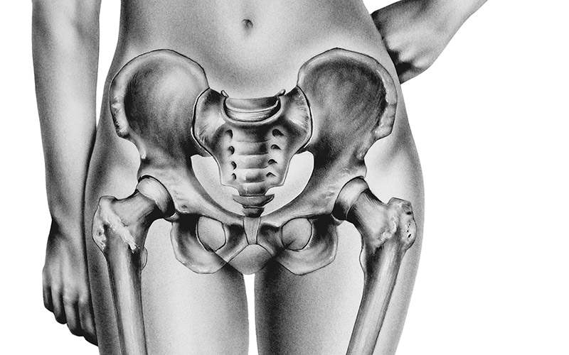 It is connected to the lower part of the sacrum and is supported by several ligaments.
A mole consists of four vertebrae that merge into a triangular shape.
Female pelvic floor muscles
Levators or muscles
The levator ani muscles are the largest muscle group in the pelvis. They have several functions, including helping to support the pelvic organs.
The levator ani is composed of three separate muscles:
- Puborectalis. This muscle is responsible for holding urine and feces. It relaxes when you urinate or defecate.
- pubococcygeal. This muscle makes up the bulk of the anil's muscles. It is formed on the pubic bone and connects to the coccyx. nine0024
- Ileococcygeal. The iliococcygeal muscle has thinner fibers and serves to elevate the pelvic floor as well as the anal canal.
coccyx
This pelvic minor muscle originates from the ischium and continues into the sacrum and coccyx.
Female pelvic organs
material
The uterus is a hollow, thick-walled organ in which the baby develops during pregnancy.
During your reproductive years, the lining of your uterus is shed every month during your period, unless you are pregnant. nine0003
ovaries
There are two ovaries on either side of the uterus. The ovaries produce eggs and secrete hormones such as estrogen and progesterone.
Fallopian tubes
Fallopian tubes connect each ovary to the uterus. Specialized cells in the fallopian tubes use hair-like structures called cilia to help guide an egg from the ovary to the uterus.
Cervix
The cervix connects the uterus to the vagina. It can expand to allow sperm to pass into the uterus. nine0003
In addition, the thick mucus that forms in the cervix can help prevent bacteria from entering the uterus.
Vagina
The vagina connects the cervix to the female external genitalia. It is also called the birth canal because the baby passes through the vagina during childbirth.
rectum
The rectum is the lowest part of the large intestine.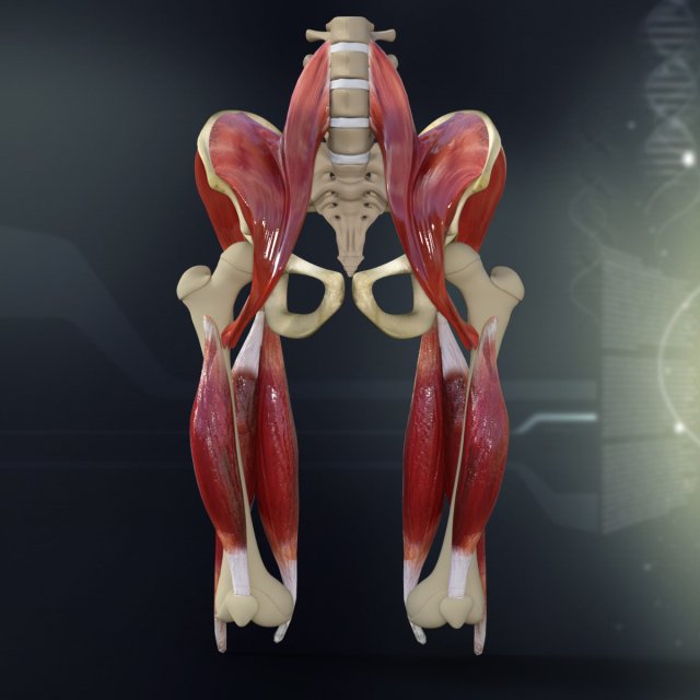 Here the stool is collected until it exits through the anus.
bladder
The bladder is the organ that collects and stores urine until it is released. Urine reaches the bladder through tubes called ureters that connect to the kidneys.
urethra
The urethra is the tube through which urine passes to exit the body from the bladder. The female urethra is much shorter than the male.
Women's Pelvic Ligaments
Broad Ligament
The broad ligament supports the uterus, fallopian tubes and ovaries. It extends to both sides of the pelvic wall. nine0003
Wide ligament can be divided into three components that are associated with different parts of the female reproductive organs:
- Mesometry, which supports the uterus
- Meserium, supporting the ovarian
- Mesosalpinx, supporting the phallopian trumpets
Material borders 9000 borders of the Higions extra support for the uterus. Some of the major uterine ligaments include:
- ligament teres
- cardinal ligaments
- pubocervical ligaments
- uterosacral ligaments
Ovarian ligaments
Ovarian ligaments support the ovaries. There are two main ovarian ligaments:
- ovarian ligament
- ovarian suspensory ligament
Diagram of a woman
Explore this interactive 3D diagram to learn more about the female pelvis:
Contains a large number of organs in the pelvic floor 9002 , bones, muscles, and ligaments, so many conditions can affect the entire pelvis or parts of it. nine0003
Some conditions that can affect a woman's pelvis in general include:
- Pelvic inflammatory disease (PID). PID is an infection that occurs in the female reproductive system. Although it is often caused by a sexually transmitted infection, other infections can also cause PID. Left untreated, PID can lead to complications such as infertility or ectopic pregnancy.
- Pelvic organ prolapse. Pelvic organ prolapse occurs when the muscles of the pelvis can no longer support its organs, such as the bladder, uterus, or rectum. Therefore, one or more of these organs may be pressed against the vagina.
 In some cases, this can lead to the formation of a protrusion outside the vagina. nine0024
 - Endometriosis. Endometriosis occurs when the tissue that lines the inside of the uterus (endometrium) begins to grow outside the uterus. The ovaries, fallopian tubes, and other pelvic tissues are usually affected. Endometriosis can lead to complications, including infertility or ovarian cancer.
Pelvic symptoms
Some common symptoms of pelvic disease may include:
- pain in the lower abdomen or pelvis
- Sensation of pressure or completeness in the pelvis
- Unusual or fetid vagina
- Pain during sex
- Bleeding between menstruation
- Painful seizures during or before the period of
- pain during defecation
- Burning
The female pelvis is a complex and important part of the body. Follow these tips to stay healthy:
Look after your reproductive health
Get your annual medical check-up with your gynecologist. Things like a pelvic exam and a Pap test can help you catch pelvic disease or infections earlier.
You can get a free or low-cost pelvic exam at your local Family Planning Clinic.
Practice safer sex
Use barriers such as condoms or dental pads during intercourse, especially with a new partner, to avoid infections that can lead to PID. nine0003
Try Pelvic Floor Exercises
These types of exercises can help strengthen your pelvic muscles, including those around your bladder and vagina.
Stronger pelvic floor muscles can help prevent things like urinary incontinence or organ failure. Here's how to get started.
Never ignore unusual symptoms
If you experience anything unusual in your pelvis, such as bleeding between periods or unexplained pelvic pain, make an appointment with your doctor. If left untreated, some pelvic conditions can permanently affect your health and fertility. nine0003
Scientists told how the shape of the female pelvis changes throughout life
Scientists told how the shape of the female pelvis changes throughout lifeTwo points from Kaprizov helped the Minnesota beat the Islanders 06:13
Kasatkina reached the final of the tournament in Adelaide due to the absence of her opponent for the match 06:10
In the Zaporozhye region, car passes were introduced for the movement of.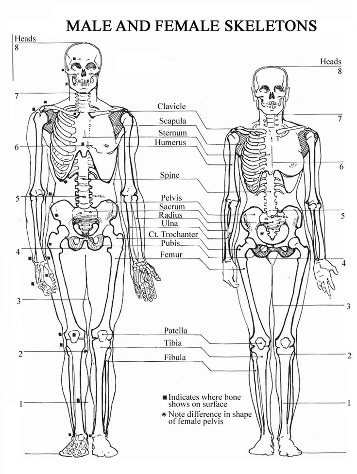 .. 05:58
Kucherov's goal helped Tampa beat Vancouver 05:50
Panarin's pass in overtime brought the Rangers victory... 05:44
The musicians who left the Russian Federation recorded a collection of songs After Russia on verses ... 05:44
Before the alleged "leaving" from the Comedy Club, Pavel Volya called the girl... 05:37
Ural coach Goncharenko explained a series of seven matches... 05:31
Elvis Presley's Daughter Lisa Marie Passed Away 05:20
The Foreign Ministry said that possible negotiations between Russia and Ukraine will be direct... 05:10
Science
close
100%
At the optimal age for the birth of a child, the structure of the female pelvis may change somewhat, scientists have found. Is there a solution to the "obstetric dilemma" and when the female body is most prepared for procreation, says the science department of Gazeta.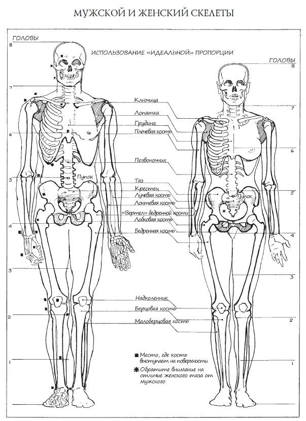 Ru.
Ru.
No matter how much scientists struggle to study nature, constantly improving their methods, it still remains the biggest mystery. Researchers still do not cease to be amazed at how everything is thought out and organized in the living world. nine0003
Jealousy is a reason for a medical examination
A 43-year-old Turkish woman unreasonably began to be jealous of her faithful husband, with whom she was married 20...
April 25 11:10
Anthropologists and biologists from Switzerland and Belgium, in a collaborative study, have found evidence that the shape and size of a woman's pelvis can change to adapt to having offspring. Research progress and data analysis published in the latest edition of Proceedings of the National Academy of Sciences. nine0003
The structure of the human pelvis is classified as sexually dimorphic, that is, it distinguishes males and females from each other. As a rule, the hips of women are wider and more rounded than those of men.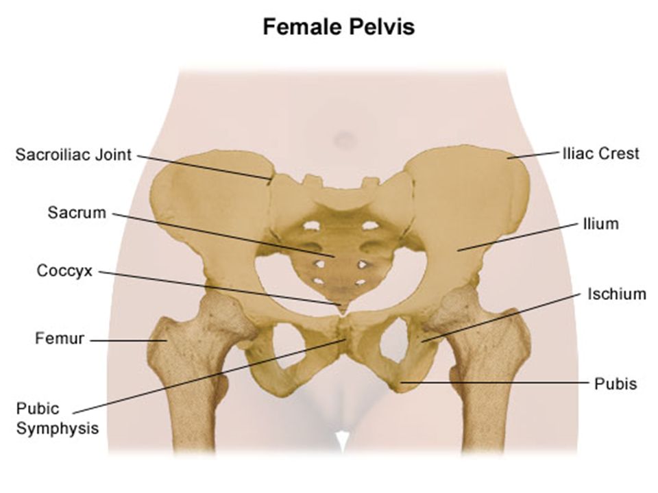
Studying the structure of the female skeleton, scientists put forward the hypothesis of the so-called obstetric dilemma. On the one hand, the female pelvis should be relatively wide in order to facilitate the birth of babies, which, when properly positioned in the womb, go head first. On the other hand, a narrow pelvis makes it easier for a person to walk upright. Recently, this hypothesis has been challenged on the basis of biomechanical and biocultural parameters, as well as metabolic processes. However, until the emergence of recent studies by Swiss and Belgian scientists, it remained unclear which factors are responsible for the differences in the shape and size of the pelvis in adult men and women. nine0003
Weekends are not for childbirth
Tuesday is the most favorable day for the birth of a child, but on Thursday, Saturday and Sunday babies ...
November 26 14:28
Starting their scientific work, the researchers put forward the following assumption: during the course of life, the structure of the pelvis in women specially changes, facilitating the birth of children.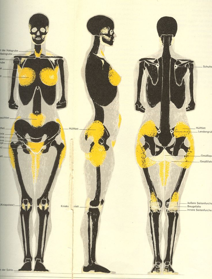 To test their own hypothesis, a group of scientists invited 275 women of different ages up to 95 years to participate in the research. The authors of the work used the methods of computed tomography, and also engaged in geometric morphometry, that is, they analyzed the ratio of the sizes and shapes of the parts of the pelvis. The obtained data were compared with the already available data on the structure of the pelvis in men. nine0003
The results obtained showed that before the onset of puberty, the structure of the pelvis in boys and girls practically does not differ, and all changes proceed in the same way.
With the onset of active sexual development, which in some girls occurs already at the age of 10 years, the change in the shape and size of the female pelvis occurs according to its own unique laws.
The processes that prepare the female body for the birth of children are completed by about 25 years. Scientists note that the period when differences in the structure of the pelvis in women and men are most noticeable coincides with the peak of fertility.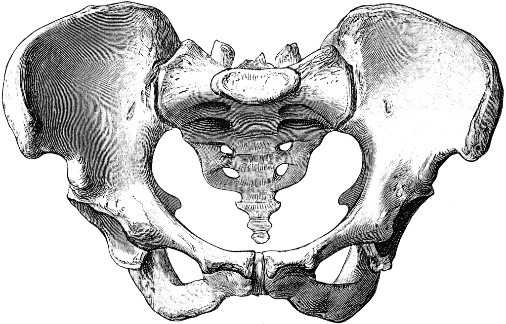 According to the researchers, in women this age occurs on average from 25 to 30 years. Biologists have noticed a curious feature: at the age of 40 to 45 years, the structure of the female pelvis continues to develop, but the changes again proceed by analogy with men. Transformations lead to a reduction in the size of the birth canal (the birth canal is a canal formed by the bones of the small pelvis and soft tissues located in it, through which the fetus and placenta pass during childbirth). nine0003
Swiss and Belgian scientists suggest that all changes in the shape and size of the female pelvis, depending on age, may be associated with the production of hormones during puberty and before menopause. According to biologists, estradiol, which is part of the group of female sex hormones - estrogen, takes the most active part. It depends on him how rounded the outlines of the female figure will be. Often, estradiol is called the "hormone of pregnancy", as it regulates blood circulation in the pelvic cavity, directs the increase in the size of the uterus, and is also responsible for the formation of the placenta. nine0003
Subscribe to Gazeta.Ru in News, Zen and Telegram.
To report a bug, select the text and press Ctrl+Enter
News
Zen
Telegram
Picture of the day
Obama-era classified documents. What was found in Biden's garage
The White House admitted that Joe Biden's lawyers found classified papers at his home
"Hegumen, pedophile and swindler". Details of the case against the abbot of the monastery of the Holy Trinity
The 85-year-old hegumen of the "True Orthodox Church" was suspected of pedophilia
The State Duma argued whether it is time to "drive all the youth" through military training
Head of the State Duma Defense Committee Kartapolov did not support military training for all youth
Advisor to the head of the DPR Kimakovsky: Artemovsk is in the operational encirclement of the Russian troops
nine0002 Matvienko stated that a federal statehood without vulnerabilities was built in RussiaA Ukrainian soldier told CNN that the command abandoned the military near Soledar
Congress said that Republicans were denied access to data on Biden's classified papers
News and materials
Kasatkina reached the final of the tournament in Adelaide due to the absence of her opponent for the match
Legendary guitarist Jeff Beck dies
“Not fascism? Fascism". The State Duma demands to check Boris Grebenshchikov's statements
Deputy Khamzaev asked the Investigative Committee to check Grebenshchikov's words for "assistance to terrorism"
"We were hostages." Sailors detained in Ukraine returned to Russia
Ombudsman Moskalkova: all Russian civilian sailors returned to their homeland from Ukraine
Rospotrebnadzor announced the detection of a subvariant of the “kraken” coronavirus in Russia
 01.2023, 15:33
 "Yerevan's statements are absurd." The Russian Foreign Ministry denied the danger of the presence of Russian troops in Armenia
Zakharova called Pashinyan's words about the threat to Armenia from Russia
"Hidden with secret documents": in the USA they found a reason to impeach Biden
Snowden: Biden seems to have taken more classified documents than many others
"Disgusting behavior." The State Duma decides how to punish artists who "betrayed" Russia
Deputy Stenyakina proposed to deprive artists who "betrayed the country" of state funding
Anastasia Mironova
The watchman knows all the parents by sight
About studying in a modern village school

