Do pelvic ultrasounds hurt
Pelvic Ultrasound | Johns Hopkins Medicine
What is a pelvic ultrasound?
(Ultrasound-Pelvis, Pelvic Ultrasonography, Pelvic Sonography, Pelvic Scan, Lower Abdomen Ultrasound, Gynecologic Ultrasound, Transabdominal Ultrasound, Transvaginal Ultrasound, Endovaginal Ultrasound)
A pelvic ultrasound is a noninvasive diagnostic exam that produces images that are used to assess organs and structures within the female pelvis. A pelvic ultrasound allows quick visualization of the female pelvic organs and structures including the uterus, cervix, vagina, fallopian tubes and ovaries.
Ultrasound uses a transducer that sends out ultrasound waves at a frequency too high to be heard. The ultrasound transducer is placed on the skin, and the ultrasound waves move through the body to the organs and structures within. The sound waves bounce off the organs like an echo and return to the transducer. The transducer processes the reflected waves, which are then converted by a computer into an image of the organs or tissues being examined.
The sound waves travel at different speeds depending on the type of tissue encountered - fastest through bone tissue and slowest through air. The speed at which the sound waves are returned to the transducer, as well as how much of the sound wave returns, is translated by the transducer as different types of tissue.
An ultrasound gel is placed on the transducer and the skin to allow for smooth movement of the transducer over the skin and to eliminate air between the skin and the transducer for the best sound conduction.
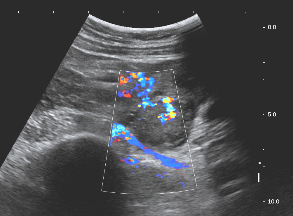
Another type of ultrasound is Doppler ultrasound, sometimes called a duplex study, used to show the speed and direction of blood flow in certain pelvic organs. Unlike a standard ultrasound, some sound waves during the Doppler exam are audible.
Pelvic ultrasound may be performed using one or both of 2 methods:
-
Transabdominal (through the abdomen). A transducer is placed on the abdomen using the conductive gel
-
Transvaginal (through the vagina).
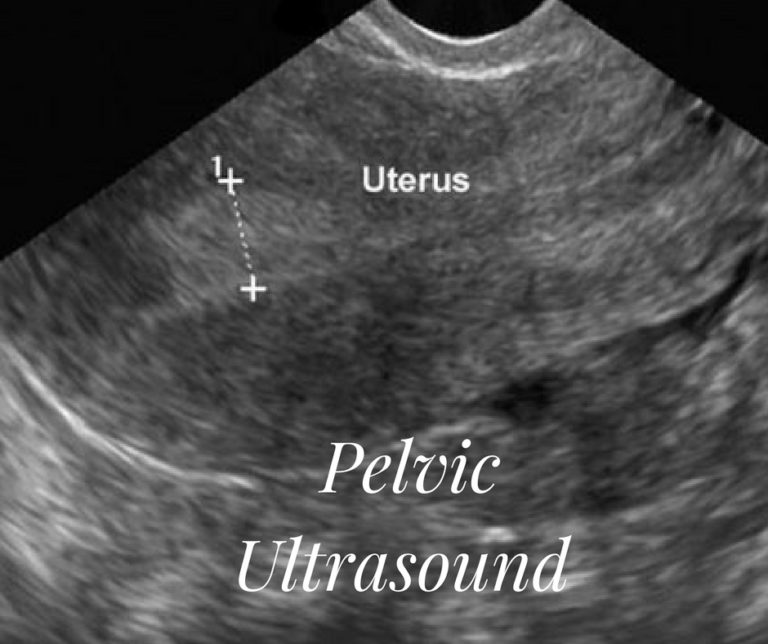 A long, thin transducer is covered with the conducting gel and a plastic/latex sheath and is inserted into the vagina
The type of ultrasound procedure performed depends on the reason for the ultrasound. Only one method may be used, or both methods may be needed to provide the information needed for diagnosis or treatment.
Other related procedures that may be used to evaluate problems of the pelvis include hysteroscopy , colposcopy , and laparoscopy .
What are female pelvic organs?
The organs and structures of the female pelvis are:
-
Endometrium. The lining of the uterus
-
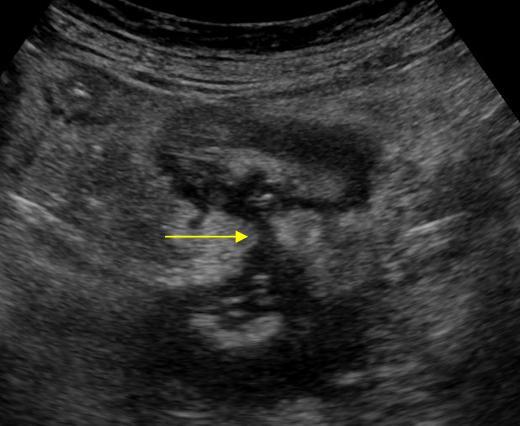
Uterus (also known as the womb). The uterus is a hollow, pear-shaped organ located in a woman's lower abdomen, between the bladder and the rectum. It sheds its lining each month during menstruation, unless a fertilized egg (ovum) becomes implanted and pregnancy follows.
-
Ovaries. Two female reproductive organs located in the pelvis in which egg cells (ova) develop and are stored and where the female sex hormones estrogen and progesterone are produced.
-
Cervix. The lower, narrow part of the uterus located between the bladder and the rectum, forming a canal that opens into the vagina, which leads to the outside of the body.
-
Vagina (also known as the birth canal). The passageway through which fluid passes out of the body during menstrual periods. The vagina connects the cervix and the vulva (the external genitalia).
-
Vulva. The external portion of the female genital organs
What are the reasons for a pelvic ultrasound?
Pelvic ultrasound may be used for measurement and evaluation of female pelvic organs. Ultrasound assessment of the pelvis may include, but is not limited to, the following:
-
Size, shape, and position of the uterus and ovaries
-
Thickness, echogenicity (darkness or lightness of the image related to the density of the tissue), and presence of fluids or masses in the endometrium, myometrium (uterine muscle tissue), fallopian tubes, or in or near the bladder
-
Length and thickness of the cervix
-
Changes in bladder shape
-
Blood flow through pelvic organs
Pelvic ultrasound can provide much information about the size, location, and structure of pelvic masses, but cannot provide a definite diagnosis of cancer or specific disease. A pelvic ultrasound may be used to diagnose and assist in the treatment of the following conditions:
-
Abnormalities in the anatomic structure of the uterus, including endometrial conditions
-
Fibroid tumors (benign growths), masses, cysts, and other types of tumors within the pelvis
-
Presence and position of an intrauterine contraceptive device (IUD)
-
Pelvic inflammatory disease (PID) and other types of inflammation or infection
-
Postmenopausal bleeding
-
Monitoring of ovarian follicle size for infertility evaluation
-
Aspiration of follicle fluid and eggs from ovaries for in vitro fertilization
-
Ectopic pregnancy (pregnancy occurring outside of the uterus, usually in the fallopian tube)
-
Monitoring fetal development during pregnancy
-
Assessing certain fetal conditions
Ultrasound may also be used to assist with other procedures such as endometrial biopsy . Transvaginal ultrasound may be used with sonohysterography, a procedure in which the uterus is filled with fluid to distend it for better imaging.
There may be other reasons for your doctor to recommend a pelvic ultrasound.
What are the risks of a pelvic ultrasound?
There is no radiation used and generally no discomfort from the application of the ultrasound transducer to the skin during a transabdominal ultrasound. You may experience slight discomfort with the insertion of the transvaginal transducer into the vagina.
Transvaginal ultrasound requires covering the ultrasound transducer in a plastic or latex sheath, which may cause a reaction in patients with a latex allergy.
During a transabdominal ultrasound, you may experience discomfort from having a full bladder or lying on the examination table.
If a transabdominal ultrasound is needed quickly, a urinary catheter may be inserted to fill the bladder.
There may be risks depending on your specific medical condition. Be sure to discuss any concerns with your doctor prior to the procedure.
Certain factors or conditions may interfere with the results of the test. These include, but are not limited to, the following:
-
Severe obesity
-
Barium within the intestines from a recent barium procedure
-
Intestinal gas
-
Inadequate filling of bladder (with transabdominal ultrasound). A full bladder helps move the uterus up and moves the bowel away for better imaging.
How do I prepare for a pelvic ultrasound?
EAT/DRINK : Drink a minimum of 24 ounces of clear fluid at least one hour before your appointment. Do not empty your bladder until after the exam.
Generally, no fasting or sedation is required for a pelvic ultrasound, unless the ultrasound is part of another procedure that requires anesthesia.
For a transvaginal ultrasound, you should empty your bladder right before the procedure.
Your doctor will explain the procedure to you and offer you the opportunity to ask any questions that you might have about the procedure.
Based on your medical condition, your doctor may request other specific preparation.
What happens during a pelvic ultrasound?
A pelvic ultrasound may be performed in your doctor’s office, on an outpatient basis, or as part of your stay in a hospital. Procedures may vary depending on your condition and your hospital’s practices.
Generally, a pelvic ultrasound follows this process:
For a transabdominal ultrasound
-
You will be asked to remove any clothing, jewelry, or other objects that may interfere with the scan.
-
If asked to remove clothing, you will be given a gown to wear.
-
You will lie on your back on an examination table.

-
A gel-like substance will be applied to your abdomen.
-
The transducer will be pressed against the skin and moved around over the area being studied.
-
If blood flow is being assessed, you may hear a "whoosh, whoosh" sound when the Doppler probe is used.
-
Images of structures will be displayed on the computer screen. Images will be recorded on various media for the health care record.
-
Once the procedure has been completed, the gel will be removed.
-
You may empty your bladder when the procedure is completed.
For a transvaginal ultrasound
-
You will be asked to remove any clothing, jewelry, or other objects that may interfere with the scan.
-
If asked to remove clothing, you will be given a gown to wear.

-
You will lie on an examination table, with your feet and legs supported as for a pelvic examination.
-
A long, thin transvaginal transducer will be covered with a plastic or latex sheath and lubricated. The tip of the transducer will be inserted into your vagina. This may be slightly uncomfortable.
-
The transducer will be gently turned and angled to bring the areas for study into focus. You may feel mild pressure as the transducer is moved.
-
If blood flow is being assessed, you may hear a "whoosh, whoosh" sound when the Doppler probe is used.
-
Images of organs and structures will be displayed on the computer screen. Images may be recorded on various media for the health care record.
-
Once the procedure has been completed, the transducer will be removed.
What happens after a pelvic ultrasound?
There is no special type of care required after a pelvic ultrasound. You may resume your normal diet and activity unless your doctor advises you differently.
There are no confirmed adverse biological effects on patients or instrument operators caused by exposures to ultrasound at the intensity levels used in a diagnostic ultrasound.
Your doctor may give you additional or alternate instructions after the procedure, depending on your particular situation.
Is it painful, purpose, and results
A transvaginal, or endovaginal, ultrasound is a safe, straightforward way for doctors to examine the internal organs of the female pelvic region.
An ultrasound uses high-frequency sound waves to produce detailed images of internal organs.
Unlike X-rays, ultrasound scanning does not involve radiation, which means that it has no known side effects and is very safe.
In this article, we explain the uses of transvaginal ultrasounds and how to prepare for one. We also describe what to expect during the scan.
A healthcare professional uses an ultrasound to create images of the inside of the body. High-frequency sound waves bounce off the internal organs and form these images.
There are two ways to perform an ultrasound — abdominally and transvaginally.
A transvaginal ultrasound is an internal scan of the female reproductive organs. It involves inserting a small ultrasound probe, called a transducer, into the vagina to produce incredibly detailed images of the organs in the pelvic region.
It may be necessary to use a transvaginal ultrasound to examine the:
- vagina
- cervix
- uterus
- fallopian tubes
- ovaries
- bladder
Transvaginal ultrasounds can check for:
- the shape, position, and size of the ovaries and uterus
- the thickness and length of the cervix
- blood flow through the organs in the pelvis
- the shape of the bladder and any changes
- the thickness and presence of fluids near the bladder or in the:
- fallopian tubes
- myometrium, the muscle tissue of the uterus
- endometrium
Doctors may request a transvaginal ultrasound for a variety of reasons. For example, it might be necessary to identify the cause of:
- pelvic pain
- unexplained vaginal bleeding
- infertility
- abnormal results of a pelvic or abdominal exam
These scans can also help diagnose:
- benign growths, such as fibroids, cysts, and masses
- pelvic inflammatory disease
- endometriosis
- postmenopausal bleeding
In addition, it can check for the presence of an intrauterine contraceptive device.
Doctors may request a transvaginal ultrasound during pregnancy, as it can help:
- check the heartbeat of the fetus
- confirm the date of delivery
- assess the condition of the placenta
- check for an ectopic pregnancy
- monitor pregnancies with a higher risk of pregnancy loss
A transvaginal ultrasound is not typically painful, but the insertion of the probe may be uncomfortable.
The healthcare professional performing the scan first covers the probe in a sheath and lubricating gel before inserting it slowly into the vagina to a depth of around 5–8 centimeters (cm).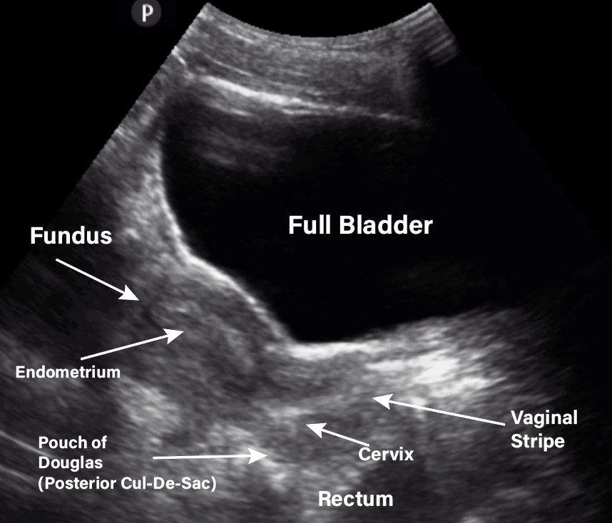 There may be mild pressure or discomfort at this stage.
There are no after-effects of a transvaginal ultrasound, and a person can return to their regular activities afterward.
A pelvic ultrasound is a noninvasive exam that produces images of the internal reproductive organs to help healthcare professionals diagnose certain conditions.
Doctors may use the term “pelvic ultrasound” to describe both a transvaginal and a transabdominal ultrasound. During a transabdominal ultrasound, the person performing the scan uses the probe outside the abdomen.
Another name for a transabdominal ultrasound is an external pelvic ultrasound. A person lies on their back on an examination table, and the healthcare professional applies a warm gel to the lower section of the person’s abdomen. Then, they move a probe over the area.
The person might feel slight pressure on their abdomen, but the scan is not painful. The probe uses sound waves to form an image of the internal organs and structures of the pelvic area.
By comparison, a transvaginal ultrasound can provide more close-up images of the internal organs than an external pelvic ultrasound.
A transvaginal ultrasound is a simple, painless scan that requires very little preparation. A person should be able to eat and drink normally before the appointment.
At the appointment, a healthcare professional will describe any necessary steps. A person needs to empty their bladder right before the scan, and anyone using a tampon should remove it.
A doctor or a specially trained technician, called a sonographer, performs most transvaginal ultrasounds.
A person needs to undress from the waist down and put on a hospital gown. Next, they lie on an examination table with their knees bent. The healthcare professional uses a sheet to cover the person’s lower body.
The transducer resembles a wand and is slightly larger than a tampon. The sonographer or doctor covers the transducer with a sheath and lubricating gel before inserting it 5–8 cm into the vagina.
Once the transducer is in place, it produces sound waves that bounce off of the internal organs and relay information. To create a complete picture and bring different areas into focus, the sonographer or doctor rotates the transducer. This tool transmits the information directly to a screen.
The images display immediately on the screen, making it possible for the person and the healthcare professional to monitor the scan in real-time.
The whole process may last 15–30 minutes.
Unlike a traditional X-ray, a transvaginal ultrasound does not use radiation. As a result, it is very safe — there are no known risks.
It is also safe to perform transvaginal ultrasounds during pregnancy — there is no risk to the fetus.
During the insertion of the transducer, there may be some pressure and minimal discomfort. This feeling should go away after the scan.
It is essential to let the sonographer or doctor right away if anything feels particularly uncomfortable.
As the Miscarriage Association confirms, there is no evidence that a transvaginal ultrasound can harm a fetus or cause pregnancy loss.
If a person notices bleeding after a transvaginal ultrasound, this may be because blood has collected higher up in the vagina, and the transducer may have dislodged it.
People may have a transvaginal ultrasound in the first 11–12 weeks of pregnancy. After 11–12 weeks, it may be more common to have a transabdominal ultrasound. However, in some cases, doctors may still request a transvaginal ultrasound, which is a more accurate and detailed scan.
Transvaginal ultrasounds are safe up until a person’s water breaks.
If a specialist is present at the ultrasound, the person may hear the results immediately. If a sonographer does the scan, they send the images to a radiologist for analysis. The radiologist then sends a written report of the results to the person’s doctor.
Either way, it is important to discuss the results with a doctor, who can provide more information.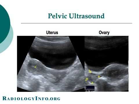 This discussion can take place in person or over the phone.
A transvaginal ultrasound is a safe scan, with no known risks. People might experience some light discomfort during it, but this should go away afterward.
The ultrasound may take 15–30 minutes, and the results may be available immediately or within a few days, depending on whether a doctor was present during the scan.
If a person experiences extreme discomfort during the ultrasound, the doctor or sonographer may perform a transabdominal ultrasound instead. This type does not involve inserting the scanning tool into the vagina.
The effect of ultrasound on the fetus: is ultrasound harmful to the baby
Table of contents
- Ultrasound principle
- Effect of ultrasound on the fetus
- Effect of ultrasound on women
- Conclusion
Ultrasound has been used in the diagnosis of pregnancy since the 1960s. During this time, this method constantly confirms its reliability and effectiveness in terms of monitoring the condition of the baby and identifying possible violations. At the same time, many expectant mothers naturally have a question - what is the effect of ultrasound on the fetus and is there a risk to his health from exposure to ultrasonic waves?
How ultrasound works
To understand whether there is a risk of ultrasound affecting the baby or the mother's body, you need to consider the very principle of ultrasound. Sonography (or echography) is based on the high penetrating power of ultrashort sound waves. Penetrating into various tissues of the human body, they change their characteristics. These changes are captured by a sensor that picks up the reflected signal and converts it into a visual image. To date, there are 3 types of ultrasound:
- 2D - displays a two-dimensional image;
- 3D - displays a 3-dimensional model of the object under study;
- 4D - Shows a 3D model in motion.
The latter method is the most informative, as it allows assessing not only the morphological features of the fetus (size, structure of the body and limbs, the width of the cervical-collar space, etc. ), but also its motor activity, heart rate. Using it, the doctor can determine such life-threatening conditions for the child as hypoxia, breech presentation, etc. Ultrasound also allows you to assess the condition of the amniotic membranes and the uterus itself.
As part of the management of pregnancy in accordance with the order of the Ministry of Health of the Russian Federation “On approval of the Procedure for the provision of medical care in the profile “Obstetrics and Gynecology”), a planned ultrasound is performed 3 times:
- 1 screening - at 11-14 weeks;
- 2 screening - at 19-21 weeks;
- 3 screening - at 30-34 weeks.
Also, the doctor may prescribe additional unscheduled ultrasounds if there are certain indications - for example, for women at risk.
Effect of ultrasound on the fetus
Numerous medical studies have been devoted to the impact of ultrasonic waves on the condition of the fetus, designed to objectively assess the safety of this method. None of the scientific papers available today provide evidence in favor of the fact that ultrasound negatively affects the child.
However, it cannot be argued that ultrasound has no effect on the human body. High-frequency sound waves heat tissues, for 1 hour of continuous exposure their temperature can rise by 2-5ᵒС. Hyperthermia potentially has a teratogenic effect - that is, it is capable of causing fetal developmental disorders when certain factors are combined.
Ultrasound carries the greatest potential risk for the baby at an early stage of pregnancy, when embryonic cells are actively dividing, and the organs and systems of the unborn child are just beginning to form. During this period, prolonged exposure to ultrasound and heating of tissues can disrupt the process of this formation and provoke congenital anomalies of the fetus.
The FDA (US Federal Food and Drug Administration) guidelines indicate an upper allowable threshold for ultrasonic exposure of 720 mW/cm 2 . In obstetric practice, ultrasound is used with a power of more than 2 times lower than this threshold of 310 mW/cm 2 . In other words, when examined in licensed medical institutions, overheating of the fetus during ultrasound with long-term consequences for its body is excluded.
The effect of ultrasound on a woman
The mother's body is obviously more resistant to external influences than the fetus. However, intense or prolonged exposure to ultrasound in early pregnancy can provoke contractile activity of the uterine muscles, and this creates a risk of miscarriage.
At the same time, ultrasound used in diagnostics does not reach values at which such an outcome is likely. Therefore, for the body of the woman herself, diagnostic ultrasound in a licensed medical institution does not carry any risk.
Conclusion
Based on the data of available studies, it can be argued that it was not possible to trace the relationship between ultrasound and abnormalities in the development of the fetus. Evidence that sometimes appears in the media that ultrasound causes autism or other developmental problems is not confirmed by other researchers.
To completely eliminate the possible negative impact of ultrasound on the fetus, it is recommended:
- undergo an examination in a medical institution that has a state license to provide obstetric and gynecological care;
- to undergo an examination only as prescribed by a doctor - if a woman is not at risk and she has no problems with bearing, 3 fetal screening ultrasounds are enough for her during the entire period of pregnancy.
All pregnant women undergo ultrasound, and in the entire world practice of using this method, not a single case has been registered that clearly indicates its danger. But there is a lot of evidence that a timely examination helps to detect pregnancy anomalies and take appropriate measures.
Ultrasound has been used in the diagnosis of pregnancy since the 1960s.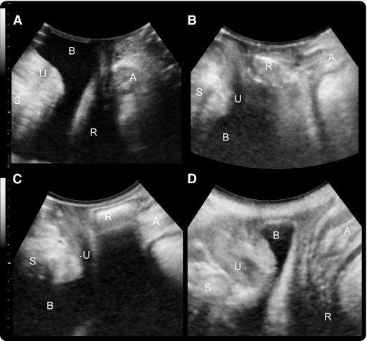 During this time, this method constantly confirms its reliability and effectiveness in terms of monitoring the condition of the baby and identifying possible violations. At the same time, many expectant mothers naturally have a question - what is the effect of ultrasound on the fetus and is there a risk to his health from exposure to ultrasonic waves?
Is ultrasound (ultrasound) harmful to the body
Ultrasound (ultrasound) in modern medicine is used as a diagnostic tool. It allows you to identify a wide variety of diseases and pathologies. Also, this method is often preferred for monitoring the condition of pregnant women, because it allows you to track the stages of fetal development.
Therefore, many patients, and sometimes even specialists, ask themselves: can ultrasound have a negative impact on human health?
If the volume of ultrasound does not exceed 120 dB, its influence will not harm the body. Today, ultrasound is used for diagnosis with a maximum of 90 dB, so this method is completely safe.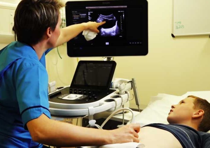 Moreover, not a single fact has been officially recorded, indicating the negative consequences of this procedure.
Without ultrasound, it is impossible to diagnose a number of serious diseases, monitor their development and, therefore, prescribe adequate treatment.
Ultrasound during pregnancy is used to monitor the health and development of the fetus. This method allows you to detect dangerous pathologies in the baby in advance. However, future parents sometimes doubt whether to give preference to this procedure. What if it harms the unborn baby?
Is it harmful to do ultrasound during pregnancy
Using ultrasound, specialists detect pregnancy at an early stage. In addition, with the help of ultrasound, developmental delays or dangerous pathologies are diagnosed. For example, carrying out this procedure will ensure the timely detection of a non-developing pregnancy. This will save the patient from complications caused by intrauterine death of the embryo.
Official medicine claims that ultrasound is absolutely safe for the mother and fetus!
Before the baby is born, such an examination is necessary to find out about the position of the fetus and its condition. Depending on the results of the ultrasound, doctors make recommendations for natural childbirth or decide to have a caesarean section.
Practical experiments
A number of experiments carried out by PP Garyaev, a specialist in wave genetics, testify to the negative effect of ultrasound on the fetus. According to the results of these studies, ultrasound is able to provoke cell mutations by acting directly on DNA. Professor Levashov also spoke about the dangers of this diagnostic method. This conclusion is confirmed by the restless behavior of the baby during the diagnostic process: the fetus begins to move and tries to close itself from the scanner. But such behavior is not characteristic of all embryos, and the reason for the activity may be an interest in ultrasound, and not its rejection. Moreover, other experiments that were conducted by competent scientific and medical organizations did not confirm that ultrasound is harmful to the child.
When to do ultrasound during pregnancy
In Russia and the CIS countries there is a normalized plan that dictates ultrasound diagnostics during pregnancy:
- the first screening to establish, in fact, the fact of pregnancy
- examination after a visit to the antenatal clinic and registration (11-14 weeks)
- Ultrasound to detect fetal malformations, if present (22-24 weeks)
- dopplerometry - assessment of embryo development (32-34 weeks)
Additional diagnostics is carried out exclusively for medical reasons:
- suspected non-developing pregnancy
- the age of the patient is over 35 years old
- the future mother has any diseases
- severe toxicosis
The final decision whether or not to have an ultrasound is made by the woman.


