Baby brain map
3-D mapping babies’ brains - The Source
During the third trimester, a baby’s brain undergoes rapid development in utero. The cerebral cortex dramatically expands its surface area and begins to fold. Previous work suggests that this quick and very vital growth is an individualized process, with details varying infant to infant.
Research from a collaborative team at Washington University in St. Louis tested a new, 3-D method that could lead to new diagnostic tools that will precisely measure the third-trimester growth and folding patterns of a baby’s brain.
The findings, published online March 5 in PNAS, could help to sound an early alarm on developmental disorders in premature infants that could affect them later in life.
“One of the things that’s really interesting about people’s brains is that they are so different, yet so similar,” said Philip Bayly, the Lilyan & E. Lisle Hughes Professor of Mechanical Engineering at the School of Engineering & Applied Science. “We all have the same components, but our brain folds are like fingerprints: Everyone has a different pattern. Understanding the mechanical process of folding — when it occurs — might be a way to detect problems for brain development down the road.”
Working with collaborators at Washington University School of Medicine in St. Louis, engineering doctoral student Kara Garcia accessed magnetic resonance 3-D brain images from 30 pre-term infants scanned by Christopher Smyser, MD, associate professor of neurology, and his pediatric neuroimaging team. The babies were scanned two to four times each during the period of rapid brain expansion, which typically happens at 28 to 38 weeks.
Using a new computer algorithm, Bayly, Garcia and their colleagues — including faculty at Imperial College and King’s College in London — obtained accurate point-to-point correspondence between younger and older cortical reconstructions of the same infant. From each pair of surfaces, the team calculated precise maps of cortical expansion. Then, using a minimum energy approach to compare brain surfaces at different times, researchers picked up on subtle differences in the babies’ brain folding patterns.
“The minimum energy approach is the one that’s most likely from a physical standpoint,” Bayly said. “When we obtain surfaces from MR images, we don’t know which points on the older surface correspond with which points on the younger surface. We reasoned, that since nature is efficient, the most likely correspondence is the one that produces the best match between surface landmarks, while at the same time, minimizing how much the brain would have to distort while it is growing.
“When you use this minimum energy approach, you get rid of a lot of noise in the analysis, and what emerged were these subtle patterns of growth that were previously hidden in the data. Not only do we have a better picture of these developmental processes in general, but doctors should hopefully be able to assess individual patients, take a look at their pattern of brain development, and figure out how it’s tracking. ”
”
It’s a measurement tool that could prove invaluable in places such as neonatal intensive care units, where preemies face a variety of challenges. Understanding an individual’s precise pattern of brain development also could assist physicians trying to make a diagnosis later in a patient’s life.
“You do also find folding abnormalities in populations that have cognitive issues later in life, including autism and schizophrenia,” Bayly said. “It’s possible, if medical researchers understand better the folding process and what goes on wrong or differently, then they can understand a little bit more about what causes these problems.”
This work was supported by National Institute of Health grants R01 NS055951, R01 NS070918, T32 EB018266, K02 NS089852, K23 Mh205179, UL1 TR000448
Kara E. Garcia, Emma C. Robinson, Dimitrios Alexopolous, Donna L. Dierker, Matthew F. Glasser, Timothy S. Coalson, Cynthia M. Ortinau, Daniel Rueckert, Larry A. Taber, David C. Van Essen, Cynthia E.
 Rogers, Christopher D. Smyser, and Philip V. Bayly, “Dynamic patterns of cortical expansion during folding of the preterm brain.” DOI 10.1073/pnas.1715451115
How your baby’s brain develops
How your baby’s brain develops | Pregnancy Birth and Baby beginning of content5-minute read
Listen
The experiences and relationships your baby has in their early years help shape the adult they will become. Creating a supportive, loving environment filled with warm, gentle interactions helps your baby’s brain to develop. It lays the foundation for your baby’s future development and learning.
Play is the main way your baby learns and develops. It helps them solve problems, explore and experiment. Other factors that can influence healthy brain development are:
- your baby’s health
- the food your baby eats
- genes
- the quality of the relationship your baby has with you and other carers
- how active your baby is
- the experiences your baby has
While babies develop skills at different times, development happens in the same order in most children.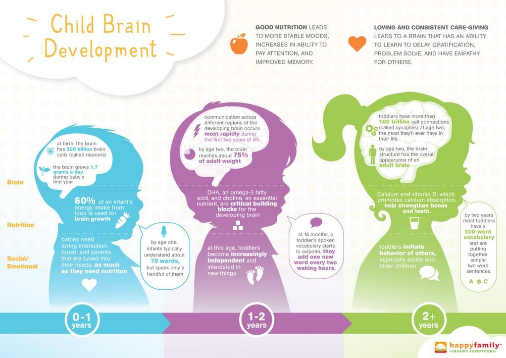 For example, children learn to walk before they learn to run.
Your baby’s brain development
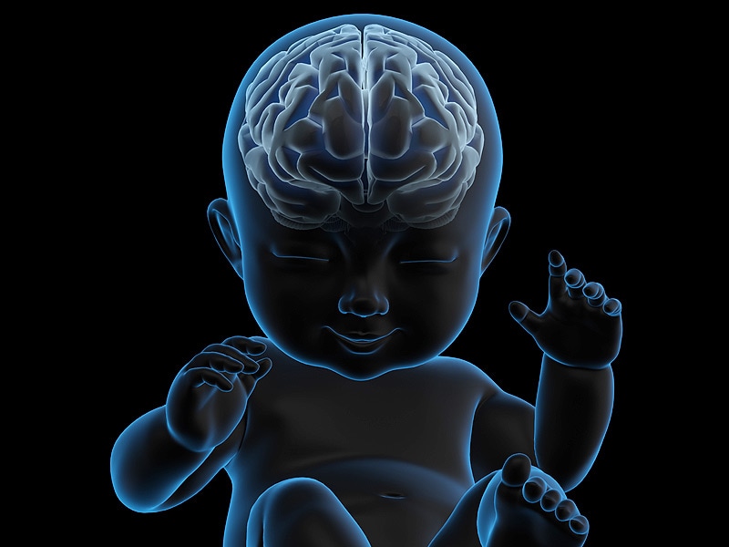
Your baby’s brain has been developing since they were in your womb. In the first trimester of pregnancy nerve connections are built that enable your baby to move around in the womb. While in the second trimester, more nerve connections and brain tissue are formed.
In the third trimester, the cerebral cortex starts to take over from the brain stem, preparing your baby for future learning.
By the time your baby arrives, they can hear (they will know your voice!) and also see a little. Their brain will then continue to grow and develop for many years.
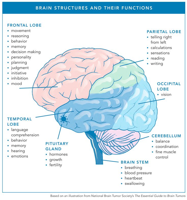
The human brain has 3 main parts:
Brain stem and cerebellum — these connect the brain to the spinal cord and control the body's breathing, heart rate, blood pressure, balance and reflexes.
Limbic system — this sits on top of the brain stem and looks after many different functions including emotion, thirst, hunger, memory, learning, and the body's daily rhythms.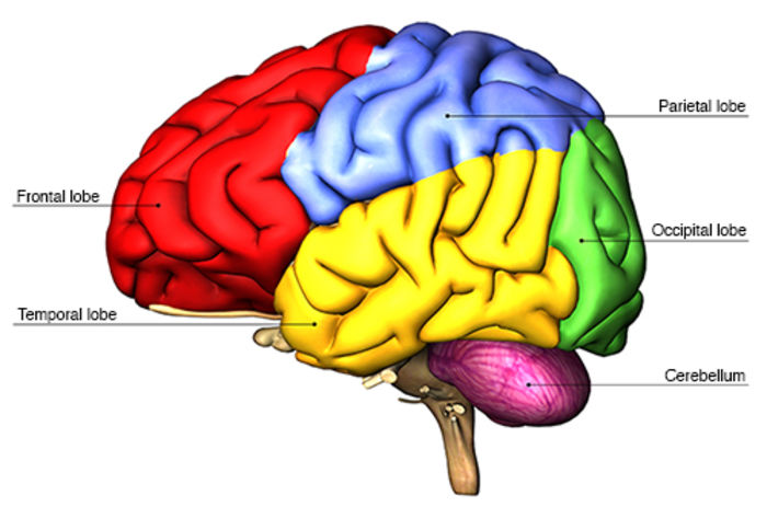
Cerebral cortex — this consists of a left and right hemisphere and sits on top of the limbic system. The cerebral cortex contains:
- occipital lobe — for vision
- temporal lobe — for hearing, language and social interaction
- frontal lobe — for memory, self-regulation, planning and problem solving
- parietal lobe — for bodily sensations like pain, pressure, heat and cold
Diagram showing different parts of the brain.
Loving relationships and stimulating experiences are vital for your baby's development since they give your baby opportunities to communicate, move and learn about their world.
How you can help your baby’s brain develop
Your baby's brain develops through use — by your baby interacting, observing and doing things.
You can help your baby's development by creating an interesting environment with different types of activities that offer your baby the chance to play. It's through play that your baby will learn important skills like talking, listening, moving, thinking, solving problems and socialising.
You can play and spend time with your baby by:
- singing together
- reading books
- talking about what you’re doing and seeing
- playing games like peekaboo
- giving lots of cuddles
Creating a warm, loving environment helps your baby feel safe and loved. This promotes brain development. Everyday moments, such as having a bath and eating, are great opportunities for you to get to know each other and build your relationship.
It's these moments that help your baby's brain form new connections. This in turn prepares your baby for the next stage in their development.
Other things your baby needs include:
- Healthy food to help them grow. Good foods for your baby are breast milk (or formula). Once your baby is ready for solid foods, iron rich foods and a balanced diet of fresh vegetables, fruit, grains, dairy and proteins (such as meat, chicken and eggs) is important.
- Moving and being active to develop their motor skills.
 This allows them to explore their surroundings, which helps them think and learn.
 - Loving relationships and interactions with others. This will boost your baby’s communication skills and understanding about the world around them.
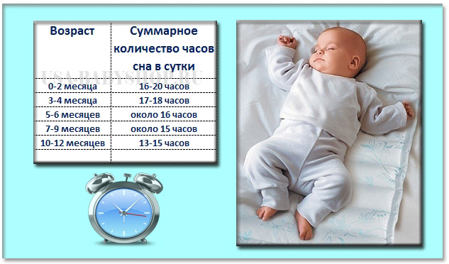
- Sleep helps your baby’s development. While all babies sleep in different ways at different times, you can help them sleep to help their development.
Milestones
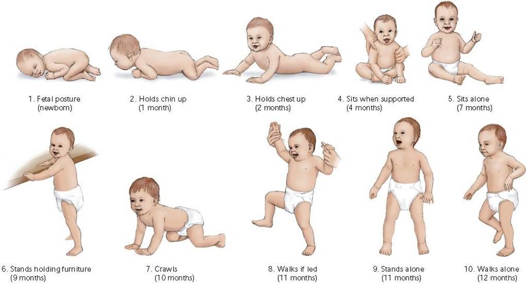
Milestones are developmental achievements and they are helpful for keeping track of your baby’s development. They can be grouped into 6 categories:
- Gross motor skills — the control and coordination of large muscles. For example: walking, lifting, throwing and sitting.
- Fine motor skills — the control and coordination of small muscles. For example: holding or picking up items.
- Vision
- Hearing
- Speech and language
- Social and emotional development — learning to interact with others and to understand and control your emotions.
You can find more information about each milestone and when to expect them in your baby’s infant health record (Child Health Record Book). This book records important information about your baby from birth. You can also speak to your child health nurse.
Where to get help and support
See your doctor or child and family health nurse if:
- your baby isn’t meeting the milestones listed in their child health record book
- you think that there’s something wrong with your baby’s vision, hearing, communication, behaviour, movement or growth
Speak to a maternal child health nurse
Call Pregnancy, Birth and Baby to speak to a maternal child health nurse on 1800 882 436 or video call. Available 7am to midnight (AET), 7 days a week.
Sources:
Murdoch Children’s Research Institute (Bonding for Brain Development), Australian Early Development Census (Brain development in children), Acta Paediatrica (Nutritional influences on brain development), Australian Children's Education and Care Quality Authority (Developmental milestones and the Early Years Learning Framework and the National Quality Standards)Learn more here about the development and quality assurance of healthdirect content.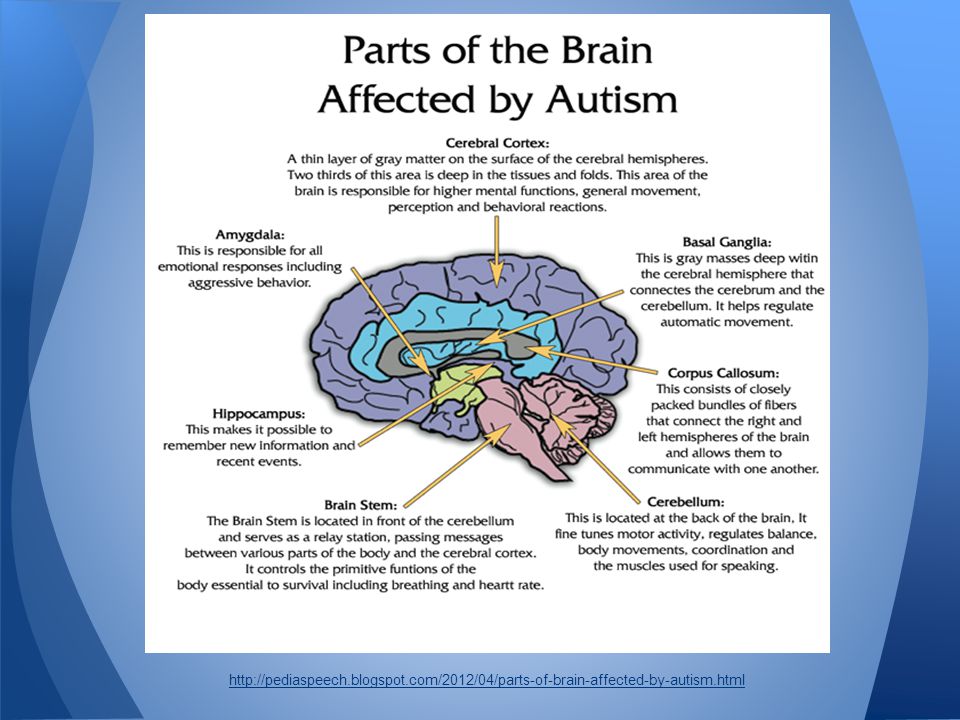
Last reviewed: June 2022
Back To Top
Related pages
- Your baby’s growth and development – first 12 months
Need more information?
Baby development milestones | Raising Children Network
Babies develop through relationships and play. Developmental milestones track changes in babies as they learn to move, see, hear, communicate and interact.
Read more on raisingchildren.net.au website
Reading to your child
Reading regularly with children from a young age stimulates their brain development and strengthens parent-child relationships. This helps to build language, literacy, and social-emotional skills
Read more on Pregnancy, Birth & Baby website
Crying Baby | Sydney Children's Hospitals Network
Crying is a normal part of your baby’s development and is normal for all babies from all cultural backgrounds
Read more on Sydney Children's Hospitals Network website
Physical activity and exercise for children
Here is trusted advice about how much daily exercise your child needs for good health and fitness and how you can encourage your child to be more active.
Read more on Pregnancy, Birth & Baby website
Child development: the first five years | Raising Children Network
The first five years of life are critical for child development. Find out how your child’s experiences and relationships shape the way your child develops.
Read more on raisingchildren.net.au website
Learn about children's development | StartingBlocks.gov.au
Get trusted information about your child’s developmental milestones from birth to 5 years.
Read more on Starting Blocks website
Toddler development - motor skills
Toddlers develop fast, exploring their world and doing things independently. Here's how to help your toddler develop fine and gross motor (movement) skills.
Read more on Pregnancy, Birth & Baby website
The first 1,000 days
The first 2 years of a baby's life can impact their health and wellbeing later on. Here's what you can do to give your child the best possible start.
Read more on Pregnancy, Birth & Baby website
Role modelling - Ngala
There are many wonderful developmental milestones during the toddler years and you will be surprised how fast they grow
Read more on Ngala website
Helping your baby grow from 0 to 5 years
Understanding how your baby will grow is important for all Aboriginal and Torres Strait Islander parents.
Read more on Pregnancy, Birth & Baby website
Disclaimer
Pregnancy, Birth and Baby is not responsible for the content and advertising on the external website you are now entering.
OKNeed further advice or guidance from our maternal child health nurses?
1800 882 436
Video call
- Contact us
- About us
- A-Z topics
- Symptom Checker
- Service Finder
- Linking to us
- Information partners
- Terms of use
- Privacy
Pregnancy, Birth and Baby is funded by the Australian Government and operated by Healthdirect Australia.
Pregnancy, Birth and Baby is provided on behalf of the Department of Health
Pregnancy, Birth and Baby’s information and advice are developed and managed within a rigorous clinical governance framework. This website is certified by the Health On The Net (HON) foundation, the standard for trustworthy health information.
This site is protected by reCAPTCHA and the Google Privacy Policy and Terms of Service apply.
This information is for your general information and use only and is not intended to be used as medical advice and should not be used to diagnose, treat, cure or prevent any medical condition, nor should it be used for therapeutic purposes.
The information is not a substitute for independent professional advice and should not be used as an alternative to professional health care. If you have a particular medical problem, please consult a healthcare professional.
Except as permitted under the Copyright Act 1968, this publication or any part of it may not be reproduced, altered, adapted, stored and/or distributed in any form or by any means without the prior written permission of Healthdirect Australia.
Support this browser is being discontinued for Pregnancy, Birth and Baby
Support for this browser is being discontinued for this site
- Internet Explorer 11 and lower
We currently support Microsoft Edge, Chrome, Firefox and Safari. For more information, please visit the links below:
- Chrome by Google
- Firefox by Mozilla
- Microsoft Edge
- Safari by Apple
You are welcome to continue browsing this site with this browser. Some features, tools or interaction may not work correctly.
The most detailed map of neural connections in the human brain has been created
02 June 2021 16:52 Olga Muraya
Color image of 4,000 axons transmitting nerve impulses to one neuron. nine0005 Google/Lichtman Laboratory illustration.
After long and painstaking work, researchers from Google and Harvard have published one of the most detailed 3D models of the human brain in the public domain.
The human brain has 86 billion neurons that communicate with each other through hundreds of trillions of synapses. It is a tangled network of neural connections, in the depths of which our consciousness, thoughts, feelings, memories and individuality are hidden. nine0003
The human brain is more complex than any computer that exists today. Therefore, the visualization of the full structure of all its connections looks, at first glance, a completely impossible task.
However, researchers around the world are working hard, literally picking up small areas of the human brain piece by piece. By the way, such a map of neural connections is called a "connectome", and the science that deals with the "cartography" of the nervous system is called connectomics.
In 2020, Google researchers collaborated with colleagues at the Howard Hughes Medical Institute to create a fruit fly brain connectome. Sounds like a pretty simple task, but so far they've only been able to map about half of the insect's brain. nine0003
Google and Harvard researchers recently released a similar model of the human brain. More precisely, its tiny area.
The researchers used a sample from the temporal lobe of the cerebral cortex as small as 1 mm 3 . It was dyed with special substances and cut into 5,300 layers about 30 nanometers thick. Then each of these layers was scanned using an electron microscope.
So the scientists obtained 225 million two-dimensional images, which were then "stitched" into a 3D model. nine0003
Different cells and their structures within the sample were identified using machine learning algorithms. The researchers only occasionally manually tested the accuracy with which the machines determined which cells belonged.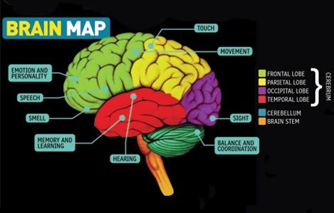
The end result has been named data set H01 and is one of the most complete maps of the human brain ever created. It contains information about 50,000 nerve cells, 130 million synapses, and, in addition, visualizes additional details: axons and dendrites of neurons, myelin and ciliary epithelial cells. nine0003
The most impressive thing about this dataset was that it took up a whopping 1.4 petabytes of memory. That's over a million gigabytes.
At the same time, Google claims that this is only one millionth of a complete map of the human brain.
It turns out that not only the work of mapping this entire volume, but also the search for a place to store this incredible array of information, will present a huge challenge. In addition, researchers have yet to find a way to organize the data obtained and provide convenient access to them. nine0003
In the meantime, the currently collected H01 dataset is available for online review.
The scientific paper accompanying this achievement was published on the bioRxiv preprint site.
Recall that earlier we wrote about creating a map of the brain showing where individual words are "stored" in it (by the way, it is also available online). We also reported that scientists have found similarities between the human brain and a swarm of bees.
More news from the world of science and technology can be found in the "Science" section of the "Looking" media platform. nine0003
science 3D nerve cells brain neurobiology society news
Previously related
-
How to get rid of an obsessive melody stuck in my head?
-
Strange voids first seen in the brains of people with migraines
-
Language proficiency can be determined by eye movement
-
Just one sound saves people from nightmares
-
Current and light: a way to restore brain cells was tested in Russia
-
Brain cells played a popular video game right in the test tube
Scientists have created a new map of brain development at an early age
The surface area of the human cerebral cortex increases very dynamically and inhomogeneously, starting from the third trimester of pregnancy and up to the age of 2 years. However, until now, little has been known about developmental patterns at this stage due to the lack of high-quality imaging data and accurate computational tools for processing brain tomograms in children. To fill this knowledge gap, researchers at the University of North Carolina School of Medicine used 1,037 high-quality MRI scans of children aged 29weeks of fetal development and up to 24 months after birth, created a map of brain development exactly according to models of cortical surface development. The results of a new mapping of cerebral cortex development, published in the Proceedings of the National Academy of Sciences, can be considered an important starting point for the study and understanding of dynamic early brain development in both normal and pathological conditions.
Map of the regionalization of the cerebral cortex, based on the dynamics of the development of the cortical surface, starting from 2
The development of the human brain is a complex and lengthy process that begins at conception and continues actively until the end of adolescence.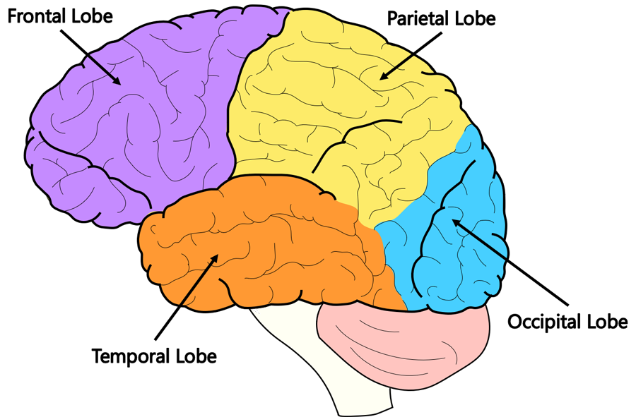 In particular, during the third trimester of pregnancy and in the early postnatal stages, the human brain undergoes exceptionally intensive development of cortical volume and cortical folding, which are associated with an increase in both surface area and cortical thickness. In fact, compared with an increase in the thickness of the cortex, its surface area expands much more dynamically and largely determines the growth of the brain in the perinatal and early postnatal periods. The early dynamic development of cortical surface area is critical to the development of cognitive abilities and behaviors that can be maintained throughout life. nine0003
Compared to the thickness of the cortex, its surface area has its own distinct genetic and cellular mechanisms and growth patterns. From this, the researchers hypothesized that the surface area of the cortex would form its own distinct regional area of development. In addition, abnormal early development of the cortical surface is closely associated with neurodevelopmental disorders and neuropsychiatric disorders, such as autism spectrum disorders and schizophrenia.
In their study, the researchers combined two widely recognized and complementary large-scale neuroimaging datasets (Developing Human Connectome Project and UNC/UMN Baby Connectome Project) containing a total of 1,037 high-resolution MRI scans from 735 children from 29 weeks gestation to age 2 years old The team analyzed the images using modern computer-aided image processing techniques. They divided the crustal surface into a virtual grid containing thousands of tiny regions and calculated the surface expansion rate for each of them. nine0003
Ultimately, the scientists were able to identify 18 areas that are bilaterally symmetrical and largely correspond to structurally and functionally significant areas, according to current knowledge in the field of neuroscience. They then applied the GAMM method to model the pattern of surface area development in each area.
They were able to show that the general developmental trajectories of both the left and right hemispheres in the same area are largely similar.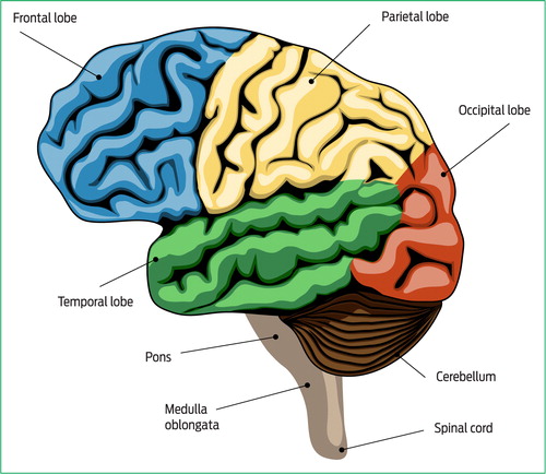 All regions show a dramatic increase in surface area with age, with each region showing its distinct development trajectory. Moreover, the growth rate of the surface area in the early stages is much higher than in the later ones. nine0003
Significant gender differences appear only in a few areas of both hemispheres, but males have, on average, slightly more surface area than females. In particular, for the left hemisphere in the caudal middle frontal, medial prefrontal, and inferior temporal gyrus, significant gender differences are present between 3.8 and 6.5 months of postnatal development, as well as in the isthmus of the cingulate gyrus, starting from 20.9 months of the postnatal period.
For the right hemisphere, significant gender differences exist in the posterior cingulate gyrus, isthmus of the cingulate gyrus, and dorsal superior frontal gyrus, starting at ages 6.5, 9.3 and 5.7 months after birth, respectively. Although the surface area of each region shows a dynamic increase with age in the perinatal and early postnatal stages, the rate of development differs sharply across regions.