Where is your pelvic area located
The pelvic area | Macmillan Cancer Support
The pelvis is the area of the body between the hip bones, in the lower part of the tummy (abdomen).
It contains:
- the sex organs
- the bladder
- a section of the small bowel
- the lower end of the large bowel (colon, rectum and anus).
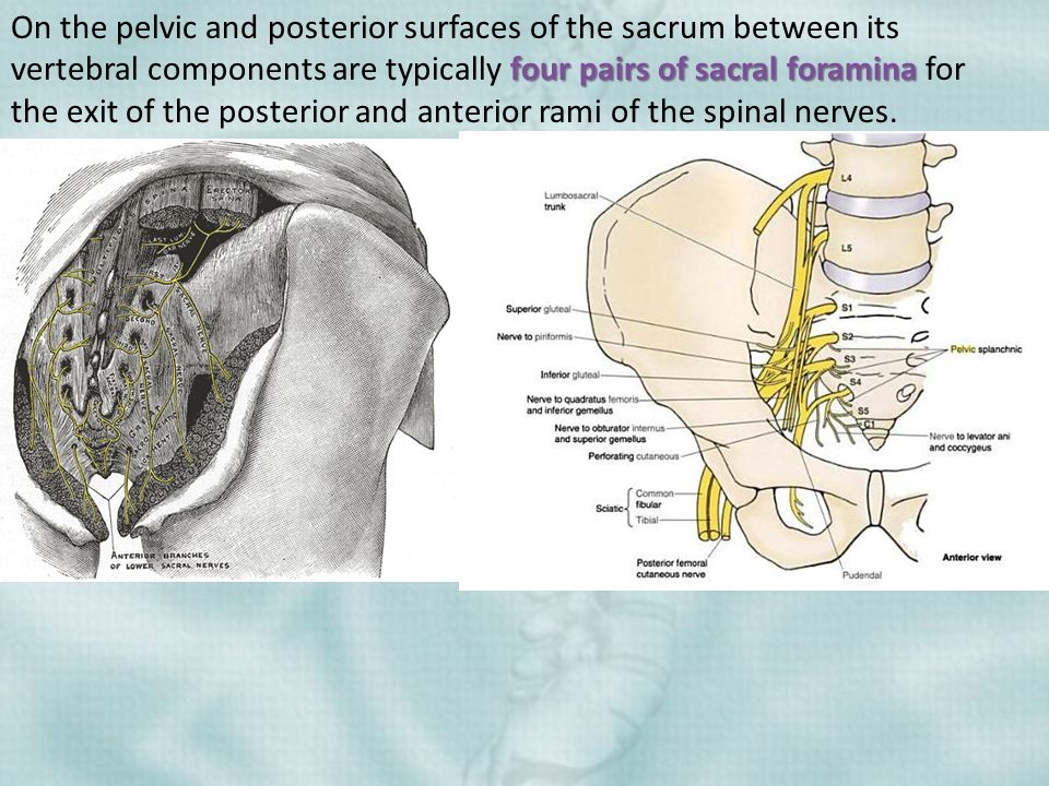
The pelvis also contains bones, lymph nodes (glands), blood vessels and nerves.
The male pelvis
In men, and people assigned male at birth, the sex organs include the prostate gland, testicles and penis.
Image: The pelvis
The female pelvis
In women, and people assigned female at birth, the sex organs include the ovaries, fallopian tubes, uterus (womb) and vagina.
Image: The pelvis
If you are transgender
Not all transgender (trans) people have had genital gender-affirming surgery. But if you have, you may not have all the sex organs you were born with. If you are not sure how this affects your symptoms, talk to your doctor or nurse. They can give you more information.
See also
- Pelvic radiotherapy
The bladder is in your pelvis and its job is to collect, store and pass urine (pee).
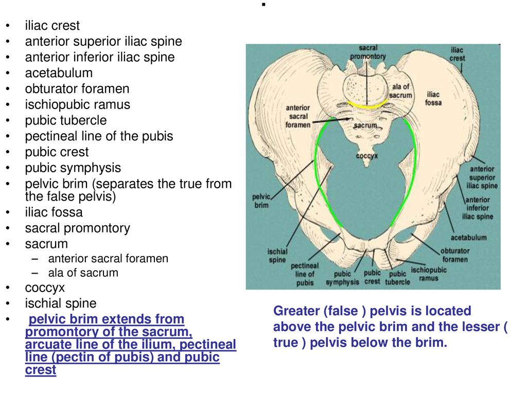
The kidneys produce urine. The kidneys are connected to the bladder by muscular tubes called ureters. The bladder is muscular and stretchy so that it can hold the urine until you feel the urge to go to the toilet.
When you need to pee, the urine exits your bladder through a tube called the urethra.
The bladder and urethra are supported by the pelvic floor muscles. The muscle that wraps around the urethra is called the urethral sphincter. It works like a valve to keep the opening at the bottom of the bladder closed until you want to pass urine.
When your bladder is full, it sends a signal to your brain that you need to go to the toilet. The pelvic floor muscles relax to open your urethral sphincter. At the same time, the bladder muscles tighten to push the urine out.
Image: The bladder and kidneys
See also
- The bladder
The bowel is part of the digestive system. It is divided into 2 parts:
- the small bowel
- the large bowel.
The large bowel is made up of the colon, rectum and anus.
Image: The bowel
When you swallow food, it passes down the gullet (oesophagus) to the stomach. This is where digestion begins.
The food then enters the small bowel, where nutrients and minerals are absorbed. The digested food then moves into the colon. This is where water is absorbed. The remaining waste matter (stool, or poo) is held in the rectum (back passage).
Nerves and muscles in the rectum help to hold onto stools until they are passed out of the body through the anus. The anus is the opening at the end of the large bowel. It contains a ring of muscle called the sphincter. This muscle helps to control when you empty your bowels.
-
Below is a sample of the sources used in our pelvic radiotherapy information. If you would like more information about the sources we use, please contact us at [email protected]
Andreyev HJN, Muls AC, Norton C, et al. Guidance: The practical management of the gastrointestinal symptoms of pelvic radiation disease. Frontline Gastroenterology, 2015; 6, 53-72.Dilalla V, Chaput G, Williams T and Sultanem K. Radiotherapy side effects: integrating a survivorship clinical lens to better serve patients.
 Current Oncology, 2020; 27, 2, 107-112.
 The Royal College of Radiologists. Radiotherapy dose fractionation. Third edition. 2019. Available from: www.rcr.ac.uk/system/files/publication/field_publication_files/brfo193_radiotherapy_dose_fractionation_third-edition.pdf [accessed March 2021].
-
This information has been written, revised and edited by Macmillan Cancer Support’s Cancer Information Development team. It has been reviewed by expert medical and health professionals and people living with cancer. It has been approved by Chief Medical Editor, Professor Tim Iveson, Consultant Medical Oncologist.
Our cancer information has been awarded the PIF TICK. Created by the Patient Information Forum, this quality mark shows we meet PIF’s 10 criteria for trustworthy health information.
Print page
Macmillan Cancer Support Line
The Macmillan Support Line is a free and confidential phone service for people living and affected by cancer.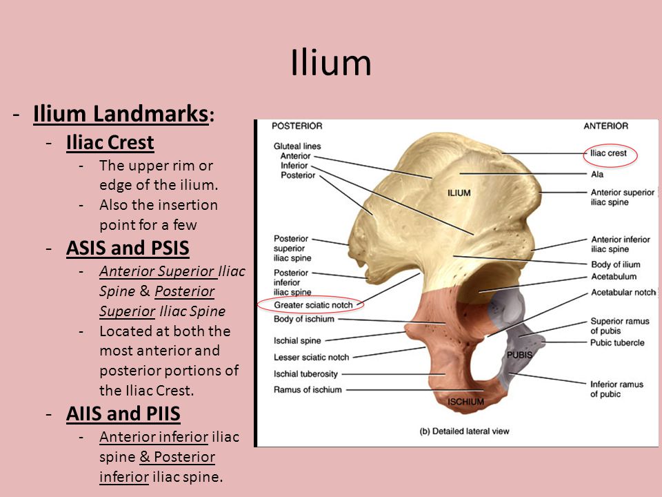 If you need to talk, we'll listen.
0808 808 00 00
Every day 8am - 8pm
Chat online
Every day 8am - 8pm
Get help
Anatomy, Function of Bones, Muscles, Ligaments
What is the female pelvis?
The pelvis is the lower part of the torso. It’s located between the abdomen and the legs. This area provides support for the intestines and also contains the bladder and reproductive organs.
There are some structural differences between the female and the male pelvis. Most of these differences involve providing enough space for a baby to develop and pass through the birth canal of the female pelvis. As a result, the female pelvis is generally broader and wider than the male pelvis.
Below, learn more about the bones, muscles, and organs of the female pelvis.
Female pelvis anatomy and function
Female pelvis bones
Hip bones
There are two hip bones, one on the left side of the body and the other on the right.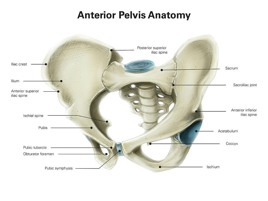 Together, they form the part of the pelvis called the pelvic girdle.
The hip bones join to the upper part of the skeleton through attachment at the sacrum. Each hip bone is made of three smaller bones that fuse together during adolescence:
- Ilium. The largest part of the hip bone, the ilium, is broad and fan-shaped. You can feel the arches of these bones when you put your hands on your hips.
- Pubis. The pubis bone of each hip bone connects to the other at a joint called the pubis symphysis.
- Ischium. When you sit down, most of your body weight falls on these bones. This is why they’re sometimes called sit bones.
The ilium, pubis, and ischium of each hip bone come together to form the acetabulum, where the head of the thigh bone (femur) attaches.
Sacrum
The sacrum is connected to the lower part of the vertebrae. It’s actually made up of five vertebrae that have fused together. The sacrum is quite thick and helps to support body weight.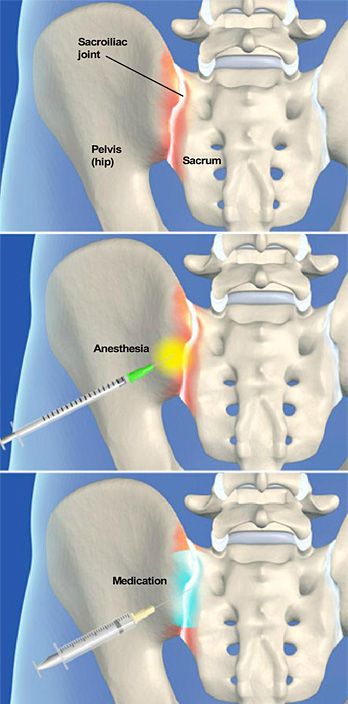
Coccyx
The coccyx is sometimes called the tailbone. It’s connected to the bottom of the sacrum supported by several ligaments.
The coccyx is made up of four vertebrae that have fused into a triangle-like shape.
Female pelvis muscles
Levator ani muscles
The levator ani muscles are the largest group of muscles in the pelvis. They have several functions, including helping to support the pelvic organs.
The levator ani muscles consist of three separate muscles:
- Puborectalis. This muscle is responsible for holding in urine and feces. It relaxes when you urinate or have a bowel movement.
- Pubococcygeus. This muscle makes up most of the levator ani muscles. It originates at the pubis bone and connects to the coccyx.
- Iliococcygeus. The iliococcygeus has thinner fibers and serves to lift the pelvic floor as well as the anal canal.
Coccygeus
This small pelvic floor muscle originates at the ischium and connects to the sacrum and coccyx.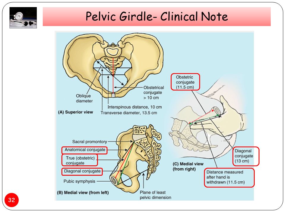
Female pelvis organs
Uterus
The uterus is a thick-walled, hollow organ where a baby develops during pregnancy.
During the reproductive years, the lining of the uterus sheds every month during menstruation if you don’t become pregnant.
Ovaries
There are two ovaries located on either side of the uterus. The ovaries produce eggs and also release hormones, such estrogen and progesterone.
Fallopian tubes
The fallopian tubes connect each ovary to the uterus. Specialized cells in the fallopian tubes use hair-like structures called cilia to help direct eggs from the ovaries toward the uterus.
Cervix
The cervix connects the uterus to the vagina. It’s able to widen, allowing sperm to pass into the uterus.
In addition, thick mucus produced in the cervix can help to prevent bacteria from reaching the uterus.
Vagina
The vagina connects the cervix to the exterior female genitalia. It’s also called the birth canal, as the baby passes through the vagina during delivery.
Rectum
The rectum is the lowest part of the large intestine. Feces collects here until exiting through the anus.
Bladder
The bladder is the organ that collects and stores urine until it’s released. Urine reaches the bladder through tubes called ureters that connect to the kidneys.
Urethra
The urethra is the tube that urine travels through to exit the body from the bladder. The female urethra is much shorter than the male urethra.
Female pelvis ligaments
Broad ligament
The broad ligament supports the uterus, fallopian tubes, and ovaries. It extends to both sides of the pelvic wall.
The broad ligament can be further divided into three components that are linked to different parts of the female reproductive organs:
- mesometrium, which supports the uterus
- mesovarium, which supports the ovaries
- mesosalpinx, which supports the fallopian tubes
Uterine ligaments
Uterine ligaments provide additional support for the uterus. Some of the main uterine ligaments include:
- the round ligament
- cardinal ligaments
- pubocervical ligaments
- uterosacral ligaments
Ovarian ligaments
The ovarian ligaments support the ovaries. There are two main ovarian ligaments:
- the ovarian ligament
- the suspensory ligament of the ovary
Female pelvis diagram
Explore this interactive 3-D diagram to learn more about the female pelvis:
Female pelvis conditions
The pelvis contains a large number of organs, bones, muscles, and ligaments, so many conditions can affect the entire pelvis or parts within it.
Some conditions that can affect the female pelvis as a whole include:
- Pelvic inflammatory disease (PID). PID is an infection that occurs in the female reproductive system. While it’s often caused by a sexually transmitted infection, other infections can also cause PID. Untreated PID can lead to complications, such as infertility or ectopic pregnancy.
- Pelvic organ prolapse. Pelvic organ prolapse occurs when the muscles in the pelvis can no longer support its organs, such as the bladder, uterus, or rectum. This can cause one or more of these organs to press down on the vagina. In some cases, this can cause a bulge to form outside of the vagina.
- Endometriosis. Endometriosis occurs when the tissue that lines the inside walls of the uterus (endometrium) begins to grow outside of the uterus. The ovaries, fallopian tubes, and other tissues in the pelvis are typically affected by the condition. Endometriosis can lead to complications, including infertility or ovarian cancer.
Symptoms of a pelvic condition
Some common symptoms of a pelvic condition can include:
- pain in the lower abdomen or pelvis
- a feeling of pressure or fullness in the pelvis
- unusual or foul-smelling vaginal discharge
- pain during sex
- bleeding in between periods
- painful cramping during or before periods
- pain during bowel movements or when urinating
- a burning feeling when urinating
Tips for a healthy pelvis
The female pelvis is a complex, important part of the body. Follow these tips to keep it in good health:
Stay on top of your reproductive health
See a gynecologist for a yearly health screen. Things like pelvic exams and Pap smears can aid in identifying pelvic conditions or infections early.
You can get a free or low-cost pelvic exam at your local Planned Parenthood clinic.
Practice safe sex
Use barriers — such as condoms or dental dams — during sexual activity, especially with a new partner, to avoid infections that could lead to PID.
Try pelvic floor exercises
These types of exercises can help to strengthen the muscles in the pelvis, including those around the bladder and vagina.
Stronger pelvic floor muscles can aid in preventing things like incontinence or organ prolapse. Here’s how to get started.
Never ignore unusual symptoms
If you’re experiencing anything unusual in your pelvic area, such as bleeding between periods or unexplained pelvic pain, make an appointment with your doctor.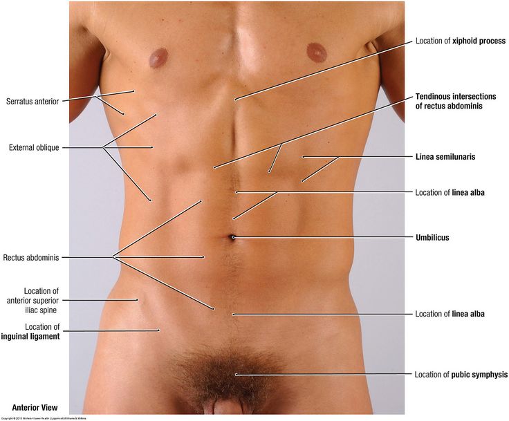 Left untreated, some pelvic conditions can have lasting impacts on your health and fertility.
Pelvis / Female Anatomy
Pelvis
The small pelvis is the lower half of the pelvis of a woman, in which the uterus with appendages, the bladder and rectum, blood vessels and nerves are located. The organs of the small pelvis are fixed to the walls of the pelvis with the help of ligaments and fascia. For ease of understanding, this design can be compared to a suspension bridge.
Front and back - bridge supports (pubis and sacrum). A bridge (vagina) is stretched between them. Ropes (bundles) stretch from the upper part of the supports to the bridge. From the back support (sacrum) - sacro-uterine ligaments. From the anterior support (pubis) - pubo-urethral ligaments.
The bridge (vagina) can be compared to the membrane of a trampoline, and the ligaments of the pelvis to its springs.
The bladder lies on the anterior wall of the vagina like in a hammock.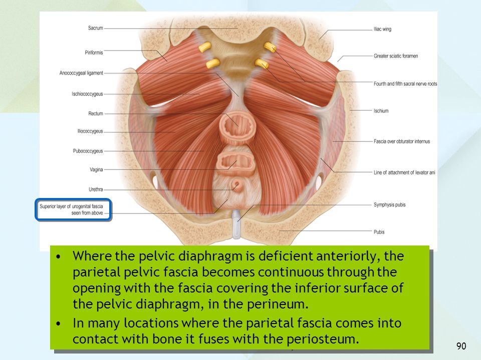
The main pelvic fascia is the pubocervical fascia. This is a connective tissue sheet between the anterior wall of the vagina and the bladder. This fascia is the hammock, trampoline, or suspension bridge. She fascia is stretched from front to back with the help of the described ligaments, and on the sides is attached to the walls of the pelvis.
Defects in the ligamentous and/or fascial structures that occur during childbirth lead to various types of prolapse of the pelvic organs.
The most important supporting ligament is the sacro-uterine, which fixes the entire structure from above. With its defect, the connection between the bridge and its rear support is lost. The pubocervical fascia, having lost support from above, breaks away from the walls of the pelvis. There is a prolapse of the uterus and the anterior wall of the vagina. Therefore, the most common type of prolapse (omission) is anteroapical (a combination of prolapse of the uterus and the anterior wall of the vagina).
The pubo-urethral ligament plays an important role in the urinary continence mechanism. It fixes the urethra in the middle third, limits its mobility. A defect in this ligament causes abnormal mobility of the urethra and disrupts the mechanism of urinary retention.
Corresponding to this anatomical organ:
- Diseases
- Endometriosis
- Uterine prolapse
- Rectocele
- Operations
- Infiltrate resection
- Surgery for complications
- Enterocele correction
- Rectocele correction
- Correction of the entrance to the vagina
- Laparoscopic sacrovaginopexy
- OPUR
- Sacrospinal hysteropexy
How to correct pelvic tilt back
November 6, 2017HealthSports and fitness
Back tilt of the pelvis often occurs due to long sitting and entails pain, tension and various diseases of the spine. A detailed guide with exercises will help correct this posture disorder.
Iya Zorina
Author of Lifehacker, athlete, CCM
Share
0Why is this a problem
The condition of your lower back depends on the tilt of the pelvis. When the pelvis is in a neutral position, the back retains normal physiological curves, when the pelvis is tilted forward, an excessive deflection is created in the lower back, and when the pelvis is tilted back, the lower back becomes flat.
Neutral position and various pelvic tiltsAll physiological curves of the spine are necessary for a healthy back, and if one of them disappears, this negatively affects all departments, including the chest and cervical.
A flat lower back impairs cushioning, so stress on the spine can result in soreness, protrusion and herniation, nerve root problems, stiffness and muscle pain.
Why there is a backward tilt of the pelvis
The main causes of this disorder are a long time spent sitting and incorrect body position.
If you keep the wrong position for 6-8 hours a day, your body will adapt to it. As a result, some muscles become too stiff, others too stretched and weak.
Stiff muscles Weak musclesStiff muscles pull the pelvis along and tilt it back, not only when you are sitting, but also when you stand up, walk or squat.
How to tell if you have a posterior pelvic tilt
Two finger test
Stand up straight, place one finger on the protruding pelvic bone in front and the other finger on the pelvic bone in the back. If your pelvis is tilted back, the toe on the anterior pelvic bone will be significantly higher than the toe on the back.
Movement test
Stand in front of a mirror or have a partner take a picture of you to assess your posture from the side. Lean forward, do a squat, or just sit on a chair.
Rounded back during squatsIf your lower back rounds while doing this, the cause may be a backward tilt of the pelvis.
Wall test
Wall test Sit next to a wall with your back pressed against it and legs extended forward.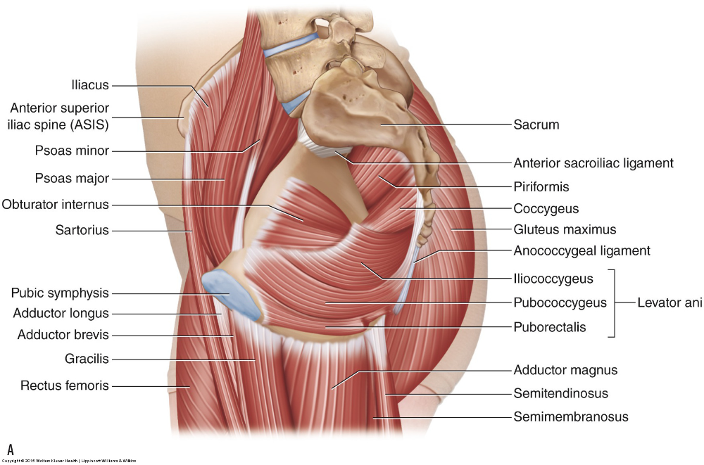 If you can't straighten your legs without twisting your lumbar spine, you have a posterior pelvic tilt.
How to correct the tilt of the pelvis back
Correction of posture requires complex measures. We will show you how to stretch and relax tight muscles, how to activate and strengthen weak ones, open your hips and find the right sitting position.
Stretching and Relaxing
1. Biceps Stretch
Left lower biceps stretch, right upper- Stand up straight with hands on hips.
- Extend your leg forward, either straight or slightly bent at the knee.
- Lean forward with a straight back.
- Hold the position for 60 seconds and repeat on the other leg.
If you bend your knee while stretching, the upper part of the hamstring is stretched, if you fully extend the leg, the lower part is stretched.
2. Glute Stretch
Glute Stretch- Lie on your back, bend your knees and place your feet flat on the floor.

- Place your right ankle on your left knee.
- Grab your right knee and pull it closer to your chest.
- Feel the stretch in the right gluteal muscle.
- Arch your back slightly to increase the stretch.
- Hold the position for 60 seconds on each side.
3. Rectus Abdominis Stretch
Abdominal Stretch
Rectus Abdomen StretchIf you have lower back problems, skip this exercise and move on to the next one.
- Lie on your stomach, place your hands on the floor under your shoulders, and straighten your elbows.
- Arch your back.
- Feel the stretch in the abdominal muscles.
- You can rotate the body slightly from side to side to improve stretch.
- Use diaphragmatic breathing during the exercise.
- Hold the position for 60 seconds.
Standing Stretch
Raised Arm Pull This exercise is safer for the lower back.
- Stand up straight, raise your arms and join your palms.
- Tighten the gluteal muscles and hold the tension until the end of the exercise: this will protect the lower back from excessive arching.
- Arch your chest and move your arms as far back as possible.
- Gently return to the starting position and repeat five times.
Roller Roller
1. Biceps Femoris
Roller Roller Hammer Roller- Place a massage roller or ball under the back of the thigh of one leg, place the other leg on top to increase pressure.
- Lean on a roller or ball with body weight and slowly roll your thigh from knee to pelvis.
- Do this for 60 seconds, then switch legs.
2. Gluteal Muscles
Glute Roller Roller- Sit on the massage ball or roller and press it to the floor with your body weight.
- Place the ankle of one foot on the knee of the other.
- Roll the muscle for 60 seconds.

- Change sides and repeat.
Muscle Activation
To return the pelvis to a neutral position, weak "sleeping" muscles must be activated to pull the pelvis forward.
1. Seated Knee Raise
Seated Knee RaiseThis exercise activates the hip flexors.
- Sit on a chair or fitness ball with your back straight.
- Raise one knee.
- Hold it down for five seconds.
- Lower and repeat with the other leg.
- Do 30 reps on each leg.
To make the exercise more difficult, you can use an expander.
2. Superman
Superman ExerciseThis exercise will help activate the muscles in your lower back.
- Lie on your stomach.
- Extend your arms out in front of you.
- Raise your upper body and legs.
- Hold the position for 5-10 seconds.
- Repeat 30 times.
Muscle strengthening
1. Back arch exercise
Back arch exercise- Get on all fours.
- Arch your back so that your pelvis rolls forward.
- Hold the position for 10 seconds.
- Return to neutral position.
- Repeat 30 times.
2. Sitting back arch exercise
Seated back arch exercise- Sit on a chair or fitness ball with your back straight.
- Arch your back at the waist and twist your pelvis forward.
- Hold the position for 10 seconds, then relax and repeat.
- Perform the exercise 30 times.
This exercise can be done with or without a band.
3. Core Stretch
Bring Knee to Chest- Get on all fours with your pelvis in a neutral position.
- Tighten your abdominal muscles.
- Keeping the natural arch of the lower back, try to bring the knee to the chest. Stop the movement as soon as the lower back begins to round.
- Hold this position for 5 seconds, then lower your leg.
- Repeat 20 times and repeat with the other leg.
Hip opening
Tight hips make it difficult to maintain a neutral pelvis. Therefore, if you do not have enough mobility of the hip joint, you need to develop it.
1. Hip stretch
All four hip stretch- Get on all fours.
- Place your right ankle behind your left knee as shown in the photo.
- Maintain a natural arch in your lower back throughout the exercise.
- Pull your pelvis back, stretching your hip.
- Hold the position for 20 seconds and then relax.
- Repeat five times.
2. Joint Mobilization
Joint Mobilization- Loop the resistance band over the thigh close to the pelvis.
- Hook the other end to a stable object.
- Lie on your back away from the object to create resistance.
- Bring knee to chest, hold for 60 seconds, then switch legs and repeat.
- Perform 10 repetitions on each leg.
3. Butterfly Stretch
Wall Stretch- Sit on the floor next to a wall with your back against it.


