Signs of blindness in newborn babies
Red flags: Signs that your baby may have a vision problem
- Community
- Getting Pregnant
- Pregnancy
- Baby names
- Baby
- Toddler
- Child
- Health
- Family
- Courses
- Registry Builder
- Baby Products
Advertisement
Photo credit: Thinkstock
It takes your baby's eyes some time to adjust to the world, so at first they might not always look or function the way you expect.
For example, it's perfectly normal in the first three months of life for your infant's eyes to be crossed, or for him not to be able to see much past your face when you're holding him.
Certain signs could indicate a problem. Talk with your baby's doctor if you notice any of the following:
- Your baby's eyes don't move normally. One moves and the other doesn't, for example, or one looks different from the other when moving.
- Your baby is older than 1 month, but lights, mobiles, and other distractions still don't catch his attention.
- One of your baby's eyes never opens.
- Your baby has a persistent, unusual spot in her eyes in photos taken with a flash. Instead of the common red-eye caused by camera flash, for example, there's a white spot.
- You notice white, grayish-white, or yellow material in the pupil of your baby's eye. (His eyes look cloudy.)
- One (or both) of your baby's eyes is bulging.
- One or both of your baby's eyelids seem to be drooping.
- Your baby squints often.
- Your baby rubs her eyes often when she's not sleepy.
- Your baby's eyes seem sensitive to light.
- One of your baby's eyes is bigger than the other, or the pupils are different sizes.
- You notice any other change in his eyes from how they usually look.
In addition, once your baby is 3 months old, talk with the doctor if you notice any of the following:
- Your baby's eyes turn way in or out, and stay that way.
- Your baby's eyes don't follow a toy moved from side to side in front of her.
- Your baby's eyes seem to jump or wiggle back and forth.
- Your baby seems to consistently tilt his head when he looks at things.
You'll also want to have the doctor check your baby's eyes if they show any signs of a blocked tear duct or infection, such as pinkeye. These signs include excessive tearing, redness that lasts more than a few days, or pus or crust in her eyes.
Your baby's doctor can help you determine whether you should be concerned. The doctor may examine your child's eyes, screen his vision, or refer you to a medical eye specialist (ophthalmologist). If vision problems run in your baby's family, be sure to mention it.
See our complete article on strabismus (misaligned eyes) and amblyopia (lazy eye).
Before the next well-baby checkup, check out our complete article on what to expect when the doctor examines your baby's eyes.
Karen Miles
Karen Miles is a writer and an expert on pregnancy and parenting who has contributed to BabyCenter for more than 20 years.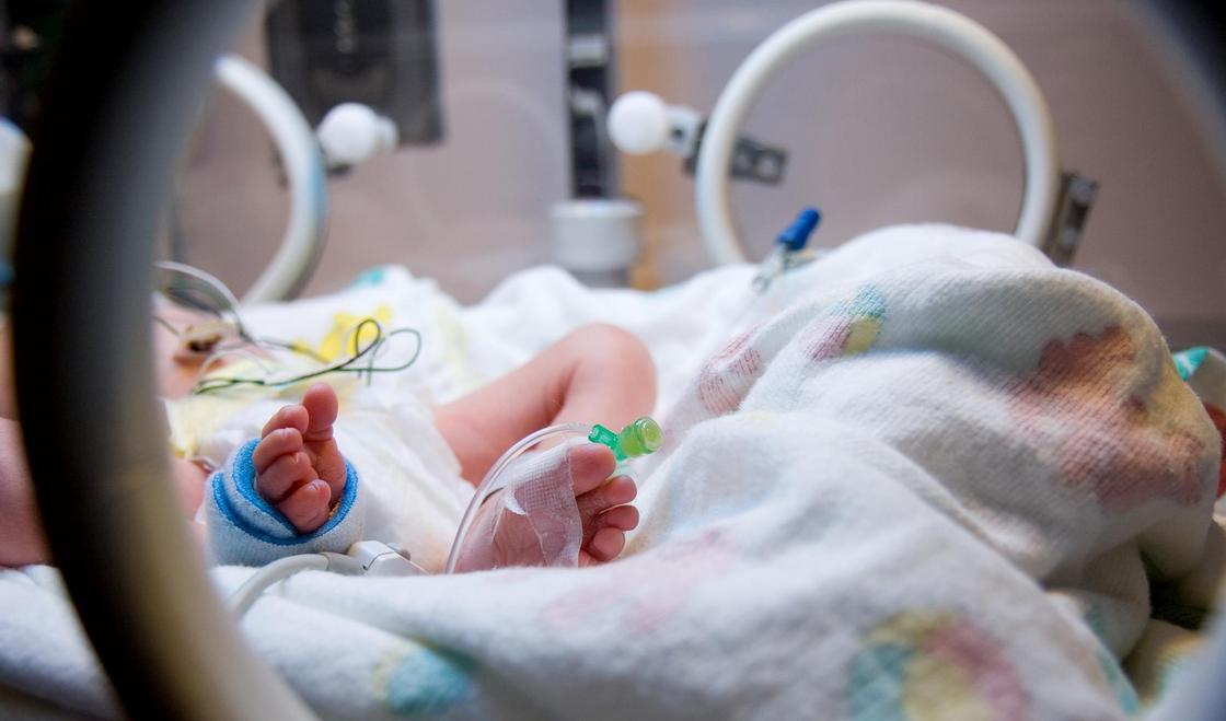 She's passionate about bringing up-to-date, useful information to parents so they can make good decisions for their families. Her favorite gig of all is being "Mama Karen" to four grown children and "Nana" to eight grandkids.
Warning Signs of Vision Problems in Infants & Children
Log in | Register
Health Issues
Health Issues
Listen
Español
Text Size
Eye exams by your child's doctor are an important way to identify problems with your child's vision. Problems that are found early have a better chance of being treated successfully.
What are warning signs of a vision problem?
Babies up to 1 year of age:
Babies older than 3 months should be able to follow or track an object, like a toy or ball, with their eyes as it moves across their field of vision.
 If your baby can't make steady eye contact by this time or seems unable to see, let your child's doctor know. See Infant Vision Development: What Can Babies See? for more information.
Before 4 months, most babies' eyes occasionally look misaligned (strabismus). However, after 4 months, inward crossing or outward drifting that occurs regularly is usually abnormal. If one of these is present, let your child's doctor know.
Preschool age:
If your child's eyes become misaligned, let your child's doctor know right away. However, vision problems such as a lazy eye (amblyopia) may have no warning signs, and your child may not report vision problems. That is why it's important at this time to have your child's vision checked. There are special tests to check your child's vision even if he cannot yet read.
All children:
If you notice any of the following signs or symptoms, let your child's doctor know:
Eyes that are misaligned (look crossed, turn out, or don't focus together)
White or grayish white color in the pupil
Eyes that flutter quickly from side to side or up and down
Eye pain, itchiness, or discomfort reported by your child.
Redness in either eye that doesn't go away in a few days
Pus or crust in either eye
Eyes that are always watery
Drooping eyelids
Eyes that often appear overly sensitive to light
Additional Information:
Vision Screenings
Specific Eye Problems in Children
Visual System Assessment in Infants, Children, and Young Adults by Pediatricians (AAP Policy Statement)
Procedures for the Evaluation of the Visual System by Pediatricians (AAP Clinical Report)
- Last Updated
- 7/19/2016
- Source
- Your Child’s Eyes (Copyright © 2011 American Academy of Pediatrics, Updated 05/2016)
The information contained on this Web site should not be used as a substitute for the medical care and advice of your pediatrician. There may be variations in treatment that your pediatrician may recommend based on individual facts and circumstances.
Vision in infants: the formation of the visual system after birth.
From All About Vision
Your heart skips a beat when your newborn baby opens its eyes and looks at you for the first time.
Don't worry if it doesn't happen right away. In newborns, the visual system develops gradually.
In the very first week of life, a child sees the world differently: indistinctly and in shades of gray.
Only a few months after birth, the baby's visual system will work to its full potential. Knowing the key milestones in the development of the eyes of a newborn baby (and how to help them along) will help you understand that your baby is developing normally and enjoying life.
Baby's eye development begins during pregnancy
Your baby's visual system begins to develop even before birth. Therefore, how you take care of yourself and your body during pregnancy is important and affects the development of the body and mental abilities of the child, including the eyes and visual centers in the brain.
Be sure to follow the nutritional advice your doctor gives you. Be sure to take your prescribed supplements and vitamins, and get enough rest.
Do not smoke or drink alcohol during pregnancy, as toxins can cause many health problems for the baby, in particular vision problems.
Smoking is especially dangerous during pregnancy. Cigarette smoke contains about 3,000 different chemicals (such as carbon monoxide or carbon monoxide) that can harm a person.
Even regular aspirin can be dangerous: it increases the risk of having a low birth weight baby and complications during childbirth. Low body weight, in turn, can contribute to vision problems.
Always check with your doctor before taking any medication during pregnancy. This also applies to over-the-counter drugs, herbal supplements, and other over-the-counter medications.
Eye Condition at Birth
Shortly after birth, your pediatrician or neonatologist will examine your baby's eyes for congenital cataracts or other serious neonatal eye problems in newborns.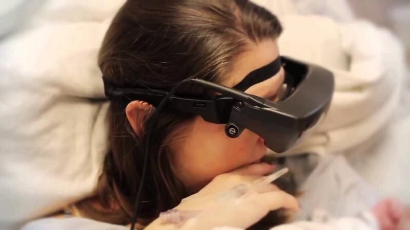
Although such diseases are rather an exception, it is better to detect and treat them at an early stage in order to reduce the pathological impact on the development of the infant's visual system.
To protect the child's eyes from pathogenic bacteria and microorganisms that could enter them during the passage through the birth canal and cause eye infection , an antibiotic ointment is placed in them. Early prevention of possible eye infections is very important for the normal development of the visual system.
ARE YOU CONCERNED WITH YOUR BABY'S VISION? Find an optometrist nearby .
At birth, your baby sees objects only in black and white and in shades of gray. This is due to the fact that the nerve cells in the retina and brain that are responsible for color perception are not yet fully developed.
A newborn baby is not yet able to focus on nearby objects (disturbance of accommodation). Don't worry if you notice that your baby is unable to "focus" on objects or on your face. It will take time for him to acquire this ability.
Despite all these features, studies have shown that just a few days after birth, the baby is able to distinguish the mother's face from the face of a stranger.
Scientists believe that the baby recognizes the mother's face thanks to the contrasting hairline. (During the study, women covered their hair with a scarf or swimming cap, and the baby could not tell the mother's face from that of a stranger.)
Therefore, to encourage eye contact, during the first weeks of a child's life, do not change the hairstyle or appearance.
You may also notice that babies have rather large eyes. This is because usually a child's head grows first, and then the rest of the body. At birth, a baby's eyes are 65% the size of an adult's eyes!
Baby's eyes in the first month of life
In the first month of life, the baby's eyes are not highly sensitive to light.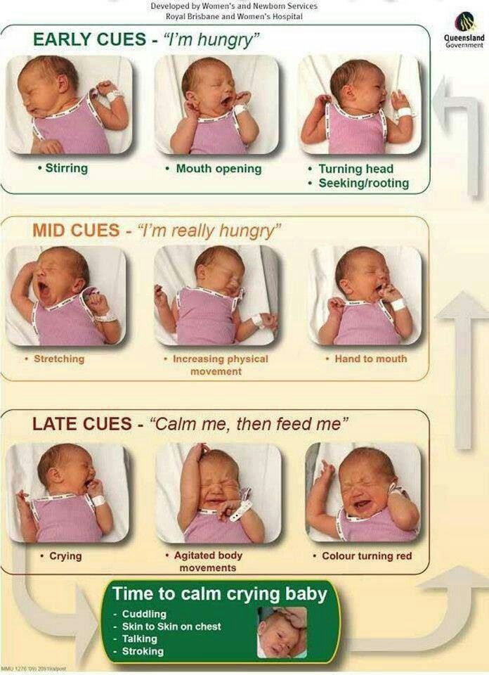 By the way, in order for a child who is 1 month old to understand that there is light in the room (the threshold of sensitivity to light), the light must be 50 times brighter than normal.
Don't be afraid to leave a light on in the nursery, because it won't disturb the baby's sleep and you won't trip over the furniture when you go to visit him at night!
Very quickly the baby acquires the ability to distinguish colors. A week after birth, the baby can see red, orange, yellow and green. A little later, he will be able to distinguish between blue and purple. This is because blue light is the shortest wavelength, and there is only one type of receptor in the retina that can "see" blue light.
Don't worry if you notice that your baby's eyes are looking in different directions. One eye may sometimes be slightly averted to one side or the other. This is fine. But if the baby's eyes squint to the side, consult an optometrist immediately.
Tips: Paint your baby's room in bright, cheerful colors to stimulate his eyesight. Furniture or interior details should be of contrasting colors and shapes. Hang a bright, colorful hanging piece above or next to your crib. It is important that it be multi-colored and consist of different geometric shapes.
Development of the organs of vision: 2nd and 3rd month of life
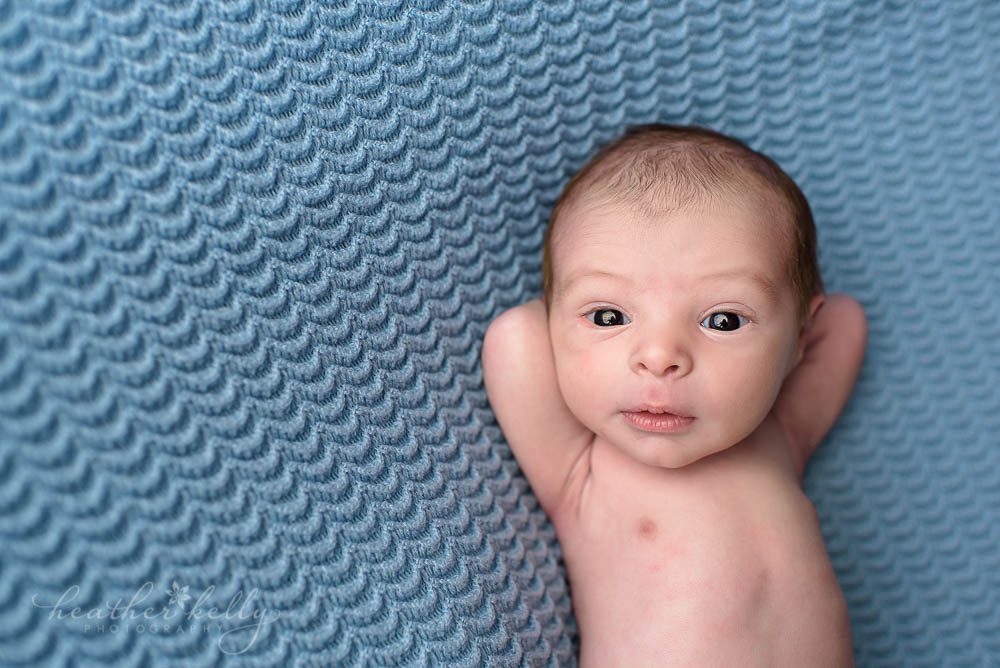
Significant changes occur in the baby's visual system during the second and third months of life. During this period, visual acuity increases , functional strabismus (if it was) disappears. Now your child is able to follow moving objects and tries to fix his eyes on them.
A bright, bright room with lots of colors and shapes to help stimulate your baby's vision development.
Moreover, the baby learns to look from one object to another only with his eyes, without moving his head. Also, the child's eyes become more sensitive to light: at the age of 3 months, the threshold of sensitivity to light decreases. Therefore, during sleep, the light should be dimmed.
Tips: To stimulate the eyesight of a 2-3 month old baby:
-
Add new objects to the room or change their position frequently to enrich the baby's experience.
-
Leave a night light on to stimulate vision when your baby is awake in the crib.
-
Place the baby on its back during sleep to reduce the risk of sudden infant death syndrome (SIDS). However, when the baby is awake and under supervision, put it on the tummy. This posture stimulates the development of visual perception and motor skills.
Development of the organs of vision: Months 4-6
By 6 months, the visual centers of the baby's brain are quite well developed. The child sees more clearly and can follow moving objects with quick and precise eye movements.
Visual acuity improves from about 20/400 (6/120) at birth to 20/25 (6/7.5) at 6 months. Color perception reaches the level of an adult; the child can distinguish all the colors of the rainbow.
At the age of 4-6 months, the child's hand-eye coordination becomes more perfect, which allows him to quickly find and pick up objects with his hands, as well as accurately guide the bottle (and more!) into his mouth.
Six months is an important milestone in life, as this is when you should have your baby's first eye exam.
If necessary, a qualified eye specialist will examine a child at 6 months of age. But routine eye exams are usually recommended after kindergarten age (3-4 years and older).
If you have any concerns, you can also consult your doctor for further advice.
For a thorough examination of your six-month-old baby's eyes, see an optometrist who specializes in children's vision and eye development.
Eye Development: Months 7-12
Your baby is now moving around a lot, crawling and covering distances you never dreamed possible. He is getting better at estimating the distance to objects, more precisely grabbing and throwing them. (Beware!)
This is an important period in your child's development. At this stage, the baby feels his whole body better and learns to coordinate vision and movement.
Now you have to pay more attention to the baby to keep him out of harm's way.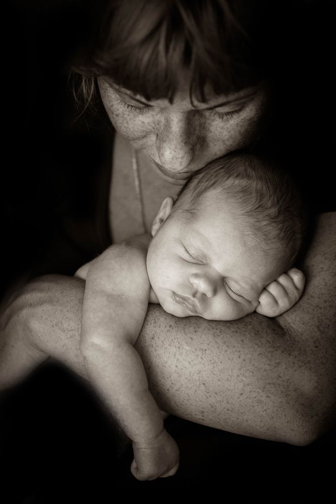 It is not uncommon for bumps, bruises, eye injuries and other serious injuries when the baby explores the world around him.
Close cabinets with household chemicals with a padlock, place special railings in front of stairs.
Don't worry if your child's eyes start to change color. Most babies are born with blue eyes because at birth there are not enough dark pigments in the iris. Over time, there will be many more of them, so your baby's eyes may change color from blue to brown, green, gray or swampy.
Tips: To stimulate your child's hand-eye coordination, you can lie on the floor with him and invite him to crawl to an object. Put your baby's favorite toy away and invite him to get to it. You can also offer to disassemble and assemble various objects and toys.
Strabismus problems
Pay close attention to how your child's eyes move, either together or separately. Strabismus is a term for a misalignment of the eyes. It is very important to detect and treat it at an early stage so that the child's vision develops properly.
Left untreated, strabismus can lead to amblyopia or "lazy eye."
It may take months for a child to achieve joint eye movement. But if you notice that one of the baby's eyes is squinting or moving separately from the other, contact an optometrist immediately.
Vision problems in premature babies
The average duration of a normal pregnancy is approximately 40 weeks (280 days). According to the WHO, a baby born before 37 weeks is considered premature.
Compared to full-term babies, premature babies are at greater risk of developing eye problems. And the shorter the period, the more serious the complication.
The following visual problems associated with preterm birth are characteristic:
Retinopathy of prematurity (RP)
This is a disease in which fibrous changes and blood vessels develop in the thickness and on the surface of the retina. ROP is often accompanied by retinal scarring, poor vision and retinal detachment . In severe cases, retinopathy of prematurity can lead to blindness.
All premature babies are at risk of ROP. Extremely low birth weight is an additional risk factor, especially when the baby is placed in a high oxygen incubator immediately after birth.
If your baby was born prematurely, ask your obstetrician for a referral to a pediatric ophthalmologist for an eye exam to rule out ROP.
Nystagmus
These are involuntary oscillatory eye movements.
In most cases, nystagmus manifests itself in the form of involuntary eye movements in various directions, with different frequencies and amplitudes of "oscillation". Eye oscillations are pendulum in nature, horizontal, diagonal and rotational movements are observed.
Nystagmus may be congenital or may develop over several weeks or months. Risk factors include optic nerve hypoplasia, albinism, and congenital cataracts. By the amplitude of eye oscillations, you can determine how badly the baby's visual system is damaged.
If the child shows signs of nystagmus, consult an optometrist immediately.
Remember that smoking during pregnancy greatly increases the chance of preterm labor.
Page published on Monday, November 16, 2020
norm and pathology - Articles
Vision is the most informative and at the same time the most fragile external analyzer. It is with the help of the eyes that a person receives most of the information about the world around him. The visual centers are connected and have a strong influence on almost all vital structures of the brain (vision is involved in the digestive, motor, vestibular, sexual and other activities of the body). Especially important in the formation and development of vision is the first year of life, when the eyes and the child's body as a whole are easily susceptible to various harmful effects of both internal and external factors.
If at this age the visual organ is damaged, then the child develops movement coordination disorders, the baby experiences fear of the outside world, which often leads to a significant lag in the development of the child, since the rest of the senses are not able to fully compensate for the lack of information.
In the first year, the child's vision develops very intensively. The almost complete blindness of a newborn baby (non-directional light perception) in a few months develops into the ability to analyze objects and their movement, evaluate and compare objects according to their various characteristics, including color. Therefore, it is especially important for parents to understand the basic principles of vision development in children of the first year of life and to be aware of the early signs of the onset of eye pathology.
Development of the visual system
There are several periods in the development of the organ of vision. The most important of them is the laying and intrauterine formation. At this stage, the action of damaging factors can lead to catastrophic consequences (developmental anomalies - hypoplasia of the optic nerves, congenital cataract, glaucoma; inflammation of the membranes of the eye; etc.). The next period is from birth to 1 year. At this time, areas of the visual cortex of the brain are actively developing, receiving information about the world around them. The simultaneous movement of the eyes is trained, the visual control of the movement of the hands is formed, the "library" of visual images is filled. If at this stage there is a restriction in the flow of light to the retina (violation of the transparency of the optical media of the eye), a violation of the focus of objects (the presence of myopia or a high degree of hyperopia) or a deterioration in the perception of visual images (damage to the optic nerves, visual centers of the brain), then vision may stop at the initial stage of development and not form to a normal level.
Immediately after birth, the child is only able to perceive the presence or absence of a light source. In the first months of life, various objects of the surrounding world appear in front of the child, as if from a fog. At first, the baby only fixes his gaze on large objects (the first month), then he tries to trace their movement in space - he studies the parents passing by, follows the moving toys (3-4 months). At this age, you should not hang toys directly in front of your eyes - place them on the sides of the child or on your feet. At 6 months, the child's visual acuity allows him to observe small objects, visually recognize "their own", grab and throw toys, while learning the three-dimensionality of space. Place rattles and "rattles" in the area of the movement of the child's hands to facilitate their capture.
A one-year-old baby is already collecting “little rubbish” on the floor, actively moving towards a bright toy. Use distant objects to attract attention. Receiving powerful visual stimuli, the baby begins to strive for the objects of interest to him, makes attempts to stand up and takes his first steps.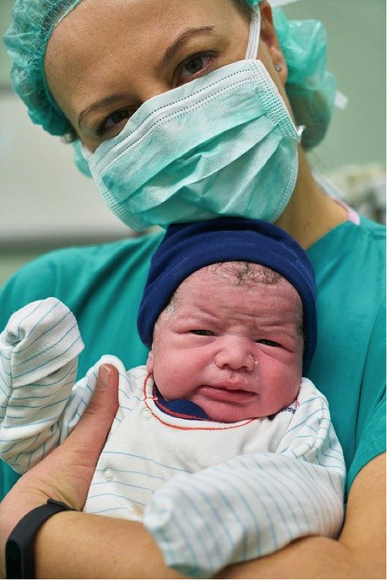 Only by the age of 6-7 years does the child's vision reach the level of an adult (according to special tables, he names the 10th line).
Parental control
Even in the maternity hospital, a visual examination of a newborn can reveal signs of some congenital eye diseases. A cataract is a clouding of the lens that appears as a grayish glow instead of a black pupil. The most common treatment is surgical removal of the cloudy lens. The prolonged existence of interference with the passage of light into the eye will lead to a significant delay in the development of vision (obscurative ambiopia). After such an operation, the child wears special glasses or a contact lens that replaces the lens. Recently, the technique of early implantation of an artificial lens has been widespread. Some types of translucent cataracts are not operated on in early childhood. In such cases, periodic courses of stimulating treatment are carried out (exposure to the eye with light and laser radiation, electric and magnetic fields, classes on special computer programs) and delayed surgical intervention is performed at an older age.
External manifestations similar to cataracts can be detected in a more dangerous disease - retinoblastoma (retinal tumor). In the early stages, the tumor can be affected with the help of special radiation applicators - plates with a radioactive substance applied to them. They are sutured directly to the sclera at the site of the projection of the tumor, the shadow of which is determined during the operation, transilluminating the sclera with a diaphanoscope (a device similar to a flashlight). The radioactive material of the applicator destroys the tumor through the sclera. In the later stages, when there is a danger of the tumor spreading outside the eye, there is only one way - removal of the affected eye.
Congenital glaucoma is an eye disease characterized by an increase in intraocular pressure due to congenital disorders in the formation and outflow of intraocular fluid. As a result, the child's eye stretches and increases in size, moving forward (up to limiting the complete closure of the eyelids). Also, with glaucoma, there may be clouding of the cornea (leukoma). Since this disease is associated with structural changes in some parts of the eye, the treatment is mainly surgical. The purpose of the operation is to ensure the normal outflow of intraocular fluid from the eye cavity. If by the time of the operation the cornea and optic nerve are not affected, then it is possible to preserve and develop full vision.
Inflammatory diseases (conjunctivitis - inflammation of the outer shell of the eyes, covering the back surface of the eyelids and the anterior surface of the eyeball to the cornea, dacryocystitis - inflammation of the lacrimal sac, uveitis - inflammation of the choroid, etc.). The main signs of this group of eye diseases are redness of the eye, watery eyes, swelling of the eyelids and conjunctiva, and profuse discharge from the eyes. In such cases, only an ophthalmologist should determine the means and methods of treatment, since improper treatment can aggravate inflammation and complicate the process. Unreasonably prescribed antibiotics often lead to allergization of the mucous membrane of the eye, and their long-term use disrupts the vital activity of the normal bacterial flora.
In infants during the first months of life, mucous discharge from the eyes, similar to pus, may appear. There is a blockage of the lacrimal system. Often, in order to cope with the banal "festering of the eyes", ordinary hygiene procedures in the form of washing and massaging the area of the lacrimal ducts are enough. First, using a cotton pad moistened with boiled water, remove mucous membranes and crusts from the surface of the eye. Then use your little finger to massage several movements of the inner corner of the eye towards the nose. After that, pour a puddle of boiled water into the inner corner of the eye (the child should lie on his back) and try to make the child blink. When blinking, there is an active washing of the nasolacrimal ducts, which helps to improve the outflow of tear fluid. If necessary, repeat this procedure after each sleep, when the outflow of tears is blocked by tightly squeezing the eyelids.
In case of inflammatory diseases of the eyes, breast milk should not be instilled into them - this is an excellent breeding ground for harmful microorganisms, moreover, the fat contained in milk disrupts the outflow of tears.
The most common external manifestations of eye pathology that can be detected during a non-specialized examination of a newborn include: that is, the eye cannot fixate on an object and therefore sees its details as “blurry”). The cause can be both various eye diseases (high degree of myopia, lesions of the central retina, etc.) and brain damage;
 If such a problem does not interfere with the baby, he is able to view toys at different distances with this eye and he does not have strabismus, the issue of surgical intervention can be postponed to a later date, since surgical assistance in this case will be needed only for cosmetic purposes. To maintain the normal functioning of the eye in this case, it is necessary to conduct special training.
 Parents or an ophthalmologist during a physical examination may detect strabismus in a baby (a change in the correct position of one or both eyes in the palpebral fissure). It occurs due to visual impairment in one or both eyes, a change in the muscle tone of the oculomotor muscles, lesions of the oculomotor nerves, etc. The object in question is focused not on the central part of the retina, but on the neighboring area, where visual sensitivity is significantly lower, which creates a threat to the formation of binocular vision in a baby. Binocular vision - vision with two eyes with a combination of images simultaneously received by them, which allows you to localize objects in direction and in their relative distance. In this case, it is necessary to start treatment as soon as possible. A “curtain” of a gauze napkin is glued onto the non-squinting eye with the help of a patch (in the case of bilateral strabismus, a napkin with a patch is attached alternately on each eye), while the “problem” eye is trained. The only exceptions are cases when visual acuity in both eyes is sharply reduced and gluing can lead to inhibition of vision development in the eye that sees better.
If the angle of deviation of the eye is large enough, surgical correction of strabismus cannot be dispensed with. This in no way cancels the application of patches and stimulating treatment. With the help of these measures, by the time of the operation (most often it is performed at the age of 4–5 years, so that before school there is an opportunity to form binocular vision), it is possible to reduce the angle of strabismus and maintain good visual acuity. And this contributes to a smaller amount of surgical intervention, a better postoperative effect and makes it possible to further normalize visual functions.
Specialized examination by an ophthalmologist
When examining a baby in a maternity hospital (and for premature babies - in the departments of nursing premature babies), an ophthalmologist can identify other eye diseases that do not have external manifestations in the early stages. The most formidable of them today are retinopathy of prematurity and atrophy of the optic nerves.
Retinopathy of prematurity is a disease of the retina, in which the normal development and growth of its vessels stop, and pathological vessels begin to develop that do not fulfill their function of delivering oxygen to the retina. The vitreous body becomes cloudy and thickens, which causes tension and retinal detachment, and if not adequately treated, this can lead to irreversible loss of vision. Unfortunately, outwardly this disease does not manifest itself in any way, and only at the last stage, when it is no longer possible to help the child, the gray glow of the pupil becomes noticeable.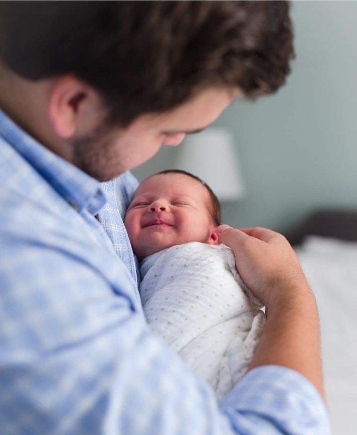 The disease in the early stages can only be diagnosed by an experienced ophthalmologist. Mild stages of retinopathy may leave behind minor changes that do not significantly affect vision. But when the 3rd threshold or 4th stage of the disease is reached, the child needs to be operated on urgently.
Optic nerve atrophy is a lesion of the nerve fibers that conduct visual signals from the eye to the visual centers of the cerebral cortex. The main reason is various lesions of the structures and the ventricular system of the brain. If the atrophy of the optic nerve is complete (which is rare), then vision may be completely absent. In the case of partial atrophy, visual acuity is determined by the degree and location of damage to the optic nerve. In case of atrophy of the optic nerves, stimulating functional treatment is used with the help of special devices, nootropic (improving metabolic processes in the brain) and vasodilating therapy.
Dynamic monitoring of the child
After the maternity hospital, parents should carefully monitor the development of their baby, paying attention to the formation of visual functions.
It is important that the first examination by an ophthalmologist is carried out in the first 3 months of a baby's life (during this period, most congenital diseases can be diagnosed in the early stages, which is the key to successful treatment). If there is no pathology at the initial examination, the next visit to the doctor is needed when the child is six months old (the maturation of the main structures of the eye responsible for the correct focusing of the image on the retina).
Primary examination is carried out according to the following algorithm:
Determination of sharpness (at 1 month - by the reaction of fixation on the object, at 2-3 months - by tracking a bright toy 15-20 cm in size on a light plain background, at 4-5 months - according to the tracking clarity up to a distance of 3-5 m) and fields of view (field of view - the maximum space inspected by one eye.). Fields of vision are determined approximately - the doctor moves the toy forward from behind the child's head until the baby's reaction to the object appears. At the same time, the appendages of the eye are examined: muscles, lacrimal ducts, eyelids (eye movements in different directions, patency of the lacrimal ducts, the fullness of opening and closing of the eyelids), as well as the optical media of the eye and fundus using an ophthalmoscope and a slit lamp (devices that send a slit-like or a round beam of light through the optical media of the eye).
The ophthalmologist also measures refraction using skiascopy (shadow test with optical rulers) or refractometry (a special device is used).
If visual acuity cannot be determined (the baby has a fuzzy fixation or tracking reaction), then a study of brain impulses in response to visual stimuli (visual evoked potential method) is performed. Based on its results, one can judge the presence of functional and structural lesions of the visual analyzer or a delay in its development.
At 6 months, at the examination by an ophthalmologist, in addition to the standard examination of the child, the dynamics of refraction of the eye is monitored, that is, the newly obtained and primary data of this study are compared. In most children at 6 months, refraction fluctuates between +1–+2.5 diopters. Sometimes at this age there may be a shift towards negative refraction, which indicates the baby's predisposition to the development of myopia. In this case, it is necessary to limit visual loads - remove small and close-hanging toys, focus on distant and moving objects. If myopia is detected more than 2 D, especially if, along with this, the child's visual acuity decreases and strabismus appears, vision correction with the help of glasses is prescribed as soon as possible. Glasses, if necessary, can be prescribed as early as 6 months (sometimes, with large degrees or severe asymmetry between the eyes, vision correction is used using contact lenses).
Even if in 6 months no pathology of the organ of vision was revealed at the preventive examination, in the future it is supposed to be examined by an ophthalmologist every six months, since during this time the refractive indices of the eyes may begin to change (myopia, astigmatism form), some genetic syndromes that occur with a sharp impairment of visual acuity.