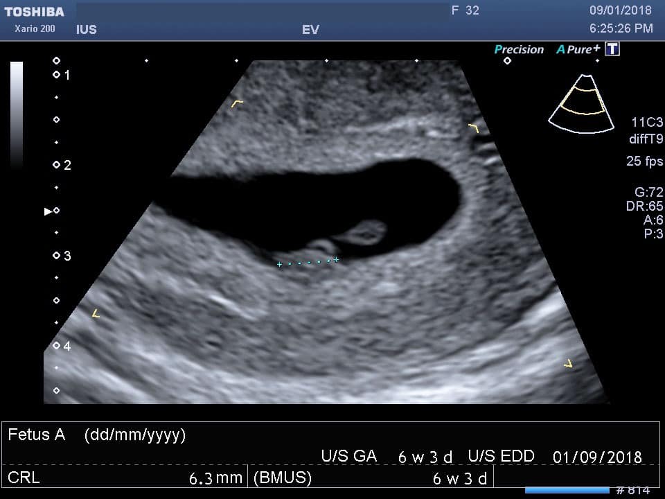
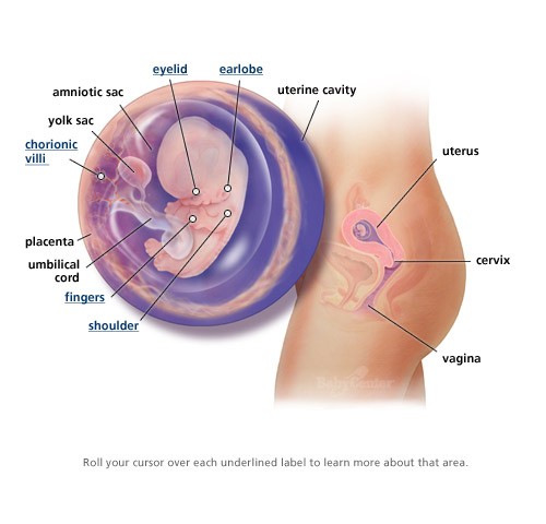
Scanning pregnancy 6 weeks
6 week Pregnancy Scan - | 6 week Ultrasound | 6 week Scan
At 6 weeks, you won’t, in general, be able to see much detail of your baby. The ultrasound scan, however, should be able to confirm the gestation age by measuring either the gestation sac or the foetal pole if visible. Sometimes but not always you will be able to see the baby’s heartbeat.
Most importantly the sonographer who will do your private ultrasound in London will be able to check that your baby is in the right place i.e. in the endometrial cavity and that you do not have an ectopic pregnancy.
Ectopic pregnancy is when the fertilized egg attaches itself outside the uterus with the most common location being the fallopian tube on the side where you ovulated from.
Everyone obviously is different and sometimes a follow-up ultrasound in a week to 10 days later might be necessary to give you more information.
Your baby at 6 weeks
How many mm is a 6-week-old foetus?
At 6 weeks, your baby should measure approximately 5-9mms in length.
Can you see the baby at 6 weeks?
6 weeks into your pregnancy is also the earliest time that you might be able to see the foetal pole and the foetal heartbeat.
Can you see the baby heartbeat at the 6-week scan?
The foetal heartbeat looks like two parallel lines flickering, and it is not always visible at the 6-week scan. The literature suggests that the foetal heartbeat should be around 90-110 beats per minute, but we have seen slower heartbeats with positive pregnancy outcomes.
What else can you see?
The yolk sac, a ring shape bright circle might also be visible. The yolk sac is where your baby is feeding on at this early stage in pregnancy.
Sometimes only the gestation sac is visible with no foetal pole or yolk sac, and you might be asked to come back in a week to 10 days. In most cases, this is because you might be earlier in your pregnancy than you think.
What is the earliest you can have a pregnancy scan?
The 6-week scan is the most common gestation age that a private ultrasound is performed.
We do not recommend a scan before the 6 weeks gestation unless you are worried about a miscarriage or an ectopic pregnancy, as at 5 weeks gestation you will possibly see the endometrium being thickened and echo bright and possibly a gestation sac. A 5-week baby scan however might help to find the cause for any early pregnancy pain or bleeding.
If you are more than 6 weeks you may also want to read more about the 7-week scan and the 8+ week scan.
What happens at a 6-week scan?
It is more likely that at 6 weeks gestation age you will need to have a transvaginal or internal ultrasound scan instead of a transabdominal scan (through the abdomen). This is because it is a very early stage and everything is still very small. The transvaginal scan probe will be able to get closer to the endometrium and produce a better clearer image of the pregnancy insitu.
Feeling nervous about having an ultrasound scan so early in your pregnancy is normal. Try to stay calm and prepare yourself for what may happen. Bringing with you your partner or a close family member for extra support might be a good idea.
We do allow another family member present in the scanning room during the private ultrasound even during Covid-19 pandemic.
How many scans will I have during pregnancy through the NHS?
You will have at least two baby scans during your pregnancy provided by the NHS:
- a 12-week dating scan and
- a 20-week anomaly scan.
The 12-week scan will provide confirmation and dating for your pregnancy. The 20-week scan will provide information about your baby’s growth and development.
About Pregnancy Scans
A pregnancy ultrasound scan is the same as a ‘normal’ scan, but it is being used to evaluate the overall health of your baby instead of looking at other organs such as gallbladder for gallstones or kidney for kidney stones. So in pregnancy, ultrasound scans are being used to visualise the baby, the placenta, the uterus and cervix and your ovaries.
Pregnancy ultrasound scans or prenatal ultrasounds are very common and being carried in any stage of the pregnancy.
If you have any questions, or you want to know more about our private ultrasound in London please get in touch with us, and we will do our best to answer.
At our private ultrasound clinic, we offer pregnancy scans from as early as 5-6 weeks gestation in times to suit you as well as private scans for men and women.
Who interprets the results of the early pregnancy scan and how do I get them?
A Sonographer, a Health Care Professional specifically trained to perform and understand the ultrasound images, will most likely perform your exam and provide you with a digital ultrasound report that you can take it to your doctor or keep it for your records.
About Ultrasound
Diagnostic Medical ultrasound scan or medical sonography as otherwise known is a painless imaging technique utilising sound waves to produce internal images of the body.
It is called ultrasound as the sound frequency being used is at the region of 1 to 20MHz. The human ear can’t hear these frequencies.
The sound waves are produced by the transducer or the probe as most commonly known. As they travel through the body they bounce back to the transducer due to various sound transmissions differences in tissues. The returning echoes are picked up by the probe and a powerful computer analyses the echoes and creates the 2d image on the screen.
There are various kinds of ultrasound scans that can be performed and each looks at different organs of the body such as tendons, muscles, joints, blood vessels, liver, kidneys, uterus and ovaries to confirm or exclude possible pathology.
Unlike CT and MRI, pregnancy ultrasound does not use radiation and therefore is pregnancy-friendly. It is also live and is ideal for musculoskeletal exams to evaluate moving joints.
You can read more about the history of ultrasound.
Other ultrasound scans related to pregnancy?
Some mothers to be will, unfortunately, get various complications during pregnancy such as high blood pressure, kidney infections and abnormal liver function tests. As ultrasound scans are pregnancy-friendly your doctor might refer you for an abdominal/liver scan or a kidney scan to check for anything that might explain your symptoms.
Although these ultrasound scans are not pregnancy scans, they are related to pregnancy.
In most cases, all the complications resolve after delivery, but like everything else related to your health and your baby’s health better safe than sorry.
What are ultrasound scans used for in pregnancy?
Depending on your stage of pregnancy, ultrasounds will be used to give you and your doctor or midwife answers about the health of your pregnancy.
First Trimester Ultrasounds
- Check that you are pregnant and that your baby has a heartbeat.
- Check if you have a singleton or twins
- Make sure that the pregnancy is not an ectopic located within the endometrial cavity and is not outside the womb such as in the fallopian tube.
- Look for the cause of any bleeding you might have.

- Date the pregnancy by measuring the crown-rump length of the foetal pole.
Second Trimester Ultrasounds
- Verify dates and growth
- Estimate the baby’s risk of Down’s syndrome by measuring the fluid at the back of your baby’s neck between about 10 weeks and 14 weeks
- Help with diagnostic tests by showing the position of the baby and placenta.
- Check your baby to see if all his organs are normal.
- Diagnose abnormalities
- Assess the amount of amniotic fluid and the location of the placenta.
- Evaluation of foetal well-being
Third-trimester Ultrasounds
- Make sure your baby is growing at the expected rate.
- Confirm if your baby is a boy or a girl.
Do you want to book your early pregnancy scan?
What You Can Expect to See
Your 6-Week Ultrasound: What You Can Expect to SeeMedically reviewed by Valinda Riggins Nwadike, MD, MPH — By Scott Frothingham on October 27, 2020
Excited and a bit scared are normal reactions when your doctor schedules you for a 6-week ultrasound. It’s exciting because you get to actually see what’s going on inside your body.
But it can also be a little anxiety-inducing, because you may know that such an early ultrasound isn’t always typical.
So what’s the reason for the early scan? And what can you expect to see?
Typically, the end of your first trimester (around 11 to 14 weeks) is when you have your first ultrasound in pregnancy.
But if your doctor wants you to have one at 6 weeks, they’ll tell you why. If not, ask them. It may be because you’ve had pregnancy complications or early pregnancy losses in the past. Or it may be due to your age or medical history.
In reality, there are many reasons your doctor may want an early scan. Typically, in this initial pregnancy ultrasound, your doctor wants to check on:
- Heartbeat. Often you can see a heartbeat after 5 weeks, although sometimes you’ll have to wait a little longer. Be prepared: This may be the first time you see your baby’s heartbeat, and it can be very emotional.

- Number. You might find out you’re having twins or higher-order multiples. (In the United States, the chance of having twins is about 3 percent.) Be aware, though, that sometimes 6 weeks is too early to tell.
- Location. The ultrasound can locate where the embryo is implanted. Your doctor wants to know if it’s high in the uterus or low. They also want to check that it’s in the uterus and not an ectopic pregnancy. It’s an ectopic pregnancy when a fertilized egg implants in a fallopian tube or elsewhere outside the uterus.
- Yolk sac. At this stage of your pregnancy, a yolk sac should be visible inside the gestational sac. It tends to look like a tiny balloon, and your doctor wants to see its size and shape, which are indicators of your pregnancy health.
Often it can be a challenge to find a heartbeat by ultrasound before you’ve reached the seventh week of pregnancy.
Also, it can be difficult to pinpoint the exact start of your pregnancy, so you might not actually be at the sixth week yet. If you go by the date of your last monthly period, keep in mind that you may have ovulated later in your cycle than you thought.
If the heartbeat can’t be picked up and you have no other symptoms, you’ll probably be scheduled for another ultrasound in a week.
Waiting for that next ultrasound can result in a stressful week. If you feel you need more support than what is being offered by your family and friends, talk about it with your doctor.
If you’re having your first pregnancy ultrasound at 6 weeks, there are some things you should be aware of. This is an exciting step, and being prepared can help you focus on the positive aspects.
- At 6 weeks, you’ll likely have a transvaginal ultrasound rather than the abdominal one you may be thinking of. Before 7 weeks, babies are often so small that the abdominal ultrasound may have trouble picking up the information the doctor wants. While the traditional abdominal ultrasound involves a wand (transducer) that’s placed on your belly, a transvaginal ultrasound involves a wand being inserted into your vagina.
 It shouldn’t hurt, but it may not be the most pleasant feeling in the world.
 - Your baby, at this stage, is only about a quarter of an inch long — so you might not see much detail. You have to wait until 11 to 12 weeks to get a 91 percent accurate identification of your baby’s biological sex, for example.
- The technician operating the ultrasound may not be permitted to answer many of your questions. Typically, the tech will get the results to your doctor for a follow-up visit (often right after the scan) where they’ll interpret the data for you in detail.
- The facility where you get your ultrasound may or may not be set up to give you a printout. If you want a picture, they may let you take a snapshot of the screen — so have your phone handy.
Share on PinterestX.Compagnion/Wikimedia Commons
6-week ultrasound showing gestational sac with yolk sac inside, which provides nutrients for a developing fetus early on in pregnancy. The fetal pole is a thickening on the edge of the yolk sac and the earliest sign of the developing fetus.
A pregnancy ultrasound uses sound waves to create a picture of your baby developing in your womb. There’s no radiation used.
According to the National Library of Medicine, ultrasounds are considered safe — there are no known risks — during all stages of pregnancy.
Prenatal care, such as medical checkups and screening tests, help keep you and your baby healthy. A 6-week ultrasound is a safe part of that process, providing important information to your doctor so they can offer you the best care.
Like many other aspects of your pregnancy, your first ultrasound is an exciting and potentially stressful part of your prenatal care. If possible, take a support person with you and try not to worry if you can’t see what you’re expecting — it may just be too early.
Last medically reviewed on October 27, 2020
- Parenthood
- Pregnancy
- 1st Trimester
Medically reviewed by Valinda Riggins Nwadike, MD, MPH — By Scott Frothingham on October 27, 2020
related stories
How Early Can You Hear Baby’s Heartbeat on Ultrasound and By Ear?
6 Weeks Pregnant: Symptoms, Tips, and More
When Does Your Baby Bump Start to Show?
What Is the Baking Soda Gender Test and Does It Work?
The Acupressure Points for Inducing Labor
Read this next
How Early Can You Hear Baby’s Heartbeat on Ultrasound and By Ear?
Medically reviewed by Valinda Riggins Nwadike, MD, MPH
You may be able to hear your baby’s heartbeat as early as 6 weeks past gestation if you have an early ultrasound.
 Hearing a baby’s heartbeat with the…
READ MORE
6 Weeks Pregnant: Symptoms, Tips, and More
Medically reviewed by Valinda Riggins Nwadike, MD, MPH
Although you won’t look pregnant yet, your body is going through many changes by week 6. Symptoms include nausea, constipation, and more.
READ MORE
When Does Your Baby Bump Start to Show?
Medically reviewed by Carolyn Kay, M.D.
One particularly common question, especially among people pregnant for the first time, is when do you start to show. We'll tell you what to expect in…
READ MORE
What Is the Baking Soda Gender Test and Does It Work?
Medically reviewed by Debra Rose Wilson, Ph.D., MSN, R.N., IBCLC, AHN-BC, CHT
Is the baking soda gender test an easy and accurate way to identify if you’re having a boy or girl?
READ MORE
The Acupressure Points for Inducing Labor
Medically reviewed by Debra Sullivan, Ph.
 D., MSN, R.N., CNE, COI
Are you pregnant and past your due date? Help induce labor naturally by pressing on these acupressure points along the body.
READ MORE
Support for the Non-Birthing Partner after Stillbirth
Medically reviewed by Matthew Boland, PhD
The grief that accompanies stillbirth or infant loss isn’t reserved for the birthing parent — partners also feel this loss deeply.
READ MORE
How to Offer Support After Infant Loss or Miscarriage: A Guide
Medically reviewed by Debra Rose Wilson, Ph.D., MSN, R.N., IBCLC, AHN-BC, CHT
We spoke with parents who offered suggestions for how to support them after miscarriage, infant loss, and stillbirth.
READ MORE
Preeclampsia and Pregnancy: Blood Pressure Drug May Help in Severe Cases
Researchers say the medication nifedipine was effective in helping pregnant women with preeclampsia avoid dangerously high blood pressure
READ MORE
Unexplained Recurrent Pregnancy Loss: My Journey to Baby Number 2
Healthline writer Ashley Marcin shares her story of multiple fetal losses and how they affected her and her family.

READ MORE
Stillbirth vs. Miscarriage: How They’re Similar and How They’re Different
Stillbirth and miscarriage are two terms that describe a painful event: pregnancy loss. Learn how they are different and how they are similar.
READ MORE
fetal ultrasound at the sixth week
- Fetal development by week
- Our apparatus
- other types...
- Pregnancy management
- Fetometry data at various times
The cost of ultrasound in the first trimester up to 10 weeks is 450 UAH. The price includes biometrics according to protocols, 3D/4D visualization.
The cost of ultrasound in the first trimester for a period of 10 weeks 6 days is 550 hryvnia. The price includes prenatal screening, biometric protocols, 3D/4D visualization.
The cost of comprehensive prenatal screening according to PRISCA (ultrasound + beta hCG + PAPP with calculation of individual risk of chromosomal pathologies (for example, Down syndrome or Edwards syndrome) - 1005 hryvnia.
At six weeks of pregnancy, the size of the embryo approaches the size of a pea. With ultrasound at six weeks pregnancy, the coccygeal-parietal size is measured, this is the length of the embryo.By this size, using ultrasound, you can clearly determine the gestational age, this is especially important if there are discrepancies with the menstrual gestational age.Continues to be visualized at Ultrasound at six weeks of pregnancy yolk sac next to the embryo and corpus luteum in the ovary at. If with ultrasound at five weeks of pregnancy there is still no reliable data on an increase in the size of the uterus, then with ultrasound at 6 weeks of pregnancy, the uterus increases in size. In standard ultrasound protocols at 6 weeks of gestation, the size of the uterus is usually not indicated, since these measurements are of no practical importance. Ultrasound at 6 weeks of gestation allows you to assess the presence of a developing pregnancy even with a general scan through the abdomen, but clearer data can be obtained with transvaginal ultrasound.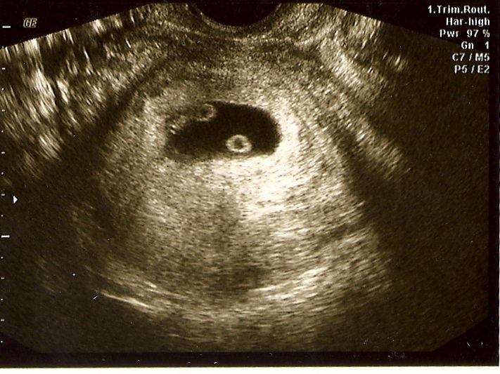
In addition to the above measurements and data, the gestational sac is measured during ultrasound at 6 weeks of gestation. The timing of pregnancy is more accurately calculated not by the average diameter of the fetal sac, but by the size of the embryo - its coccyx-parietal size (KTP). The cardiac contractility of the embryo must be assessed. It is by the presence of cardiac activity that a conclusion is made with ultrasound at six weeks of pregnancy about the development of pregnancy, about its progression.
If you could look into the uterus, at 6 weeks of pregnancy you can see the following. A fold of tissue with a protruding tubercle on top (head), which will develop the cheeks, jaws, and chin of the fetus. On the sides you can see two charming dimples - these are the emerging ear canals. Small protrusions on the front of the head are future eyes. The kidneys, liver and lungs begin organizing this week.
Outwardly, your body may not show any symptoms at all, but important changes are going on inside. You continue to feel some early pregnancy symptoms. You can mark 1 or 2 extra kilograms on the scale. This is due to the work of progesterone, which promotes fluid retention in the tissues. The external genitalia may acquire a barely noticeable cyanotic color. This is due to the increased blood supply to this area. Sometimes you may notice the presence of dizziness or fainting reactions when standing on your feet for a long time. Try to avoid such situations, be in the fresh air more often. The sense of smell is aggravated, you feel smells more acutely, some become definitely unbearable, capable of provoking nausea or vomiting. Try to avoid colliding with them.
Six weeks pregnant - it's time to go to the first appointment with an obstetrician-gynecologist and discuss the management of pregnancy from early to delivery.
read more: 7th week of pregnancy
- Hydrotubation (echohydrotubation): examination of the patency of the fallopian tubes (ultrasonic hysterosalpingoscopy)
- Transvaginal
- Ovaries
- Uterus
- Mammary glands
Female Ultrasound
- Duplex Scan
- Cerebral vessels
- Vessels of the neck (duplex angioscanning of the main arteries of the head)
- Veins of the lower extremities
Vascular ultrasound
- Transrectal (trusion): prostate
- Scrotum (testicles)
- Vessels of the penis
Male ultrasound
- Appendicitis
- Abdomen
- Gallbladder
- Stomach
- Intestines
- Bladder
- Soft tissue
- Pancreas
- Liver
- Kidney
- Joints
- Thyroid
- Echocardiography (ultrasound of the heart)
Ultrasound of organs
- Varicose veins: ultrasound diagnosis of varicose veins
- Hypertension: Ultrasound diagnosis of hypertension
- Thrombosis: ultrasound diagnosis of vein thrombosis
- Ultrasound diagnosis of chronic pancreatitis
- for kidney stones
- for cholecystitis
Ultrasound diagnostics of diseases
- Hip joints in newborns (if hip dysplasia is suspected)
Pediatric ultrasound
Early pregnancy ultrasound
In early pregnancy, ultrasound is done to identify a viable fetus in the uterine cavity, confirm the gestational age, rule out abnormalities in the fetus, or identify normal variants such as multiple pregnancies.
The initial sign of pregnancy is thickening of the endometrium, but ultrasound does not allow us to say what specifically caused this thickening.
Using a high-resolution transvaginal transducer, a 1 mm gestational sac is visualized in the uterine cavity 4 weeks and 2 days after the last menstrual period with a regular menstrual cycle.
If menstruation is delayed by 5-7 days or more (gestation period is 5 weeks), a fetal egg with a diameter of 6 mm should be clearly identified in the uterine cavity. It has a clear rounded shape with an indistinct light corolla along the periphery (hyperechoic rim - chorion). At the same time, the level of beta-hCG in the blood is 1000-1500 IU / l (see What is hCG?). At an hCG level of more than 1500 IU / l, the fetal egg in the uterine cavity should be clearly visualized.
With a lower level of hCG, the fetal egg in the uterine cavity may not be detected by transvaginal ultrasound. In a transabdominal study, the determination of a fetal egg in the uterine cavity is possible at a beta-hCG level of 3000-5000 IU / l.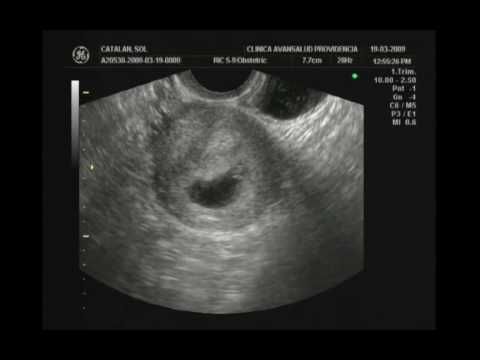
9-5 Transabdominal scan.
IMPORTANT: the gestational age cannot be accurately determined by the size of the ovum. Many tables on the Internet with the size of the fetal egg - determine the period very approximately (see table below).
From about 5.5 weeks on transvaginal ultrasound, an extraembryonic structure, the yolk sac (eng. yolk sac), begins to be visualized in the fetal egg. At the same time, the level of beta-hCG is approximately 7200 IU / l on average (see hCG norms during pregnancy).
Since the yolk sac is part of the embryonic structures, its detection makes it possible to distinguish a gestational sac from a simple accumulation of fluid in the uterine cavity between the layers of the endometrium, and in most cases, makes it possible to exclude an ectopic pregnancy. The frequency of ectopic pregnancy is 1-2 per 2000-3000 pregnancies. Its risk increases with the use of assisted reproductive technologies (ART). It is necessary to suspect an ectopic pregnancy when the hCG level is more than 1500 IU / L, and the fetal egg in the uterine cavity is not detected.
The yolk sac is determined. transvaginal scan.
From 6 weeks of gestation (sometimes a little earlier), an embryo, about 3 mm long, can be detected in the fetal egg. From the same period, most ultrasound devices allow you to determine the heartbeat of the embryo. If the heartbeat is not detected or indistinct when the length of the embryo (KTR) is 5 mm, a second ultrasound is indicated after a week. The absence of cardiac activity in this period is not necessarily a sign of fetal suffering or an undeveloped pregnancy.
Numerical values of heart rate in an embryo during uncomplicated pregnancy gradually increase from 110-130 beats/min at 6-8 weeks of pregnancy to 180 beats/min at 9-10 weeks.
The length of the embryo is measured from the head to the caudal end, and is designated under the term KTP (coccygeal-parietal size), in Eng. literature - CRL (Crown-Rump Length). It should be noted that the coccygeal-parietal size of the embryo is less subject to individual fluctuations than the average internal diameter of the fetal egg, and therefore, its use for determining the gestational age gives better results. The error in this case usually does not exceed ±3 days. With a clear visualization of the embryo, the gestational age is set depending on its length, and not on the size of the average internal diameter of the fetal egg (MID).
Accurate visualization is essential for correct measurement of the coccyx-parietal size of the embryo. In this case, one should strive to measure the maximum length of the embryo from its head end to the coccyx.
In the normal course of pregnancy, the diameter of the fetal egg increases by 1 mm per day. Lower growth rates are a poor prognostic sign. With a gestational age of 6-7 weeks, the diameter of the fetal egg should be about 30 mm.
Medvedev.
th percentile.
It should be emphasized that it is better to determine the gestational age by the length of the CTE before 12 weeks of pregnancy. At a later date, measurement of the biparietal diameter, head and abdomen circumference should be used.
Fig.3 Pregnancy 12 weeks 3 days.
The motor activity of the embryo is determined after 7 weeks of pregnancy. At first, these movements are very weak and isolated, hardly distinguishable during the study. Then, when differentiation into the head and pelvic ends of the embryo becomes possible, the movements resemble flexion and extension of the torso, then separate movements of the limbs appear.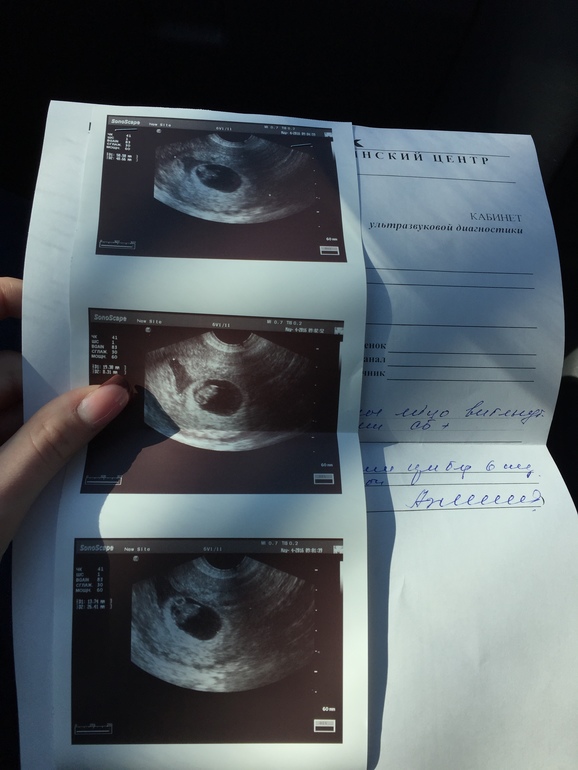 Since the episodes of the motor activity of the embryo are very short and are counted in seconds, and the periods of motor rest can be significant in time, the registration of the cardiac activity of the embryo is undoubtedly a more important criterion for assessing its vital activity.
The diagnosis of anembryony (empty gestational sac) is suspected if no yolk sac is detected in a 20 mm gestational sac. Or if a fetal egg with a diameter of more than 25 mm with a yolk sac does not contain an embryo. And also with a yolk sac size of 10 mm or more. In any case, if anembryony is suspected, all data obtained should be interpreted in favor of pregnancy, and the study should be repeated after 7 days.
A missed pregnancy should not be diagnosed if the gestational sac is less than 20 mm on ultrasound. With an embryo length of 5 mm or more, in most cases, the heartbeat should be clearly defined. If the embryo is less than 5 mm, the ultrasound should be repeated in a week. If, upon re-examination a week later, with KTP = 5-6 mm, cardiac activity is not determined, the pregnancy is not viable.