Baby heartbeat at 13 weeks
Fetal Doppler--Listening to Baby's Heartbeat
Written by R. Morgan Griffin
In this Article
- What Is a Fetal Doppler?
- How Early Will a Fetal Doppler Test Work?
- How the Fetal Doppler Test Is Done
- Fetal Doppler Test Results
- How Often Are Fetal Doppler Tests Done?
- Difference Between Fetal Doppler and Ultrasound
What Is a Fetal Doppler?
A fetal Doppler is a test that uses sound waves to check your baby’s heartbeat. It’s a type of ultrasound that uses a handheld device to detect changes in movement that are translated as sound.
How Early Will a Fetal Doppler Test Work?
Most women first hear their baby's heartbeat during a routine checkup that uses the fetal Doppler. Many ultrasound machines also allow the heartbeat to be heard even before it can be heard with a Doppler. Most women now get an ultrasound before 12 weeks.
A fetal Doppler test normally takes place during your second trimester (weeks 13 to 28 of pregnancy). Some manufacturers of at-home fetal Dopplers say you may be able to hear your baby’s heartbeat as early as 8-12 weeks of pregnancy. But professional sonographers say that they wouldn’t try to listen to a baby’s heartbeat before 13 weeks because your womb is in your pelvis during the first trimester and so the device won’t work correctly. Some at-home devices say not to use it before 16 weeks.
If you’re trying to hear your baby’s heartbeat at home, it may be best to first wait for your doctor to check it during one of your prenatal checkups. This is especially true if you’ve been pregnant for less than 12 weeks, to avoid undue concern. It’s a good idea to talk to your doctor first before buying an at home fetal Doppler. They can talk to you about the benefits and risks involved with using these devices.
How the Fetal Doppler Test Is Done
Clinical fetal Doppler test
If you go to your doctor’s office or clinic, you'll lie down and a technician will hold a small probe against your belly that makes sound waves. Technicians will ask you to stay still while this is going on. The sound waves will then be sent back to an amplifier where they can be heard. The procedure is safe and painless.
At-home fetal Doppler test
There are Dopplers that you can buy for use at home. But the FDA recommends parents not use these devices. There isn’t evidence that using them is harmful, but they haven’t been studied long term. The FDA notes that using them too much -- without medical supervision -- could pose risks to your baby’s development.
The bottom line is they should be used only if there is a medical need. The FDA does recognize that Dopplers can create bonding between parents and unborn babies, but it suggests parents can get this feeling from sessions in the doctor’s office.
Talk to your doctor before buying or using an at-home device.
Doing a fetal Doppler test at home may not run as smoothly as one done in your doctor’s office, either. You may need to watch some videos online to see how to do it correctly.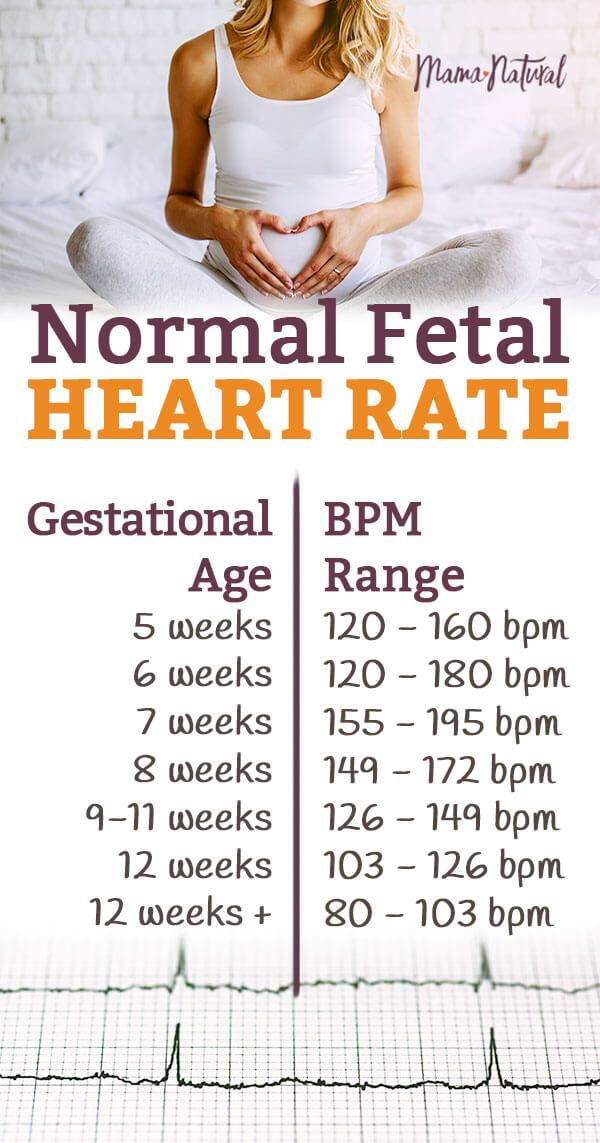 At-home devices vary from brand to brand, so it’s important to read any instructions and recommendations that come with the packaging. Some mothers find it’s easier to find the heartbeat when they do the test with a full bladder.
Fetal Doppler Test Results
Hearing your baby's heartbeat for the first time can be deeply moving. Keep in mind that a baby's heartbeat is much faster than an adult's.
If you're in your first trimester and you can't hear your baby's heartbeat, don't worry. Dopplers can't reliably detect a baby's heartbeat until 10-12 weeks. Your doctor may try again on your next visit. An ultrasound may give you better results.
A fetal heart rate is between 110-160 beats per minute and can vary by 5-25 beats per minute. Your baby’s heart rate can change based on conditions in your uterus. If the heartbeat is outside of the range, it could mean that your baby isn’t getting enough oxygen or has another issue.
If your doctor is concerned about your baby’s heartbeat, they may recommend a fetal echocardiogram. This is a safe, noninvasive test and gives a detailed picture of your baby’s heart. It can help see if and what kind of irregular heartbeat (or arrhythmia) your baby may have so it can get the right treatment.
How Often Are Fetal Doppler Tests Done?
Your doctor may use the Doppler often to listen to your baby's heartbeat during routine checkups, starting at about 8-10 weeks. Handheld Dopplers won’t work this early.
How often fetal Doppler tests are done can vary, but they may be part of routine second trimester checkups. Your doctor may even use them to monitor your baby’s heartbeat during labor and birth.
Difference Between Fetal Doppler and Ultrasound
Parents who use a fetal Doppler at home may not get as good results as they do from the ultrasound in the doctor’s office. For one thing, if you use it too early or place it in an incorrect position, you might hear no heartbeat and worry. Or you might hear a heartbeat (possibly your own or your pulse) and think everything is OK when it’s not. Also, the at-home fetal Doppler mainly picks up a baby’s heartbeat (although some companies are offering 3D and 4D ultrasounds, which allow you to see pictures and videos of your fetus.)
The most common medical ultrasound is the transvaginal ultrasound, where a wand (called a transducer) is placed in your vagina to send out sound waves and gather information. Another popular ultrasound, the transabdominal ultrasound, is done by moving the device over your stomach, similar to what you do in an at-home Doppler.
Professional ultrasounds are used for a variety of things. Ultrasound may confirm your pregnancy, determine how long you've been pregnant, hear a baby's heartbeat, as well as check for any abnormalities in your baby. It can also determine placenta and amniotic fluid levels and even what position your baby may be born in. A professional fetal Doppler can give details about a baby's blood flow.
Fetal Heartbeat By Week Chart
Let’s explore the fetal heartbeat by week chart. We also answer some of the commonly asked questions when it comes to your baby's heartbeat.
The first time you hear your baby’s heartbeat is a magical moment, whether you are a first-time parent or not.
But listening to a baby’s heartbeat is not just for parents to be excited over. It’s also essential for determining the baby’s health. A fetal heartbeat can reveal crucial information about your baby.
Keep reading to find out more about the fetal heartbeat by week chart for your baby.
Fetal Heart Monitoring
Fetal heart monitoring refers to measuring a fetus's heart rate and rhythm to ensure things are on track for a healthy pregnancy. Your doctor will record your baby’s heartbeat to see how they’re development is tracking.
Your baby’s average heart rate will vary based on fetal age and the conditions inside the uterus.
There are two fetal heart monitoring methods: external fetal heart monitoring and internal fetal heart monitoring.
External fetal monitoring is done using a device that detects a baby’s heartbeat through the mother’s abdomen. The most common device used for external fetal heart monitoring is a fetal doppler. This device detects and records the rate and rhythm of the baby’s heartbeat.
Internal fetal monitoring uses an electrode wire from the cervix to the baby’s head to monitor its heartbeat. This method is only done when the fluid-filled amniotic sac has broken. At this point, the cervix is open to allow the wire to enter the uterus.
Normal Fetal Heart Beat Chart by Week
Your baby's heartbeat will keep changing week by week, so it's essential to know the correct heart rate range depending on what week you are on.
The fetal heart rate starts at a slower rate but keeps increasing every day until stabilising at the 12th week. The average heart rate upon stabilisation is 120 to 160 beats per minute (bpm.)
Fetal heartbeat by week chart
Tracking a baby’s heart rate helps doctors determine the fetus’s age and growth. The general fetal age by weeks can be tracked by the heart rates below:
What is the normal fetal heart rate?
A baby's heart starts beating at five weeks. At this stage, the heartbeat would be described as galloping. It is faster than the average adult's heart rate. That said, your baby’s heart rate changes gradually throughout pregnancy. In the early stages of pregnancy, the fetal heart rate is between 110 to 180 beats per minute (bpm.)
Note that it is best to consult your doctor on the heart rate of your baby. If you feel anything is wrong, talk to your doctor.
When will the heartbeat start for a baby?
A baby’s heart will start beating at 5 to 6 weeks of pregnancy. This is around the time that the fetus begins to form from the embryo. A vaginal ultrasound will detect the fetus inside your womb at this stage.
From 6 to 7 weeks, your doctor can assess your baby's heartbeat to understand your pregnancy better.
If you plan to use fetal heart monitor dopplers to listen to your baby's heartbeat, then you may have to wait from weeks 9 to 28 weeks to hear a clear heartbeat.
Can I increase the fetal heart rate?
A normal fetal heart will beat at 110 to 160 bpm. Your baby's heart may beat at a rate of fewer than 110 beats per minute. An ultrasound exam by your doctor, midwife or OB-GYN will reveal more information about this.
A slow heart rate may quicken on its own. If you feel you need help increasing your baby's heartbeat, talk to your doctor.
Note that there is no scientific evidence to show that eating certain foods will increase the low fetal heart rate in the early pregnancy stages. You will have to talk to your doctor before making any dietary changes.
Does the heart rate vary for boys and girls?
No. The heart rate is not different in boys and girls. You might have heard the old wives tale that a heart rate over 140 bpm predicts a girl, and one lower than 140 bpm indicates a boy. There is no scientific basis for this fact.
You will start to clearly hear your baby's heartbeat at six weeks of pregnancy. Your baby hasn't developed enough to look like the fetus pictures you might have seen online at this stage.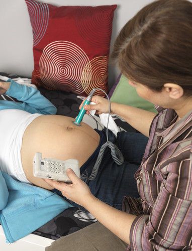 They look like a tadpole, better known as the fetal pole, and you won’t be able to know the gender yet.
The easiest way to find out the sex of your baby is via ultrasound. Your doctor will perform an ultrasound on you at 18 to 20 weeks. They will determine the sex of your baby by looking at the genitals on the baby’s image.
Is 170 bpm too high for a fetus?
Answering this question is tricky, as it depends on which stage of gestation your baby is in to know what a higher heart rate is.
When you are 9 to 10 weeks pregnant, your baby's heartbeat could average 175 bpm. At other stages, the baby's heartbeat could reduce.
If you have any concerns about your baby’s heart rate, talk to your doctor immediately. Please note that only your doctor can tell you what is expected and what is not for your baby.
How to monitor fetal heartbeat at home:
There are quite a few ways you can monitor your baby's heartbeat while at home. However, not all of them are as accurate as going to your doctor to get an ultrasound.
Here are some devices that you can use at home to listen to your baby’s heartbeat:
- Stethoscope: A stethoscope will detect your baby's heartbeat at 18 to 20 weeks.
- Fetal doppler: This is a device that allows you to listen to your baby's heartbeat by sending soundwaves through the mother's belly to the baby. A fetal doppler can be used as early as nine weeks to listen to your baby's heartbeat.
Also, note that you should not substitute at-home fetal monitoring with a real doctor’s appointment.
Final Thoughts
If you have any questions or concerns about the fetal heartbeat by week chart, talk to your doctor or OB-GYN. And remember that an at-home fetal doppler shouldn’t replace the appointments you have with medical professionals throughout your pregnancy. A fetal doppler is a tool for you to connect with your baby.
Sign up for our newsletter! We share top tips about fetal dopplers and pregnancy, so you stay informed and up to date.
13-16 weeks of pregnancy
Thirteenth week for a baby
The beginning of the week is characterized by a fetus length of 6-7 cm, and by its completion it already reaches 10 cm, the weight of the unborn child at the moment is 20-30 grams.
This week marks the start of the second trimester of pregnancy. The fetus grows intensively, its legs and arms lengthen. There is also a change in the proportions of the body, the head no longer looks as big as it used to be. The future baby already knows how to reach his mouth with his finger and sucks it, which is often seen by specialists during ultrasound diagnostics. Thus, the sucking reflex, which is so important for the baby in the first time after birth, finds its manifestation. nine0005
Intense muscle growth occurs, especially in the legs. The future baby becomes more active, his movements are now smoother. At this time, the woman is not able to feel the child, because he, being in the uterus, floats freely and does not actually come into contact with its walls. In the fetus at this stage, the formation of the rudiments of all milk teeth ends. The rudiments are located in the mucosa of both jaws.
The gastrointestinal tract is also in the stage of active growth and development. Fitting into loops, the intestine completely fills the abdominal cavity. On the inner surface of the intestine, its mucous membrane, villi are formed, which should cover the entire internal area. These villi after birth will help the baby absorb all the nutrients from the food in the intestinal cavity. The wave-like movements that the fetal intestine makes help it push through the amniotic fluid, which the unborn baby constantly swallows. These waters do not contain useful substances, they only help the intestines to train and form the necessary muscular system. nine0005
The thirteenth week for the expectant mother
A woman at the thirteenth week begins to feel much better, she finally manages to feel only positive emotions associated with the expectation of the baby. Malaise, toxicosis and the desire to sleep remain in the past. The emotional background also undergoes changes. A woman becomes calmer and more peaceful, frequent mood swings and irritability also remain in the past. This is due to the end of critical periods of pregnancy and a more stable hormonal background. nine0005
For most expectant mothers, this period is associated with a change in the shape of the abdomen. Such changes are not yet noticeable to others, but the woman herself clearly sees the roundness that has arisen, feels discomfort when wearing ordinary things. It's time to pay attention to the selection of special clothes that can create comfortable conditions for a woman and a future baby.
Fourteenth week for the baby
At this stage, the weight of the fetus is 40-45 grams, its length is 13 cm.
The distinct formation of the face leads to a change in the appearance of the fetus. The bridge of the nose becomes more pronounced, the eyebrows are outlined, the cheeks are rounded. The child moves, he feels himself on the tummy and cheeks, holds on to the umbilical cord. The embryonic fluff, which covers the entire body, is tightly attached to the skin of the fetus. This gun has a protective function - it retains a special lubricant that will allow the baby to easily pass through the birth canal during the birth process. Over time, the almost transparent birth fluff will be replaced by thicker hairs. nine0005
Important processes take place in the respiratory organs of the fetus precisely at the fourteenth week. Also, muscle tissues develop not only of the motor system, but also of the respiratory system. The fetus begins to imitate breathing with its movements. Such training is of great importance for the future baby, since he must take his first breath immediately after birth. His respiratory organs, under the influence of signals from the brain, should open up and begin to fully function. At this time, there is a partial opening of the glottis, so amniotic fluid penetrates into the respiratory organs on inspiration. Then there is an intense exhalation, and they are pushed out of the body of the unborn child. Rinsing with amniotic fluid of the lung tissues contributes to their proper maturation. nine0005
There are changes in the structure of the genital organs. In boys, the intensive formation of the prostate begins, in girls, the ovaries move to the pelvic area, which were previously located in the abdominal cavity. There is a differentiation of the external genitalia, but when examining ultrasound at this stage, it is not always possible to establish the sex of the unborn child.
The organs of the endocrine system also develop. An important event is the start of the pancreas. Insulin, an important hormone that regulates glucose levels in the body, also begins to be produced. This week, the pituitary gland begins to function, regulating the work of all the glands of the body, responsible for their coherence, interaction, as well as the process of growth of the fetus and child in the future.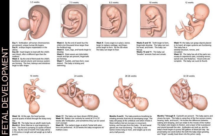 The pituitary gland is located in the most protected part - surrounded by the bones of the skull, in the thickness of the brain. nine0005
Fourteenth week for the expectant mother
A woman's uterus grows intensively, it becomes possible to independently palpate through the anterior abdominal wall its uppermost part - the bottom, which will be 10-15 cm above the pubis. It's time for a woman to purchase special care products that will help nourish her skin and make her able to calmly endure the upcoming changes.
Stretch marks on the skin, which occur in many women, are the result of microcracks, i.e. ruptures of connective fibers. The elasticity and firmness of the skin are significantly reduced, so even a small increase in weight can cause microcracks. There is a redistribution of subcutaneous fat, swelling may occur, so special care is simply necessary. Creams, lotions and other products for pregnant women are characterized by nutritionally safe formulations, they will help the skin restore elasticity and firmness, and prevent the appearance of stretch marks. Even if striae occur due to excessive weight gain, their number will be minimal. nine0005
Fifteenth week for a baby
At the end of the fifteenth week, the weight of the fetus is already about 50-70 grams, its length is 14-15 cm. The limbs grow, they can outstrip the size of the head due to the intensive development of the bones of the legs and arms. The future baby begins to actively bend his fingers and elbows, a unique skin pattern appears on the palms and fingers, and the process of nail formation begins.
At this stage, the cardiovascular system is being improved. The number of heart beats per minute is twice the mother's and is 140-160. A tiny heart pumps 20 liters of blood per day, contributing to the intensive development of the body. The fetus actively grows veins and arteries, there is a general improvement of the circulatory system. Own systems of veins and arteries are formed in each internal organ - kidneys, lungs, heart, brain, etc. The fetus acquires an unusual red color instead of pink, because it has very thin skin, and the circulatory system, developing, affects its color. nine0005
The nervous system at this time is undergoing important stages of formation. The mass of the brain increases, its furrows and convolutions increase. All muscles, internal organs and bones are braided with nerve fibers. A strong and stable connection is being established between the central nervous system and the peripheral one. Impulses begin to flow in both directions: from the brain to the organs and back. An important system of "reverse response" is emerging for the human body.
If until the fifteenth week red blood cells (erythrocytes) were produced by the gall sac and liver, then after this stage, this function begins to be performed by the red bone marrow located inside the bones. nine0005
It is already possible to determine the blood type and Rh factor of the unborn baby. The laying of the group and Rhesus occurs at the time of conception of the baby, but this information is realized only at the fifteenth week. There is a formation of special antigen proteins that appear on the surface of erythrocytes.
Fifteenth week for a future mother
A woman's belly grows quite smoothly, it increases gradually and does not cause discomfort in everyday life. The bottom of the uterus is already palpable through the anterior abdominal cavity at a height of 15-20 cm from the pubis. nine0005
Intensive production of melanin pigment occurs in the body, which is deposited in the skin. This process is caused by a hormonal background and causes the appearance of age spots on the surface of the skin, which can form in completely different places. Often a dark stripe appears on the abdomen, which has a brown tint and goes from the navel to the pubis. Rarely, the cause of increased melanin synthesis is a lack of vitamins in the body.
Brown spots should appear without any sensation. If their occurrence is accompanied by swelling, redness or discomfort, you should see a doctor. Such reactions may be the result of allergies or dermatosis of pregnant women - a common skin disease. Pigment spots can have a different shade - from light beige to dark brown. Almost always, such manifestations after the appearance of the baby disappear on their own. The use of whitening creams during pregnancy is possible only after consulting a doctor, because they may be unsafe for the fetus. nine0005
The sixteenth week for the baby
The formation of mimic muscles in the fetus takes place, he begins to develop them, which is expressed in the frown, opening and closing of the mouth. The fetus gets the opportunity to open its eyes for the first time, which up to this point were tightly closed for centuries, it begins to learn to blink. In place of eyelashes and eyebrows, you can see thin fluffy hairs. If the auricles before this stage were located in the wrong place, closer to the neck, now they are already placed as in an adult. Although there is a pronounced reaction of the fetus to loud sounds, at this stage, his middle ear is not yet able to hear. The reaction is due to such a way of perception as bone conduction. It turns out that the fetus hears through the dense parts of the body (bones).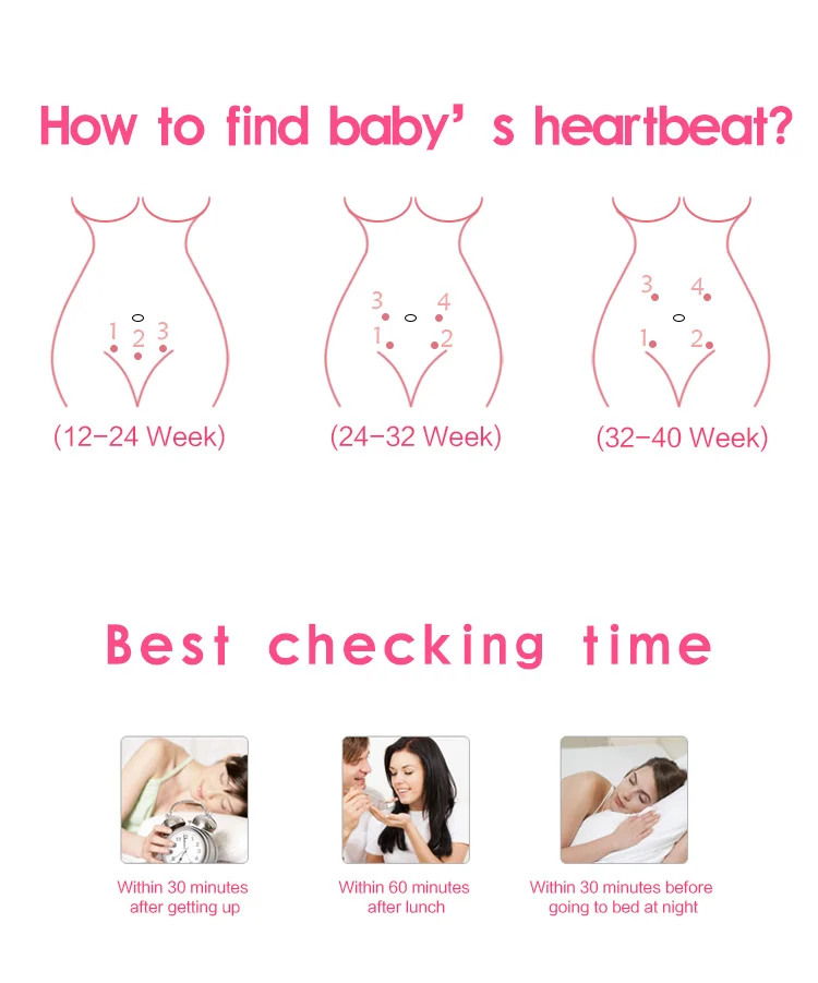 nine0005
The process of formation of all joints and bones has already been completed in the future baby. The process of ossification, i.e., compaction of bone tissue, will continue not only during the entire remaining period, but also after birth, almost until puberty.
With an ultrasound examination, it is already possible to reliably determine the sex of the child, since his external genitalia are fully formed. It is at this time or a little later (depending on the time of the appointed diagnosis) that the mother gets the opportunity to find out who will be born to her. nine0005
Kidneys work intensively in the fetus. Now they begin to perform a partially excretory function, slightly reducing the load that lies on the placenta. The urinary canal is formed, the fetus swallows 300-500 mm of amniotic fluid daily, they pass through its intestines and are excreted by the kidneys. Urination is performed in small portions every hour, urine enters the amniotic fluid.
Sixteenth week for the expectant mother
At this stage, the woman feels a significant improvement in well-being. Gradual weight gain begins, appetite normalizes. The norm at the moment is a set of 2.5-3 kg. nine0005
At the moment there is an active growth of the future baby, and a significant accumulation of amniotic fluid. Both of these factors affect the growth of the abdomen, its increasing volumes do not yet cause discomfort. However, it is time to make changes in your habits, for example, change the position of the body during sleep. Sleeping on your back is just as unindicated as sleeping on your stomach. When sleeping on the back, the uterus can put pressure on large veins, which will cause a violation of the outflow of blood, provoke the occurrence of swelling, convulsions and varicose veins. The most comfortable position for sleeping is the position on the side, in which both the unborn child and the mother will feel as comfortable as possible. nine0005
Ultrasound to determine the fetal heartbeat (6 - 32 weeks) in Chelyabinsk
Description
In the clinic in Chelyabinsk, you can sign up for an ultrasound scan with subsequent interpretation of the results: modern equipment allows you to record the child's heart rate, its frequency, the work of the heart muscle and ventricles.
When can a child's heart rate be recorded?
Transabdominal ultrasound allows you to fix heart beats at 6-7 weeks of gestation. Specialists continue to monitor the rhythm throughout pregnancy in order to notice deviations in time. nine0005
Using a vaginal probe, you can determine the heartbeat as early as the sixth week, but this method of examination can only be carried out in the early stages of pregnancy.
Norm and pathology
Let's give the rate of the pulse in the embryo in time:
● 6-8 weeks. The pulse only appears, the norm is 110-130 beats. A pulse below 85 beats per minute is a serious deviation.
● 9-10 weeks. The pulse rate develops from 170 to 190 beats per minute. Pathology is considered to be a more frequent heartbeat, a pulse of more than 200 beats.
● At 11-40 weeks, the indicators range from 140 to 160 beats. With pathology, the heartbeat is poorly distinguishable or completely absent.
If, during the examination of a fetus less than 8 mm in size, the doctor does not record heart contractions, an undeveloped pregnancy will be diagnosed.
In some cases, the heartbeat is detected at a later date. This is not considered a serious deviation. Often it is impossible to determine the exact date of conception due to an irregular menstrual cycle. Sometimes the individual characteristics of the organism do not allow to determine the exact exact period even after an ultrasound examination. nine0005
How is the examination carried out?
A transabdominal ultrasound is prescribed to check the heartbeat. The procedure does not require preliminary preparation. A special gel is applied to the patient's abdomen, which enhances contact with the sensor. After that, the doctor drives the sensor over the skin, while the image of the uterus and fetus is displayed on the monitor. The heartbeats of the embryo are also recorded. With the help of ultrasound, you can determine the following:
● Pulse rate (it should be well differentiated). nine0005
● Location of the heart (the organ is located on the left side, occupies at least one third of the chest).